Review
. 2016 Nov 17;167(5):1170-1187.
doi: 10.1016/j.cell.2016.09.018.

Ever-Changing Landscapes: Transcriptional Enhancers in Development and Evolution

Affiliations
Review

Ever-Changing Landscapes: Transcriptional Enhancers in Development and Evolution

Hannah K Long et al. Cell. .

Abstract

A class of cis-regulatory elements, called enhancers, play a central role in orchestrating spatiotemporally precise gene-expression programs during development. Consequently, divergence in enhancer sequence and activity is thought to be an important mediator of inter- and intra-species phenotypic variation. Here, we give an overview of emerging principles of enhancer function, current models of enhancer architecture, genomic substrates from which enhancers emerge during evolution, and the influence of three-dimensional genome organization on long-range gene regulation. We discuss intricate relationships between distinct elements within complex regulatory landscapes and consider their potential impact on specificity and robustness of transcriptional regulation.


Figures

Figure 1
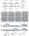
Figure 1. Nucleosome eviction, enhancer grammar and models of enhancer architecture
(A) TF mechanisms for overcoming energetic barrier to nucleosomal eviction and underlying motif requirements. (B) Parameters of motif grammar at enhancers. (C) Models of enhancer architecture and their primary mechanism of TF binding, flexibility of motif organization, and selective constraint across evolution. (D) Coactivator binding and chromatin signatures at active enhancers. Presence of cell-type specific and broadly-expressed TFs, RNA Polymerase II (RNAPII) and associated enhancer RNAs (eRNAs), coactivator complexes such as Mediator (Med), nucleosome remodeling complexes (NRCs), histone acetyltransferases (CBP/p300) and methyltransferases (MLL3/4) at tissue-specific enhancer elements is schematically depicted for a brain (top) or limb (bottom) specific enhancer. Select modifications of neighboring nucleosomes associated with active enhancer states are highlighted. An overlapping set of protein complexes and modifications is also present at promoters, with the distinction that enhancers have high ratio of H3K4me1 to H3K4me3, and the reverse is true at promoters. Both active enhancers and promoters are characterized by DNA hypomethylation, with the methylation status of enhancers being more dynamic and tracking with their cell-type specificity.
Figure 2
Figure 2. The birth and death of enhancers during evolution
(A) Following whole genome or local duplication events, enhancers and associated genes can become duplicated (left), and subsequently undergo: (i) loss of enhancer function and sub-functionalization of associated gene activity, (ii) enhancer repurposing with novel tissue or developmental stage expression for the duplicated gene or (iii) reduction in enhancer activity and gene dosage sharing between alleles. (B) Local duplication events can lead to increased enhancer copy number. (C) Novel enhancer activity can emerge from ancestral DNA through genetic drift and spontaneous appearance of transcription factor binding sites (TFBSs) and protoenhancer activity. (D) Enhancer elements can be exapted from transposable elements. For example, following endogenization and unequal homologous recombination, the long terminal repeats (LTRs) of endogenous retroviral (ERV) elements can gain tissue-specific regulatory activity through accumulation of mutations and emergence of TFBSs. (E) Enhancer activity can be transferred to a new gene target for example through a genomic inversion event.
Figure 3
Figure 3. Organization of chromatin into topologically associated domains
(A) Hi-C or 5C heatmaps visualize three-dimensional interactions, or compartmentalization of chromosomes into topologically associated domains (TADs), visible as triangular blocks of increased interaction frequencies. (B) TAD boundaries restrict the influence of regulatory elements to genes within a given TAD, and limit the spread of chromatin modifications. The boundary regions between TADs have been observed to be associated with CTCF binding sites, housekeeping genes, SINE elements and tRNA genes. (C) A model of chromosome folding corresponding to (B), with convergent CTCF sites and cohesin depicted at loop anchors. (D) Correspondence between TADs and cytological bands on polytene chromosomes, and TAD boundaries with decondensed interbands. (E) During mitosis TAD structure is lost.
Figure 4
Figure 4. Topologically associated domains define discrete units of gene regulation
(A) TAD boundaries are highly conserved across mammalian evolution, therefore structural changes tend to occur at TAD boundaries, for example (i) insertion of a new TAD or (ii) chromosomal breaks. (B) Disruption of a TAD boundary (red, dashed box) can impact gene expression. (i) Boundary deletion fuses two adjacent TADs facilitating de novo regulation of genes from one TAD by the enhancers in another. (ii) Boundary inversion can translocate genes or enhancers into an adjacent TAD where they are then incorporated into the regulatory environment of the new TAD. (iii) TAD boundary duplication can create a new TAD and potentially expose any duplicated genes to a novel regulatory environment. (C) CTCF binding sites can be DNA methylation sensitive, therefore aberrant DNA methylation can abrogate CTCF binding, causing TAD boundary defects and misregulation of gene expression. (D) Reporter constructs transposed into different regions of a TAD containing a developmental enhancer, but not into genomic regions outside the TAD, can recapitulate gene expression pattern of the endogenous gene (Gene X) controlled by this enhancer.
Figure 5
Figure 5. Regulatory crosstalk between enhancers operating within the same cis-regulatory landscape
(A-E) Potential modes of enhancer relationships and their effect on transcriptional output under wildtype or mutant conditions.

Similar articles

Cited by

References

    1. Abe N, Dror I, Yang L, Slattery M, Zhou T, Bussemaker HJ, Rohs R, Mann RS. Deconvolving the Recognition of DNA Shape from Sequence. Cell. 2015;161:307–318. - PMC - PubMed
    1. Agoglia RM, Fraser HB. Disentangling sources of selection on exonic transcriptional enhancers. Mol. Biol. Evol. 2016;33:585–590. - PMC - PubMed
    1. Allan CM, Walker D, Taylor JM. Evolutionary duplication of a hepatic control region in the human apolipoprotein E gene locus. Identification of a second region that confers high level and liver-specific expression of the human apolipoprotein E gene in transgenic mice. J. Biol. Chem. 1995;270:26278–26281. - PubMed
    1. Arnold CD, Gerlach D, Spies D, Matts JA, Sytnikova YA, Pagani M, Lau NC, Stark A. Quantitative genome-wide enhancer activity maps for five Drosophila species show functional enhancer conservation and turnover during cis-regulatory evolution. Nat Genet. 2014;46:685–692. - PMC - PubMed
    1. Arnosti DN, Kulkarni MM. Transcriptional enhancers: Intelligent enhanceosomes or flexible billboards? J. Cell. Biochem. 2005;94:890–898. - PubMed

Publication types