. 2016 Nov 17;167(5):1252-1263.e10.
doi: 10.1016/j.cell.2016.10.053.

The C. elegans Taste Receptor Homolog LITE-1 Is a Photoreceptor

Affiliations

The C. elegans Taste Receptor Homolog LITE-1 Is a Photoreceptor

Jianke Gong et al. Cell. .

Erratum in

Abstract

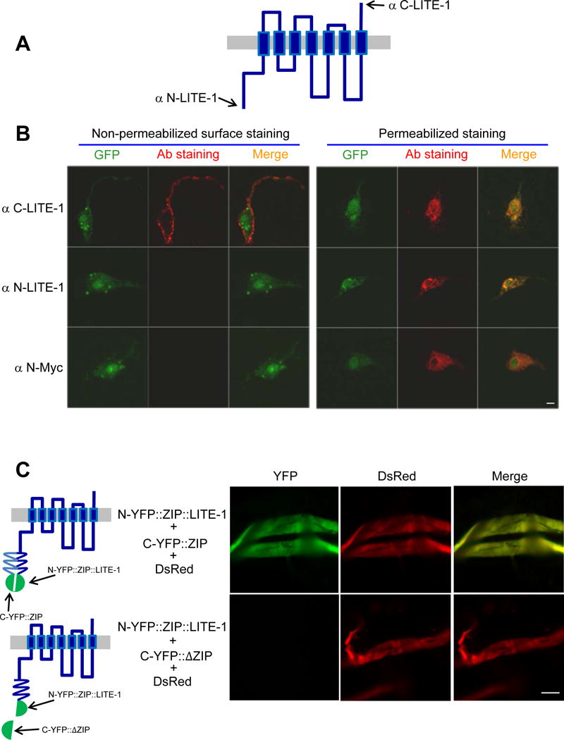
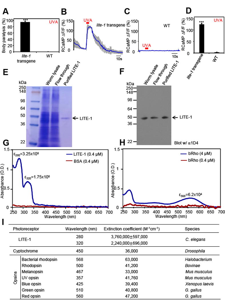
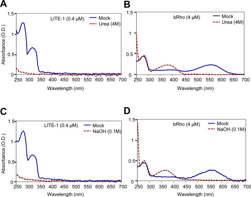
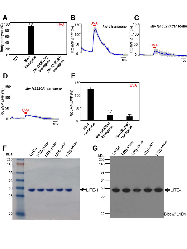
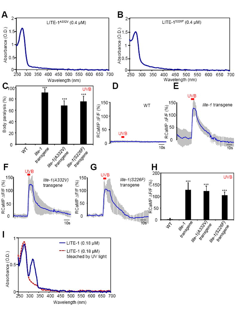
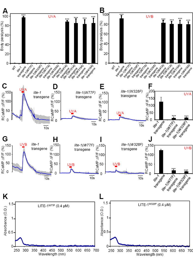
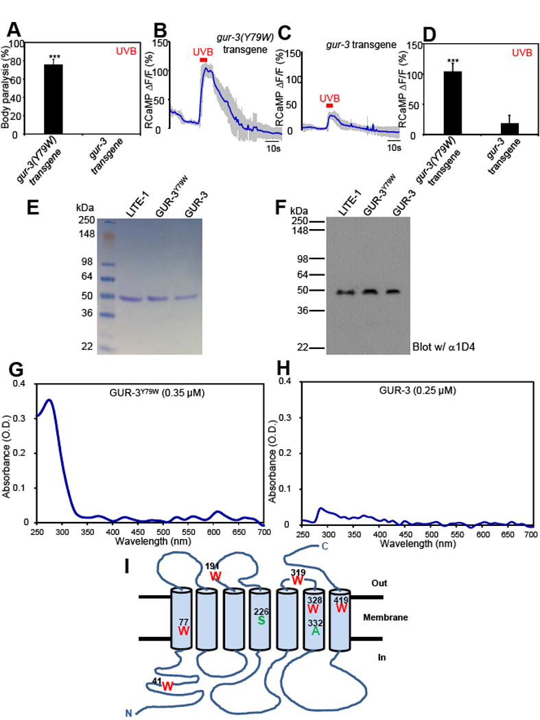
Many animal tissues/cells are photosensitive, yet only two types of photoreceptors (i.e., opsins and cryptochromes) have been discovered in metazoans. The question arises as to whether unknown types of photoreceptors exist in the animal kingdom. LITE-1, a seven-transmembrane gustatory receptor (GR) homolog, mediates UV-light-induced avoidance behavior in C. elegans. However, it is not known whether LITE-1 functions as a chemoreceptor or photoreceptor. Here, we show that LITE-1 directly absorbs both UVA and UVB light with an extinction coefficient 10-100 times that of opsins and cryptochromes, indicating that LITE-1 is highly efficient in capturing photons. Unlike typical photoreceptors employing a prosthetic chromophore to capture photons, LITE-1 strictly depends on its protein conformation for photon absorption. We have further identified two tryptophan residues critical for LITE-1 function. Interestingly, unlike GPCRs, LITE-1 adopts a reversed membrane topology. Thus, LITE-1, a taste receptor homolog, represents a distinct type of photoreceptor in the animal kingdom.

Keywords: chemosensation; chemosensory; neuron; photopigment; photosensation; photosensory.


Figures

Figure 1
Figure 1. LITE-1 adopts an unusual membrane topology with its C-terminus facing extracellularly and N-terminus located intracellularly
(A) A schematic of LITE-1 membrane topology. Antibodies were raised against the N-terminal (αN-LITE-1) and C-terminal (αC-LITE-1) peptide (15 aa) of the LITE-1 long isoform. (B) LITE-1 displays a distinct membrane topology with its C-terminus facing extracelluarly and it N-terminus located in the cytoplasm. Shown are confocal images from immunofluorescence staining. LITE-1 (long form) was co-expressed with GFP as a transgene in muscles under the myo-3 promoter. Staining was performed on primary cultured cells under non-permeabilizing conditions for surface staining or under permeablizing conditions to stain the entire cell. αN-LITE-1 and αC-LITE-1 detects the N- and C-terminal end of LITE-1, respectively. αN-Myc stains the Myc tag fused to the N-terminal end of LITE-1. See Figure S1 for controls. Scale bar: 2 μm. (C) BiFC images showing that the N-terminus of LITE-1 is located in the cytoplasm. Shown on the left are schematics describing the design of the BiFC approach. Shown on the right are fluorescence images. N-YFP∷ZIP∷LITE-1 was expressed as a transgene in muscles using the myo-3 promoter. C-YFP∷ZIP (or C-YFP∷ΔZIP that lacks a zip domain) and DsRed were co-expressed as a separate transgene in muscles using the same promoter. Two transgenes were crossed together to examine reconstitution of YFP fluorescence in muscles. Only if the N-terminus of LITE-1 is located intracellularly would one be able to detect YFP fluorescence. Muscles were acutely dissected out from transgenic worms using a protocol described previously (Liu et al., 2013). Scale bar: 100 μm. Also see Figure S1
Figure 2
Figure 2. LITE-1 absorbs UVA and UVB light, and ectopic expression of LITE-1 confers photo-sensitivity to photo-insensitive cells
(A) Transgenic expression of LITE-1 in muscle cells confers photosensitivity shown by behavioral assays. LITE-1 was expressed as a transgene in muscle cells under the myo-3 promoter. WT (wild-type) and LITE-1 transgenic worms were exposed to a 20 sec pulse of UVA light (350±20 nm, 0.8 mW/mm2). Animals showing muscle contraction-induced paralysis during light illumination were scored positive. n=20. Error bars: SEM. ***p<0.00001 (ANOVA with Bonferroni test). (B–D) Transgenic expression of LITE-1 in muscle cells confers photosensitivity shown by calcium imaging. RCaMP was expressed as a transgene in muscle cells under the myo-3 promoter. A 5 sec pulse of UVA light (340±20 nm, 0.7 mW/mm2) was applied to muscles to elicit calcium transients. Shades along the traces in (B) and (C) represent SEM. (D) Bar graph. n≥7. *p<0.0001 (t test). (E–F) Purification of LITE-1. Worm lysate, flow through, and purified LITE-1 were loaded. Shown in (E) is an SDS-PAGE gel stained with coomassie blue. Shown in (F) is a Western blot probed with anti-1D4 that recognizes the 1D4 tag attached to the C-terminal end of LITE-1. The amount of each sample loaded in (F) was 1/10 of that in (E). Samples for SDS-PAGE and Western were prepared at room temperature under non-reducing conditions (free of β-ME and DTT) to avoid aggregation of LITE-1. (G) LITE-1 shows strong absorption of UVA and UVB light while BSA does not. The same concentration of purified LITE-1 and BSA (0.4 μM) was subjected to UV-visible spectrophotometric analysis. The extinction coefficient (ε) for both peaks of LITE-1 was noted. Unit: M−1cm−1. Note: these numbers only represent the LITE-1 sample shown here and those in Figure 3, as they were from the same batch of purification. See (I) for averaged data for LITE-1 from different batches of purification. (H) Bacterial rhodopsin (bRho) shows much weaker absorption of light compared to LITE-1. The results from low and high concentrations of bRho were shown. bRho was purchased from Sigma. (I) LITE-1 is far more efficient in photon absorption than cryptochromes and opsins. The extinction coefficients for LITE-1 were averaged from samples from seven independent purifications. “±” represents SEM. The numbers for cryptochromes and opsins were from published literature: cryptochrome (Thompson and Sancar, 2002), bacterial rhodopsin (Oesterhelt and Hess, 1973), rhodopsin (Okano et al., 1992), melanopsin (Matsuyama et al., 2012), UV opsin (Insinna et al., 2012), blue opsin (Vought et al., 1999), green opsin, and red opsin (Kolesnikov et al., 2014). Also see Figure S2–3 and Supplemental movie 1–2.
Figure 3
Figure 3. Photoabsorption by LITE-1 relies on its conformation
(A) Denaturing LITE-1 with urea completely abolishes its photoabsorption. Shown are spectral data for mock- and urea-treated LITE-1. LITE-1 was treated with urea (4 M) for 5 min at room temperature prior to spectral analysis. (B) Denaturing bacterial rhodopsin (bRho) with urea does not eliminate its photoabsorption. Urea treatment shifts bRho’s 568 nm absorbance peak to 370 nm. bRho was treated with urea (4 M) for 5 min at room temperature prior to spectral analysis. (C) Denaturing LITE-1 with NaOH completely abolishes its photoabsorption. LITE-1 was treated with NaOH (0.1 M) for 5 min at room temperature prior to spectral analysis. (D) Denaturing bacterial rhodopsin (bRho) with NaOH does not eliminate its photoabsorption. NaOH treatment shifts bRho’s 568 nm absorbance peak to 370 nm. bRho was treated with NaOH (0.1 M) for 5 min at room temperature prior to spectral analysis. LITE-1 concentration: 0.4 μM. bRho concentration: 4 μM. Also see Figure S4.
Figure 4
Figure 4. Residues S226 and A332 in LITE-1 are critical for its sensitivity to UVA light in vivo
(A) S226F and A332V mutations disrupt the function of LITE-1 in vivo shown by behavioral assays. LITE-1 harboring S226F or A332V was expressed as a transgene in muscles under the myo-3 promoter. WT (wild-type) and transgenic worms were exposed to a 20 sec pulse of UVA light (350±20 nm, 0.8 mW/mm2). Animals showing muscle contraction-induced paralysis during light illumination were scored positive. Some genotypes had all data points as zero, and thus no statistical analysis was performed on them. n=20. Error bars: SEM. ***p<0.00001 (ANOVA with Bonferroni test). (B–E) S226F and A332V mutations disrupt the function of LITE-1 in vivo shown by calcium imaging. The experiments were done as described in Figure 2B. A 5 sec pulse of UVA light (340±20 nm, 0.7 mW/mm2) was applied to muscles to elicit calcium transients. Shades along the traces in (B–D) represent SEM. (E) Bar graph. n≥7. ***p<0.00001 (ANOVA with Bonferroni test). (F–G) Purification of mutant forms LITE-1. Shown in (F) is an SDS-PAGE gel stained with coomassie blue. Shown in (G) is a Western blot probed with anti-1D4 that recognizes the 1D4 tag attached to the C-terminus of LITE-1 variants. The amount of each sample loaded in (G) was 1/10 of that in (F). Samples for SDS-PAGE and Western were prepared at room temperature under non-reducing conditions (free of β-ME and DTT) to avoid aggregation of LITE-1. Also see Figure S5.
Figure 5
Figure 5. Residues S226 and A332 in LITE-1 are required for its absorption of UVA but not UVB light in vitro
(A–B) S226F and A332V mutations disrupt LITE-1’s absorption of UVA but not UVB light in vitro. The extinction coefficient at 280 nm for LITE-1A332V and LITE-1226F is: 4.0×10⁶ M−1cm−1 and 3.75×10⁶ M−1cm−1, respectively, which are similar to wild-type LITE-1 (Figure 2G). (C) S226F and A332V mutations do not disrupt the sensitivity of LITE-1 to UVB light in vivo shown by behavioral assays. LITE-1 harboring S226F or A332V was expressed as a transgene in muscle cells under the myo-3 promoter. WT (wild-type) and transgenic worms were exposed to a 20 sec pulse of UVB light (280±10 nm, 0.03 mW/mm2). Animals showing muscle contraction-induced paralysis during light illumination were scored positive. n=20. Error bars: SEM. ***p<0.00001 (ANOVA with Bonferroni test). (D–H) S226F and A332V mutations do not disrupt the sensitivity of LITE-1 to UVB light in vivo shown by calcium imaging. The experiments were done as described in Figure 2B. A 5 sec pulse of UVB light (280±10 nm, 0.02 mW/mm2) was applied to muscles to elicit calcium transients. Shades along the traces in (D–G) represent SEM. (H) Bar graph. n≥10. ***p<0.00001 (ANOVA with Bonferroni test). (I) LITE-1 absorption of UVB but not UVA light shows resistance to photobleaching. LITE-1 was pre-exposed to UV light for 5 min (17 μW/mm2, 302 nm) at room temperature prior to spectrophotometric analysis. Pre-exposure to UV light for 30 min still did not notably affect the UVB photoabsorption. The photoabsorption at 280 nm was eventually lost after 1 hour of pre-exposure, probably because LITE-1 was denatured. As a direct comparison, bRho, when tested under the same condition, showed photobleaching of its 568 nm peak in less than 5 min of pre-exposure to ambient light, and such photobleaching became complete at 10 min. Also see Figure S5.
Figure 6
Figure 6. The two tryptophan residues W77 and W328 in LITE-1 are required for LITE-1 function both in vivo and in vitro.
(A–B) Mutating W77 and W328 but not the other four W residues disrupts the sensitivity of LITE-1 to both UVA and UVB light in vivo shown by behavioral assays. LITE-1 variants harboring mutations in each W residue were expressed as a transgene in muscle cells. Wild-type (WT) and transgenic worms were exposed to a 20 sec pulse of UVA light (A), or UVB light (B). Animals showing muscle contraction-induced paralysis during light illumination were scored positive. n=20. Error bars: SEM. ***p<0.00001 (ANOVA with Bonferroni test). (C–F) W77F and W328F mutations disrupt the sensitivity of LITE-1 to UVA light in vivo shown by calcium imaging. The experiments were done as described in Figure 2B. A 5 sec pulse of UVA light (340±20 nm, 0.7 mW/mm2) was applied to muscles to elicit calcium transients. Shades along the traces in (C–E) represent SEM. (F) Bar graph. n≥6. ***p<0.00001 (ANOVA with Bonferroni test). (G–J) W77F and W328F mutations disrupt the sensitivity of LITE-1 to UVB light in vivo shown by calcium imaging. A 5 sec pulse of UVB light (280±10 nm, 0.02 mW/mm2) was applied to muscles to elicit calcium transients. Shades along the traces in (G–I) represent SEM. (J) Bar graph. n≥10. ***p<0.00001 (ANOVA with Bonferroni test). (K–L) W77F and W328F mutations disrupt LITE-1’s absorption of both UVA and UVB light in vitro. Also see Figure S6.
Figure 7
Figure 7. Genetic engineering of a photoreceptor by introducing a tryptophan residue into another GR family member GUR-3
(A) Mutating Y79 to W in GUR-3 promotes photosensitivity in vivo shown by behavioral assays. GUR-3Y79W and GUR-3 were expressed as a transgene in muscle cells. Worms were exposed to a 20 sec pulse of UVB light (280±10 nm, 0.03 mW/mm2), and those showing muscle contraction-induced paralysis during light illumination were scored positive. n=50. Error bars: SEM. ***p<0.00001 (t test). (B–D) Mutating Y79 to W in GUR-3 promotes photosensitivity in vivo shown by calcium imaging. The experiments were done as described in Figure 2B. A 5 sec pulse of UVB light (280±10 nm, 0.02 mW/mm2) was applied to muscles to elicit calcium transients. Shades along the traces in (B–C) represent SEM. (D) Bar graph. n=20. ***p<0.00001 (t test). (E–F) Purification of GUR-3Y79W and GUR-3. Shown in (E) is an SDS-PAGE gel stained with coomassie blue. Shown in (F) is a Western blot probed with anti-1D4 that recognizes the 1D4 tag attached to the C-terminus of GUR-3Y79W and GUR-3, as well as LITE-1. LITE-1 was purified side-by-side as a reference. As predicted, GUR-3 showed a slightly larger molecular weight than LITE-1. The amount of each sample loaded in (F) was 1/10 of that in (E). Samples for SDS-PAGE and Western were prepared at room temperature under non-reducing conditions (free of β-ME and DTT) to avoid aggregation of LITE-1. (G–H) Mutating Y79 to W in GUR-3 greatly potentiates the absorption of UVB light (280 nm) in vitro. (I) A schematic model denoting LITE-1 membrane topology and the position of residues investigated in this study. Also see Figure S7.

References

    1. Bellono NW, Kammel LG, Zimmerman AL, Oancea E. UV light phototransduction activates transient receptor potential A1 ion channels in human melanocytes. Proceedings of the National Academy of Sciences of the United States of America. 2013;110:2383–2388. - PMC - PubMed
    1. Benton R, Sachse S, Michnick SW, Vosshall LB. Atypical membrane topology and heteromeric function of Drosophila odorant receptors in vivo. PLoS Biol. 2006;4:e20. - PMC - PubMed
    1. Bhatla N, Horvitz HR. Light and hydrogen peroxide inhibit C. elegans Feeding through gustatory receptor orthologs and pharyngeal neurons. Neuron. 2015;85:804–818. - PMC - PubMed
    1. Christensen M, Estevez A, Yin X, Fox R, Morrison R, McDonnell M, Gleason C, Miller DM, 3rd, Strange K. A primary culture system for functional analysis of C. elegans neurons and muscle cells. Neuron. 2002;33:503–514. - PubMed
    1. Christie JM, Arvai AS, Baxter KJ, Heilmann M, Pratt AJ, O’Hara A, Kelly SM, Hothorn M, Smith BO, Hitomi K, et al. Plant UVR8 photoreceptor senses UV-B by tryptophan-mediated disruption of cross-dimer salt bridges. Science. 2012;335:1492–1496. - PMC - PubMed

Publication types

MeSH terms

LinkOut - more resources