. 2016 Dec 30;291(53):27289-27297.
doi: 10.1074/jbc.M116.744672. Epub 2016 Nov 18.

Siah2 Protein Mediates Early Events in Commitment to an Adipogenic Pathway

Affiliations

Siah2 Protein Mediates Early Events in Commitment to an Adipogenic Pathway

Gail Kilroy et al. J Biol Chem. .

Abstract

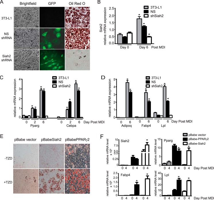
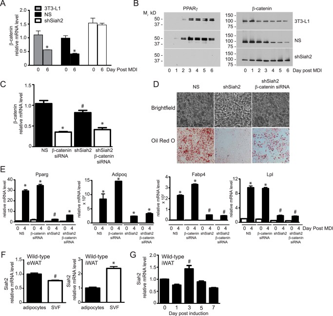
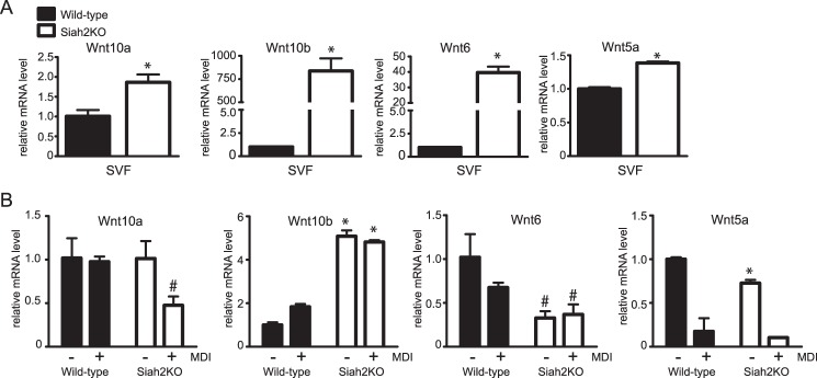
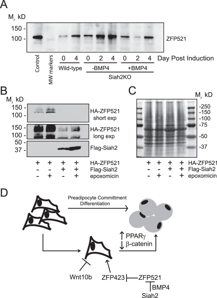
Adipose tissue expansion occurs by increasing the size of existing adipocytes or by increasing the number of adipocytes via adipogenesis. Adipose tissue dysfunction in obesity is associated with adipocyte hypertrophy and impaired adipogenesis. We recently demonstrated that deletion of the ubiquitin ligase Siah2 is associated with enlarged adipocytes in lean or obese mice. In this study, we find that adipogenesis is impaired in 3T3-L1 preadipocytes stably transfected with Siah2 shRNA and that overexpression of Siah2 in non-precursor fibroblasts promotes adipogenesis. In the 3T3-L1 model, loss of Siah2 is associated with sustained β-catenin expression post-induction, but depletion of β-catenin only partially restores PPARγ expression and adipocyte formation. Using wild-type and Siah2-/- adipose tissue and adipose stromal vascular cells, we observe that Siah2 influences the expression of several factors that control adipogenesis, including Wnt pathway genes, β-catenin, Zfp432, and Bmp-4 Consistent with increased β-catenin levels in shSiah2 preadipocytes, Wnt10b is elevated in Siah2-/- adipose tissue and remains elevated in Siah2-/- primary stromal cells after addition of the induction mixture. However, addition of BMP-4 to Siah2-/- stromal cells reduces Wnt10b expression, reduces Zfp521 protein levels, and increases expression of Zfp423, a transcriptional regulator of peroxisome proliferator-activated receptor γ expression that controls commitment to adipogenesis and is repressed by Zfp521. These results indicate that Siah2 acts upstream of BMP-4 to regulate factors that control the commitment of adipocyte progenitors to an adipogenic pathway. Our findings reveal an essential role for Siah2 in the early events that signal undifferentiated progenitor cells to become mature adipocytes.

Keywords: BMP-4; Siah2; Wnt pathway; ZFP423; adipogenesis; adipose tissue; ubiquitin; β-catenin.


Figures

FIGURE 1.
FIGURE 1.
Siah2 promotes adipogenesis in 3T3-L1 preadipocytes and non-precursor fibroblasts. A, 3T3-L1 preadipocytes did not undergo transfection (3T3-L1) or were stably transfected with non-silencing shRNA or Siah2 shRNA co-expressing GFP and induced to undergo adipogenesis. Accumulation of neutral lipids was detected using Oil Red O, and GFP was detected via fluorescence microscopy. B, Siah2 gene expression is up-regulated in untransfected and NS shRNA-transfected cells and specifically reduced in shSiah2-transfected cells during induction of adipogenesis. C and D, mRNA levels of transcription factors necessary for adipogenesis (C, Pparg and Cebpa) and late markers of adipogenesis (D; Adipoq, Fabp4, and Lpl) are not up-regulated in the absence of Siah2. E, neutral lipid accumulation is detected using Oil Red O in non-precursor NIH3T3 fibroblasts induced to undergo adipogenesis with or without rosiglitazone (2.5 μm, TZD) after ectopic expression of PPARγ or Siah2. F, mRNA levels of markers of adipogenesis (Pparg, Fabp4, and Lpl) are up-regulated in non-precursor fibroblasts with ectopic expression of Siah2. Gene expression was assayed using qRT-PCR. Statistical significance is compared with the corresponding day 0. *, p < 0.01.
FIGURE 2.
FIGURE 2.
Siah2-mediated regulation of β-catenin expression does not directly account for the effect of Siah2 on adipogenesis. A, β-catenin gene expression was assayed prior to induction of adipogenesis (day 0) or 6 days post-induction in untransfected (3T3-L1), NS shRNA transfected and shSiah2-transfected 3T3-L1 preadipocytes. B, β-catenin and PPARγ protein expression was assayed by Western blotting analysis. C, depletion of β-catenin alone (β-catenin siRNA) or in combination with shSiah2 was confirmed using real-time qRT-PCR on day 6 post-induction. D, accumulation of neutral lipids was assayed via Oil Red O staining on day 4 post-induction of Siah2-depleted or Siah2/β-catenin-depleted 3T3-L1 preadipocytes. E, adipogenic markers were assayed on days 0 and 4 post-induction in control (NS), β-catenin, Siah2, or Siah2/β-catenin-depleted 3T3-L1 preadipocytes. F, Siah2 mRNA expression was determined in mature adipocytes and SVF cells isolated from visceral (epididymal white adipose tissue, eWAT) or subcutaneous (inguinal white adipose tissue, iWAT) fat depots. G, Siah2 mRNA levels were determined during induction of adipogenesis in adherent stromal cells isolated from wild-type iWAT. Gene expression was assayed using real-time qRT-PCR. Statistical significance was compared with corresponding day 0 (A, E, and G), non-silencing preadipocytes (C), or mature adipocytes (F). #, p < 0.05; *, p < 0.01.
FIGURE 3.
FIGURE 3.
Loss of Siah2 regulates Wnt expression in adipose tissue and during adipogenesis. A and B, gene expression of Wnt5a, Wnt6, Wnt10a, and Wnt10b was assayed in inguinal adipose tissue SVF cells (A) or during induction of adipogenesis in adherent stromal cells isolated from wild-type and Siah2KO inguinal adipose tissue (B). Gene expression was assayed using real-time qRT-PCR. Statistical significance compared with the corresponding wild type. #, p < 0.05; *, p < 0.01.
FIGURE 4.
FIGURE 4.
Siah2 acts upstream of BMP-4 to promote adipogenesis. A and B, Bmp-4, Wisp2, and Zfp423 gene expression was assayed in wild-type and Siah2KO inguinal adipose tissue (A) or prior to induction of adipogenesis in wild-type and Siah2KO primary inguinal adherent stromal cells (−MDI) and on day 4 post induction (+MDI) (B). C, markers of adipogenesis (Pparg, Fabp4, and Lpl), Wnt10b, Wisp2, and Zfp423 gene expression were assayed during adipogenesis in the absence or presence of 40 ng/ml BMP-4. D, Oil Red O staining of neutral lipid accumulation on day 4 post-induction of adipogenesis in wild-type and Siah2KO stromal cells. Siah2KO stromal cells were incubated in the absence (Siah2KO/−BMP-4) or presence of 40 ng/ml BMP-4 (Siah2KO/+BMP-4). Statistical significance of Siah2KO was compared with the wild type (A), wild-type −MDI (B), or the corresponding day 0 (C). #, p < 0.05; *, p < 0.01; ∧, p < 0.01 compared with −MDI, day 0.
FIGURE 5.
FIGURE 5.
Siah2 regulates Zfp521 protein levels. A, Zfp521 protein levels were assayed during induction of adipogenesis in stromal cells isolated from wild-type and Siah2KO inguinal adipose tissue. Control Zfp521 is untagged mouse Zfp521 transiently expressed in HEK293 cells. Where indicated, BMP-4 (40 ng/ml) was added 2 days prior to induction of adipogenesis. B, HEK293 cells were transiently transfected with HA-Zfp521 and pcDNA3.1 or HA-Zfp521 and FLAG-Siah2 and treated with the proteasome inhibitor epoxomicin (1 μm) as indicated. The top panel is a short film exposure, and the bottom panel is a long film exposure to detect HA-Zfp521. Protein levels (A and B) were assayed by Western blotting analysis. C, MemCode staining of the total protein present in B. D, schematic depicting Siah2-mediated commitment to adipogenesis via regulation of a pathway that involves BMP-4, Zfp521, and Zfp423 expression in adipose tissue stromal vascular cells.

References

    1. Grundy S. M. (2000) Metabolic complications of obesity. Endocrine 13, 155–165 - PubMed
    1. Stern J. S., Batchelor B. R., Hollander N., Cohn C. K., and Hirsch J. (1972) Adipose-cell size and immunoreactive insulin levels in obese and normal-weight adults. Lancet 2, 948–951 - PubMed
    1. Lundgren M., Svensson M., Lindmark S., Renström F., Ruge T., and Eriksson J. W. (2007) Fat cell enlargement is an independent marker of insulin resistance and “hyperleptinaemia.” Diabetologia 50, 625–633 - PubMed
    1. Cinti S., Mitchell G., Barbatelli G., Murano I., Ceresi E., Faloia E., Wang S., Fortier M., Greenberg A. S., and Obin M. S. (2005) Adipocyte death defines macrophage localization and function in adipose tissue of obese mice and humans. J. Lipid Res. 46, 2347–2355 - PubMed
    1. Strissel K. J., Stancheva Z., Miyoshi H., Perfield J. W. 2nd, DeFuria J., Jick Z., Greenberg A. S., and Obin M. S. (2007) Adipocyte death, adipose tissue remodeling, and obesity complications. Diabetes 56, 2910–2918 - PubMed

Publication types

LinkOut - more resources