. 2016 Dec 15;143(24):4723-4735.
doi: 10.1242/dev.143909. Epub 2016 Nov 18.

Molecular dynamics of Dkk4 modulates Wnt action and regulates meibomian gland development

Affiliations

Molecular dynamics of Dkk4 modulates Wnt action and regulates meibomian gland development

Jian Sima et al. Development. .

Abstract

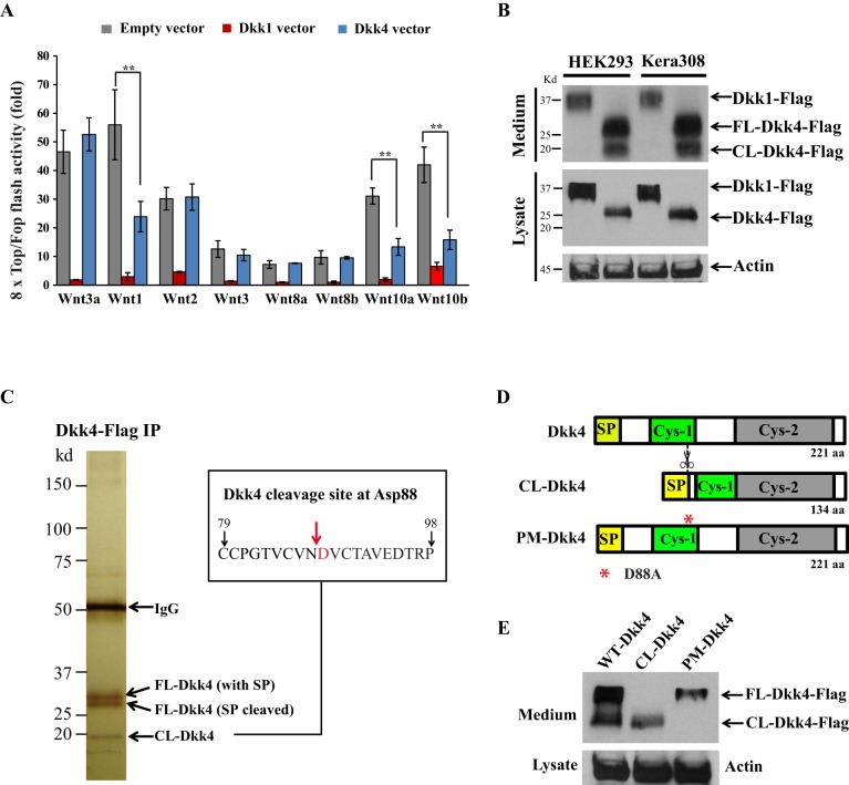
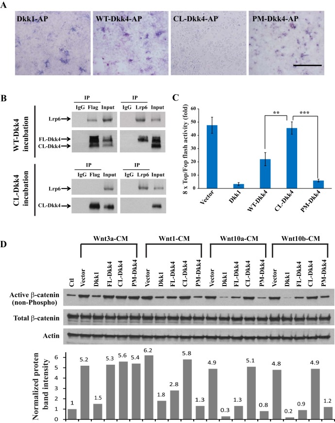
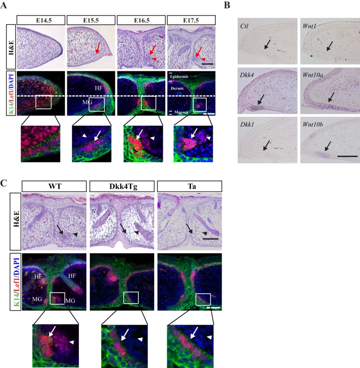
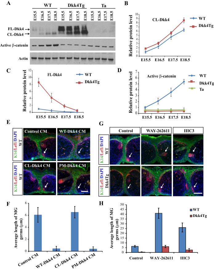
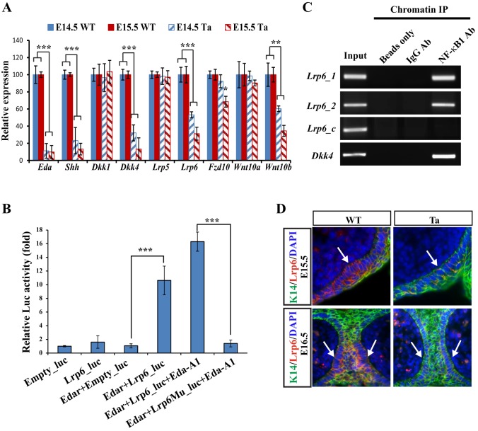
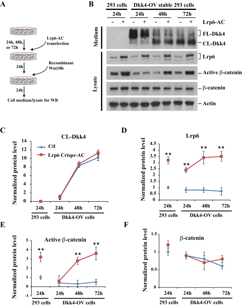
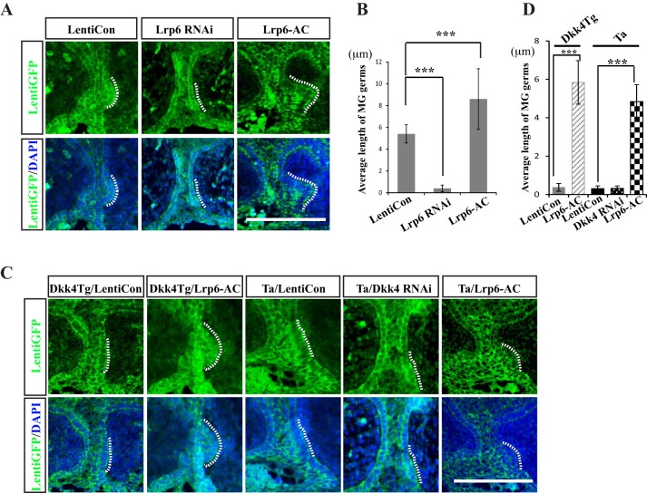
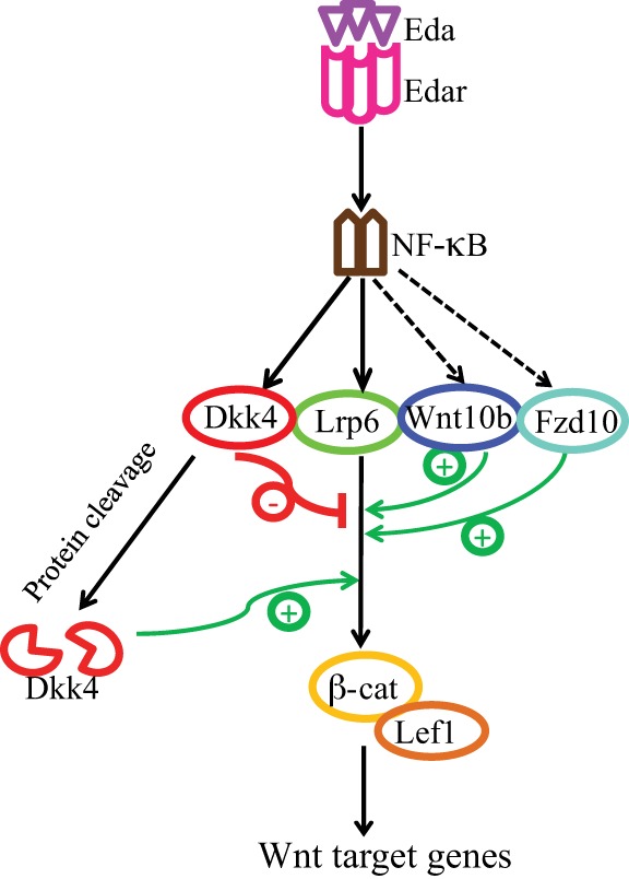
Secreted Dickkopf (Dkk) proteins are major Wnt pathway modulators during organ development. Dkk1 has been widely studied and acts as a general Wnt inhibitor. However, the molecular function of other Dkks remains largely unknown. Here, we show that Dkk4 selectively inhibits a subset of Wnts, but is further inactivated by proteolytic cleavage. Meibomian gland (MG) formation is employed as a model where Dkk4 and its Wnt targets are expressed. Skin-specific expression of Dkk4 arrests MG growth at early germ phase, which is similar to that observed in Eda-ablated Tabby mice. Consistent with transient Dkk4 action, intact Dkk4 inhibits MG extension but the cleaved form progressively increases during MG development with a concomitant upswing in Wnt activity. Furthermore, both Dkk4 and its receptor (and Wnt co-receptor) Lrp6 are direct Eda targets during MG induction. In cell and organotypic cultures, Dkk4 inhibition is eliminated by elevation of Lrp6. Also, Lrp6 upregulation restores MG formation in Tabby mice. Thus, the dynamic state of Dkk4 itself and its interaction with Lrp6 modulates Wnt function during MG development, with a novel limitation of Dkk4 action by proteolytic cleavage.

Keywords: Dkk4; Eda; Lrp6; Meibomian gland; Mouse; Proteolytic cleavage; Wnt.


Conflict of interest statement

The authors declare no competing or financial interests.

Figures

Fig. 1.
Fig. 1.
Dkk4 inhibits a subset of Wnt proteins and is sensitive to proteolytic cleavage. (A) Differential effect of Dkk1 and Dkk4 on canonical Wnt ligands. 8×Top/Fop flash assays measured Wnt/β-catenin activity. Kera308 cells were transfected with Topflash or Fopflash vector together with an empty vector, Dkk1-, or Dkk4-expressing vector. After stimulation with conditioned medium (CM) containing various Wnts for 24 h, luciferase activity of Topflash was normalized with Fopflash in all experiments. Transfection and luciferase assays were performed in triplicate. Error bars, mean±s.e.m. **P<0.01, Student's t-test. (B) Intact and cleaved forms of Dkk4 proteins. HEK293 or Kera308 cells were transiently transfected with expression vectors for Flag-tagged Dkk1 or Dkk4. The CM or cell lysate was used to detect Dkk1 or Dkk4 by western blotting with anti-Flag antibody. (C) Dkk4-Flag protein was purified from CM of HEK293 cells expressing Dkk4-Flag using anti-Flag affinity beads. Two protein bands ∼28 kDa and one lower band at 18 kDa are shown (left). Mass spectrometric analysis revealed 28 kDa bands as Dkk4 protein. Edman N-terminal sequencing of the smaller 18 kDa band identified the cleavage site at Asp88 (D88), indicated by red arrow (right). (D) Schematic structure of Dkk4 protein with signal peptide (SP), cysteine-rich domains (Cys-1 and Cys-2) and cleavage site of Dkk4 at D88 (red asterisk). (E) Plasmids expressing WT Dkk4, CL-Dkk4 and PM-Dkk4 (D88A) were transfected into Kera308 cells for 24 h culture. CM from each culture was collected for immunoblot analysis. Actin was used as a loading control.
Fig. 2.
Fig. 2.
Cleaved Dkk4 does not bind to Lrp6 and loses Wnt-inhibitory capacity. (A) Cell surface binding assays for different Dkk4 isoforms. Dkk1-AP is a positive control. HEK293 cells were transfected with Lrp6-expressing vector and cultured for 24 h, incubated with CM from each Dkk isoform for another 2 h and fixed for AP staining. Scale bar: 50 µm. (B) Lrp6 binds full-length (FL) Dkk4 but not cleaved (CL) Dkk4. Kera308 cells incubated with WT-Dkk4 (top) or CL-Dkk4 CM (bottom) for 2 h were lysed for co-IP assays. Anti-Flag antibody and anti-Lrp6 antibody were used for co-IP experiments. (C) CL-Dkk4 loses Wnt-inhibitory function. HEK293 cells transfected with TopFlash or FopFlash vector were pre-incubated with WT-Dkk4, CL-Dkk4 or PM-Dkk4 CM for 4 h, followed by Wnt10b CM for a further 24 h. Luciferase activity was measured and Top/Fop ratio calculated. Error bars, mean±s.e.m. of triplicates. **P<0.01, ***P<0.001, Student's t-test. (D) CL-Dkk4 does not reduce the protein level of active β-catenin. Kera308 cells were transfected with empty vector, Dkk1, WT-Dkk4, CL-Dkk4 or PM-Dkk4 and cultured for 24 h, followed by application of CM containing each Wnt (as indicated) for a further 24 h. Cell lysates were collected and western blotting assays carried out. The histogram (below) shows calculated density of each protein band. Average protein level of active β-catenin in naïve cells is normalized to 1 and relative density of protein bands in other lanes is reported.
Fig. 3.
Fig. 3.
Endogenous Dkk4 localizes in MG germs and epidermal expression of Dkk4 arrests the growth of MG germs. (A) Time course of early progression of MG development in WT mice. H&E staining and K14 (green)/Lef1 (red) IHC staining of WT eyelids. Bottom panels indicate amplified images of MG germs (arrows) and dermal condensation (DC) (arrowheads). (B) Expression of Dkk4, Dkk1, Wnt1, Wnt10a and Wnt10b in WT MG pre-germs at E15.5. Sense probe of Dkk4 is used as negative control. (C) K14-driven Dkk4 expression inhibits MG growth at E18.5. H&E staining shows no MG germ formed in Dkk4Tg or Tabby mice. Lef1 staining indicates MG germ growth is impaired in Dkk4Tg or Tabby (Ta) compared with WT. In Dkk4Tg and Tabby, amplified images show MG germs arrested at pre-germ stage. Dotted line separates dorsal and ventral part of skin. Arrows in A-C, MG germs; arrowheads, DC; HF, hair follicle. Scale bars: 50 µm.
Fig. 4.
Fig. 4.
Dynamic modulation of Dkk4 cleavage regulates Wnt action and affects MG formation. (A) Protein levels of FL-Dkk4, CL-Dkk4 and active β-catenin during MG development. Eyelids from WT, Dkk4Tg and Tabby (Ta) mice were separated at different stages and lysed for immunoblotting analysis. Antibodies against Dkk4 or active β-catenin were used. Actin was detected as a loading control. Experiments were performed three times. Intensity of each protein band in immunoblots was measured. (B-D) Level of each protein in WT at E15.5 was normalized to 1. Average protein level from WT, Dkk4Tg or Tabby at each time point is shown. (E) Effect of each Dkk4 isoform on MG growth in WT MG cultures. Eyelids from WT at E15.5 were collected and cultured with application of indicated CM for 48 h, and fixed for IHC staining of Lef1 (red) and K14 (green). (F) Length of MGs. (G) MG formation is augmented by the Dkk inhibitors WAY-262611 and IIIC3. Eyelids were separated from WT or Dkk4Tg mice and cultured in medium with 2 µM DMSO (control), 2 µM WAY-262611 or 10 µM IIIC3 for 48 h. Cultured tissues were fixed and double-stained with Lef1 and K14 antibodies. (H) Length of MGs from WT or Dkk4Tg. Error bars are mean±s.e.m. of at least 15 MGs. White arrows indicate MGs. Scale bars: 50 µm.
Fig. 5.
Fig. 5.
Both Dkk4 and Lrp6 are direct targets of Eda signaling during MG induction. (A) qRT-PCR assays show downregulation of Eda, Shh, Dkk4, Lrp6, Fzd10 and Wnt10b in Tabby eyelids. ***P≤0.001, **P≤0.01 by Student's t-test. Data are mean±s.e.m. for triplicate samples. (B) Luciferase reporter assays of Lrp6 WT or mutant promoter activation. Luciferase reporter assays were performed in 293 cells to measure the activity of Lrp6 WT or mutant promoter with or without recombinant Eda-A1 application. Luciferase activity from reporters of empty luciferase vector are normalized to 1. Each data point represents the mean±s.e.m. for triplicate samples. ***P≤0.001 by Student's t-test. (C) Antibody against NF-κB1 pulls down DNA fragments of Lrp6 or Dkk4 promoters containing NF-κB binding sites. Chromatin DNA input in first lane, negative controls in second (beads only) and IgG antibody in third lane. Lrp6_1 and Lrp6_2 are DNA sequences containing the first and second NF-κB binding sites in the Lrp6 promoter, Lrp6_c is the coding region of Lrp6 lacking the NF-κB binding site. (D) Lrp6 expression at E15.5 and E16.5 was reduced in Tabby. IHC staining indicates Lrp6 in red and K14 in green in WT and Tabby. Arrows indicate MG germs.
Fig. 6.
Fig. 6.
Elevated Lrp6 reverses Dkk4-mediated Wnt inhibition in cell culture. (A) Schematic of reconstructing Dkk4-Lrp6-Wnt10b signaling in 293 cells. (B) Protein levels of Dkk4-Flag, Lrp6 and β-catenin in cell cultures. Cultured medium or cell lysates at each time point were collected for immunoblotting analysis. Experiments were performed three times. Actin was detected as a loading control. (C-F) Intensity of protein bands in immunoblots. Level of each protein in 293 cells at 24 h was normalized to 1. Average protein level at each time point was calculated. Error bars are mean±s.e.m. **P≤0.01 by Student's t-test.
Fig. 7.
Fig. 7.
Lrp6 promotes MG germ growth and its activation rescues the MG defect in both Dkk4Tg and Tabby mice. (A) Effects of Lrp6 shRNA and Lrp6-AC on MG growth in WT eyelid cultures. Eyelids from WT at E15.5 were cultured with application of control, Lrp6 shRNA and Lrp6-AC lentiviral particles for 48 h. (B) Length of MGs. (C) Rescue effect of Lrp6-AC for MG formation defect. Eyelids were separated from Dkk4Tg or Tabby mice and cultured in medium with LentiCon, Dkk4 RNAi or Lrp6-AC lentiviral particles for 48 h. Cultured tissues were fixed and stained with GFP antibody. (D) Length of MGs from Dkk4Tg or Tabby. Error bars are mean±s.e.m. from at least 15 MGs. ***P≤0.001, by Student's t-test for B and D. MG germs are indicated by dotted lines. Scale bars: 20 µm.
Fig. 8.
Fig. 8.
Schematic representation of Dkk4 dynamics and function in early meibomian gland development. At early stages of MG development, Eda/Edar signaling mediates NF-κB activation and thereby induces expression of Lrp6, negative Wnt modulator Dkk4, and positive Wnt effectors, including Wnt10b and Fzd10. Eda-induced Dkk4 then limits Wnt activity at the early phase, whereas subsequent Dkk4 inactivation via proteolytic protein cleavage facilitates MG growth at later stage.

Similar articles

Cited by

References

    1. Ahn V. E., Chu M. L.-H., Choi H.-J., Tran D., Abo A. and Weis W. I. (2011). Structural basis of Wnt signaling inhibition by Dickkopf binding to LRP5/6. Dev. Cell 21, 862-873. 10.1016/j.devcel.2011.09.003 - DOI - PMC - PubMed
    1. Andl T., Reddy S. T., Gaddapara T. and Millar S. E. (2002). WNT signals are required for the initiation of hair follicle development. Dev. Cell 2, 643-653. 10.1016/S1534-5807(02)00167-3 - DOI - PubMed
    1. Bafico A., Liu G., Yaniv A., Gazit A. and Aaronson S. A. (2001). Novel mechanism of Wnt signalling inhibition mediated by Dickkopf-1 interaction with LRP6/Arrow. Nat. Cell Biol. 3, 683-686. 10.1038/35083081 - DOI - PubMed
    1. Bazzi H., Fantauzzo K. A., Richardson G. D., Jahoda C. A. B. and Christiano A. M. (2007). The Wnt inhibitor, Dickkopf 4, is induced by canonical Wnt signaling during ectodermal appendage morphogenesis. Dev. Biol. 305, 498-507. 10.1016/j.ydbio.2007.02.035 - DOI - PubMed
    1. Botchkarev V. A. and Fessing M. Y. (2005). Edar signaling in the control of hair follicle development. J. Invest. Derm. Symp. Proc. 10, 247-251. 10.1111/j.1087-0024.2005.10129.x - DOI - PubMed

Publication types

Substances