. 2017 Aug;37(8):2938-2951.
doi: 10.1177/0271678X16679671. Epub 2016 Jan 1.

Thiamet G mediates neuroprotection in experimental stroke by modulating microglia/macrophage polarization and inhibiting NF-κB p65 signaling

Affiliations

Thiamet G mediates neuroprotection in experimental stroke by modulating microglia/macrophage polarization and inhibiting NF-κB p65 signaling

Yating He et al. J Cereb Blood Flow Metab. 2017 Aug.

Erratum in

  • Corrigendum.
    [No authors listed] [No authors listed] J Cereb Blood Flow Metab. 2022 May;42(5):905-907. doi: 10.1177/0271678X211072759. Epub 2022 Feb 14. J Cereb Blood Flow Metab. 2022. PMID: 35156427 Free PMC article. No abstract available.

Abstract

Inflammatory responses are accountable for secondary injury induced by acute ischemic stroke (AIS). Previous studies indicated that O-GlcNAc modification (O-GlcNAcylation) is involved in the pathology of AIS, and increase of O-GlcNAcylation by glucosamine attenuated the brain damage after ischemia/reperfusion. Inhibition of β-N-acetylglucosaminidase (OGA) with thiamet G (TMG) is an alternative option for accumulating O-GlcNAcylated proteins. In this study, we investigate the neuroprotective effect of TMG in a mouse model of experimental stroke. Our results indicate that TMG administration either before or after middle cerebral artery occlusion (MCAO) surgery dramatically reduced infarct volume compared with that in untreated controls. TMG treatment ameliorated the neurological deficits and improved clinical outcomes in neurobehavioral tests by modulating the expression of pro-inflammatory and anti-inflammatory cytokines. Additionally, TMG administration reduced the number of Iba1+ cells in MCAO mice, decreased expression of the M1 markers, and increased expression of the M2 markers in vivo. In vitro, M1 polarization of BV2 cells was inhibited by TMG treatment. Moreover, TMG decreased the expression of iNOS and COX2 mainly by suppressing NF-κB p65 signaling. These results suggest that TMG exerts a neuroprotective effect and could be useful as an anti-inflammatory agent for ischemic stroke therapy.

Keywords: Inflammation; macrophages; microglia; stroke; thiamet G.


Figures

Figure 1.
Figure 1.
Effect of TMG on behavioral deficits and brain infarct volume in mice with cerebral I/R injury. Multiple tests were performed in mice that underwent 1 h of ischemia followed by 24 h or 72 h of reperfusion. (a) Titering assays were performed to compare changes of infarct volume caused by different doses of TMG. (b) Coronal brain sections were stained with 1.5% TTC. Mice were subjected to 1 h of ischemia followed by 72 h of reperfusion; the infarct area is white; (c) Quantitative analysis of brain infarct volume. (n = 9 per group) (d) mNSS were used to evaluate neuronal deficit after I/R with or without TMG treatment. (n = 9 per group) (e–g) Neurobehavioral tests, including foot-fault testing (e), adhesion-removal test (f), and inclined plane test (g) were applied to examine post-stroke motor coordination. (n = 9 per group) Sham: sham operation; MCAO: MCAO with placebo administration; MCAO + TMG-P: MCAO with TMG administration before surgery; MCAO + TMG-T: MCAO with TMG administration after surgery. Data are expressed as means ± SEM except for neurological testing, which is depicted with median (interquartile range). *P < 0.05, **P < 0.01, vs. the MCAO group.
Figure 2.
Figure 2.
Effect of TMG on inflammatory cytokines in ischemic brain. (a) The supernatants of brain tissues from mice that underwent 1 h of ischemia followed by 72 h of reperfusion were measured by ELISA. IL-1α, IL-1β, IL-2, IL-4, IL-6, IL-10, IL-12, IL-17A, IFN-γ, TNF-α, G-CSF, and GM-CSF were included in the panel. (n = 9 per group) (b–e) qRT-PCR were used to detect mRNA expression for TNF-α (b), IL-10 (c), IL-6 (d), and IL-1β (e) in untreated controls and TMG-treated groups. (n = 9 per group) MCAO: MCAO with placebo administration; MCAO + TMG-P: MCAO with TMG administration before surgery; MCAO + TMG-T: MCAO with TMG administration after surgery. Results are presented as mean ± SEM. *P < 0.05, **P < 0.01, vs. MCAO group.
Figure 3.
Figure 3.
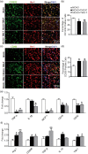
Effect of TMG on microglia/macrophage inflammatory responses in vivo and in vitro. (a) Representative images from triple independent experiments show the expression of Iba-1 in the peri-infarct area of cortex in all groups (upper panel); morphological characteristics in the MCAO group are amoeboid with small branches (lower panel: partial magnification of upper panel). (b) Quantitative analysis of the number of Iba-1-positive cells per visual field in the peri-infarct cortex of mice from the TMG-injected groups and untreated controls. (n = 9 per group) MCAO: MCAO with placebo administration; MCAO + TMG-P: MCAO with TMG administration before surgery; MCAO + TMG-T: MCAO with TMG administration after surgery. Data are expressed as mean ± SEM. *P < 0.05, **P < 0.01 vs. MCAO group. (c) qRT-PCR for mRNA expression of TNF-α, iNOS, IL-1β, IL-6 and MCP-1 in BV2 cells after stimulation with LPS. (n = 9 per group) Data are expressed as mean ± SEM. *P < 0.05, **P < 0.01 vs. Control group.
Figure 4.
Figure 4.
Co-expression of Iba-1 and M1/M2 phenotype markers. Brain slices were prepared at 72 h after MCAO. Immunofluorescent images were captured microscopically in the peri-infarct area of cortex. (a) Cortex co-stained for Iba-1 (microglia marker) (green) and CD16/32(M1 marker) (red) in the peri-infarct area. (b) Quantification of CD16/32- and Iba-1-positive cells in each group. (n = 9 per group) (c) Cortex co-stained for Iba-1 (microglia marker) (green) and CD206(M2 marker) (red) in the peri-infarct area. (d) Quantification of CD206- and Iba-1-positive cells in each group. (n = 9 per group) (e) qRT-PCR for mRNA expression of M1 cytokines (TNF-α, IL-1β, MCP-1, CD16, and CD32) on ischemic cortex. (n = 9 per group) (f) qRT-PCR for mRNA expression of M2 cytokines (Arg-1, CD206, TGF-β, IL-10, and YM-1) on the ischemic cortex in treatment groups and MCAO group. (n = 9 per group) MCAO: MCAO with placebo administration; MCAO + TMG-P: MCAO with TMG administration before surgery; MCAO + TMG-T: MCAO with TMG administration after surgery. Data are expressed as mean ± SEM. *P < 0.05, **P < 0.01 vs. MCAO group.
Figure 5.
Figure 5.
Effect of TMG on microglia polarization in vitro. BV2 cells were cultured in growth medium with LPS (100 ng/mL) or IL-4 (20 ng/mL). The phenotype of BV2 cells was examined by the co-expression of M1 marker CD16/32 (green) and M2 marker CD206 (green) with microglia/macrophage marker Iba-1 (red). Representative images of proportions of M1 (a) or M2 (c) phenotype cells in each group. (n = 9 per group). (b) and (d) Statistics for BV2 phenotypes in the presence or absence of TMG. (e–h) Amount of NF-κB p65 in cytoplasmic (e) and nuclear (f) was detected using immunoblotting and quantitated in the bar graph (g–h). (n = 9 per group) Data are expressed as mean ± SEM. *P < 0.05, **P < 0.01 vs. control group.
Figure 6.
Figure 6.
Effect of TMG on NF-κB transcriptional activity in MCAO mice. Brain sections were prepared at 72 h after stroke. Immunofluorescent images were captured microscopically. (a) Cortex splices co-stained with COX-2 (green) and DAPI (blue) in the peri-infarct area. (b) Cortex splices co-stained with iNOS (green) and DAPI (blue) in the peri-infarct area. (c) Quantification of COX-2 expression in ischemic brain. (d) Quantification of iNOS expression in ischemic brain. (n = 9 per group) *P < 0.05, **P < 0.01 vs. MCAO group. (e) and (f) mRNA expression of (e) iNOS and (f) COX-2 in mouse brain after MCAO. (n = 9 per group) *P < 0.05, **P < 0.01 vs. MCAO group. (g–j) Western blots show the distribution of p65 in cytoplasm and nuclei after stroke in the absence or presence of TMG. n = 9 per group. MCAO: MCAO with placebo administration; MCAO + TMG-P: MCAO with TMG administration before surgery; MCAO + TMG-T: MCAO with TMG administration after surgery. Data are expressed as mean ± SEM. ns: not significant, *P < 0.05, **P < 0.01 vs. MCAO group.
Figure 7.
Figure 7.
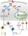
Schematic diagram of TMG effect on brain damage post stroke. TMG exerts neuroprotection by phenotypic modulation of the microglia/macrophage shift and suppression of NF-κB p65 signaling. TMG promoted O-GlcNAcylation of p65 for binding to IκB and delayed its translocation into nuclei, which activates downstream gene transcriptions. Thus, expression of pro-inflammatory cytokines was suppressed and the polarization of microglia/macrophages was shifted to the M2 phenotype. I/R: ischemic/reperfusion; OGT: O-linked N-acetylglucosaminyltransferase; OGA: β-N-acetylglucosaminidase; IKK: inhibitor of nuclear factor κB kinase; IκB: inhibitor of nuclear factor κB. “G” indicates the moiety of GlcNAc.

References

    1. ElAli A, Jean LeBlanc N. The role of monocytes in ischemic stroke pathobiology: new avenues to explore. Front Aging Neurosci 2016; 8: 29. - PMC - PubMed
    1. Emsley HC, Hopkins SJ. Acute ischaemic stroke and infection: recent and emerging concepts. Lancet Neurol 2008; 7: 341–353. - PubMed
    1. Li L, Qin JJ, Guo S, et al. Attenuation of cerebral ischemic injury in interferon regulatory factor 3 deficient rat. J Neurochem 2016; 136: 871–883. . - PubMed
    1. Ma Y, Wang J, Wang Y, et al. The biphasic function of microglia in ischemic stroke. Prog Neurobiol. Epub ahead of print 2 February 2016. pii: S0301-0082(15)30070-8. doi: 10.1016/j.pneurobio.2016.01.005. - PubMed
    1. Lefebvre T, Issad T. 30 years old: O-GlcNAc reaches the age of reason – regulation of cell signaling and metabolism by O-GlcNAcylation. Front Endocrinol 2015; 6: 17. - PMC - PubMed

MeSH terms