. 2017 Feb 1:386:141-150.
doi: 10.1016/j.canlet.2016.11.013. Epub 2016 Nov 16.

Adoptive transfer of ex vivo expanded Vγ9Vδ2 T cells in combination with zoledronic acid inhibits cancer growth and limits osteolysis in a murine model of osteolytic breast cancer

Affiliations

Adoptive transfer of ex vivo expanded Vγ9Vδ2 T cells in combination with zoledronic acid inhibits cancer growth and limits osteolysis in a murine model of osteolytic breast cancer

Aneta Zysk et al. Cancer Lett. .

Abstract

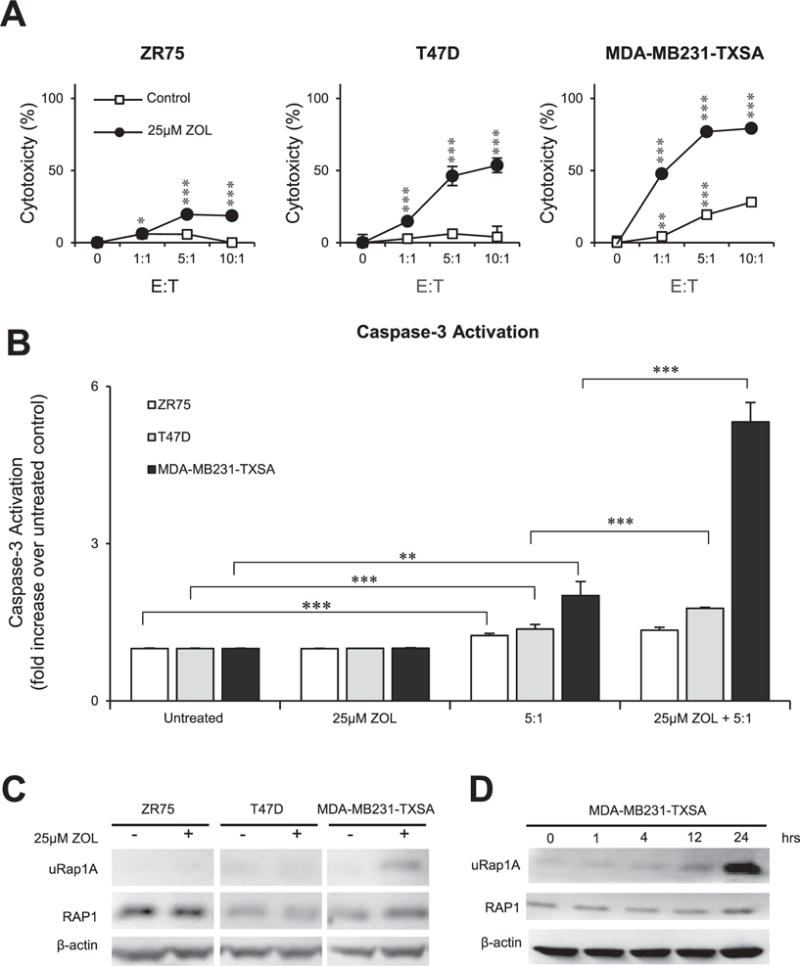
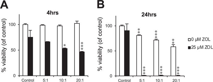
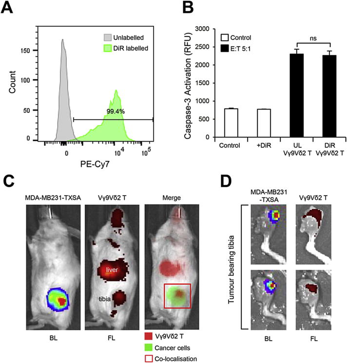
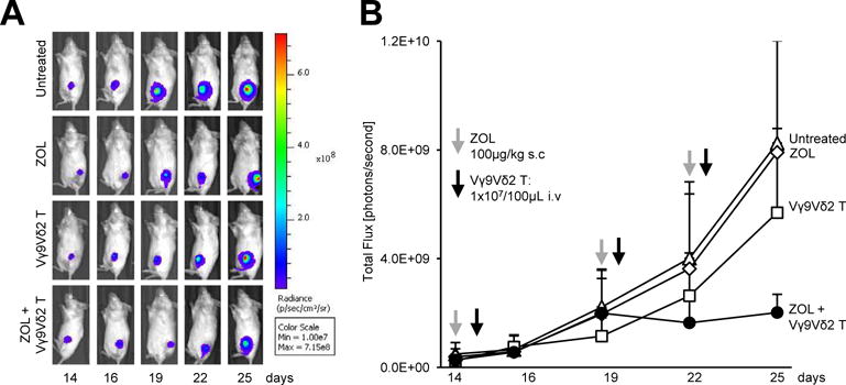
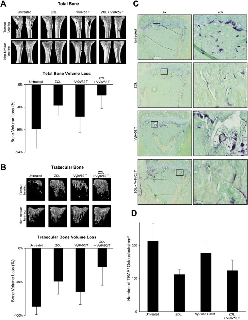
Bone metastases occur in over 75% of patients with advanced breast cancer and are responsible for high levels of morbidity and mortality. In this study, ex vivo expanded cytotoxic Vγ9Vδ2 T cells isolated from human peripheral blood were tested for their anti-cancer efficacy in combination with zoledronic acid (ZOL), using a mouse model of osteolytic breast cancer. In vitro, expanded Vγ9Vδ2 T cells were cytotoxic against a panel of human breast cancer cell lines, and ZOL pre-treatment further sensitised breast cancer cells to killing by Vγ9Vδ2 T cells. Vγ9Vδ2 T cells adoptively transferred into NOD/SCID mice localised to osteolytic breast cancer lesions in the bone, and multiple infusions of Vγ9Vδ2 T cells reduced tumour growth in the bone. ZOL pre-treatment potentiated the anti-cancer efficacy of Vγ9Vδ2 T cells, with mice showing further reductions in tumour burden. Mice treated with the combination also had reduced tumour burden of secondary pulmonary metastases, and decreased bone degradation. Our data suggests that adoptive transfer of Vγ9Vδ2 T cell in combination with ZOL may prove an effective immunotherapeutic approach for the treatment of breast cancer bone metastases.

Keywords: Bisphosphonate; Immunotherapy; Metastasis; Osteoclast; Tumour associated macrophage.


Conflict of interest statement

Conflict of interest

The authors declare no conflict of interest.

Figures

Fig. 1
Fig. 1
ZOL sensitises breast cancer cells to Vγ9Vδ2 T cell cytotoxicity in vitro. A. ZR75, T47D, and MDA-MB231-TXSA breast cancer cell lines were pre-treated with 25 μM ZOL or left untreated for 24 h. Cancer cells were then co-cultured with ex vivo expanded Vγ9Vδ2 T cells (E:T, 1:1, 5:1,10:1). After 9 h, LDH release was measured and expressed as percentage cytotoxicity compared to untreated cells. B. Caspase-3 activation of the same cell lines was measured after 24 h pre-treatment with or without 25 μM ZOL, followed by 2 h co-culture with ex vivo expanded Vγ9Vδ2 T cells (E:T, 5:1). Caspase-3 activation was expressed as a fold increase over untreated control. For the LDH and Caspase-3 activity assay, data was pooled and normalised from two separate experiments (n = 6). C. Western immunoblot analysis showing inhibition of prenylation in breast cancer cell lines after treatment with or without 25 μM ZOL for 24 h, showing unprenylated RAP1A (uRap1A), total RAP1 protein, and β-actin as loading control. D. Western immunoblot analysis showing inhibition of prenylation in MDA-MB231-TXSA treated with 25 μM ZOL over a 24 h time course (0,1, 4,12, 24 h). Images representative of n = 2–3. *p < 0.05, **p < 0.005, ***p < 0.001, ns = non-significant (two-tailed student’s t-test, data represent mean ± SEM, n = 3, unless otherwise indicated).
Fig. 2
Fig. 2
Vγ9Vδ2 T cell cytotoxicity against breast cancer cells in vitro occurs in a time dependent manner. Luciferase-tagged MDA-MB231-TXSA breast cancer cells were treated with or without 25 μM ZOL for 24 h. Cancer cells were then co-cultured with ex vivo expanded Vγ9Vδ2 T cells (E:T, 5:1,10:1,20:1) for A. 4 or B. 24 h, and luciferase activity measured and expressed as percentage viability compared to untreated control. Representative data shown from three experiments.*p < 0.05, **p < 0.005, ***p < 0.001, ns = non-significant (two-tailed student’s t-test, data represent mean ± SEM, n = 3, unless otherwise indicated).
Fig. 3
Fig. 3
Fluorescently labelled Vγ9Vδ2 T cells localise to breast cancer lesions in the bone. Ex vivo expanded Vγ9Vδ2 T cells were labelled using DiR dye as outlined in the methods. A. Flow cytometric analysis of Vγ9Vδ2 T cell DiR labelling efficacy. B. Cytotoxicity of DiR-labelled and unlabelled Vγ9Vδ2 T cells against MDA-MB231-TXSA cancer cells (E:T, 5:1) as shown by caspase-3 activation. C. In vivo localisation of DiR-labelled Vγ9Vδ2 T cells injected via the tail vein into 5-week old female NOD/SCID mice bearing luciferase-tagged osteolytic breast cancer cells (MDA-MB231-TXSA) in the left tibia. Bioluminescence and fluorescence images were acquired on the IVIS Spectrum in vivo imaging system 24 h after infusion and D. ex vivo, 6 days after infusion (representative images of n = 5). UL = unlabelled, BL = bioluminescence, FL = fluorescence. Percentages shown indicate numbers from lymphocyte population. ns = non-significant (Student’s t-test, data represent mean ± SEM, n = 3).
Fig. 4
Fig. 4
ZOL potentiates the anti-cancer efficacy of Vγ9Vδ2 T cells against osteolytic breast cancer. Luciferase-tagged MDA-MB231-TXSA breast cancer cells were injected directly into the left tibial cavity of 5-week old NOD/SCID mice. Once tumours were established, treatments were commenced as outlined in the methods. Whole body bioluminescence images were acquired on the IVIS Spectrum in vivo imaging system over the course of the study. A. A representative bioluminescence image showing a single mouse from each treatment group over the duration of the study. B. The line graph shows the quantification of bioluminescence signal over the course of the study and is expressed as total flux [photons/second] (n = 4–6 mice per group).
Fig. 5
Fig. 5
Vγ9Vδ2 T cells reduce tumour burden of lung metastases. At the time of sacrifice, lungs were removed for bioluminescence quantification of tumour burden. Bioluminescence signal was detected on the IVIS Spectrum in vivo imaging system and is expressed as total flux [photons/second]. A representative bioluminescence image of the lungs from each treatment group is shown.
Fig. 6
Fig. 6
Vγ9Vδ2 T cells in combination with ZOL reduce tumour-associated osteolysis in total and trabecular bone. The osteolytic nature of MDA-MB231-TXSA breast cancer cells can be seen in the representative qualitative μCT 3D images and in the quantitative assessment of A. total and B. trabecular bone. Bone loss percentage is calculated as the percentage difference in bone volume between the tumour bearing and contralateral non-tumour bearing control tibia. C. Representative histological sections at 4× and 40 × magnification showing decalcified tibias from each treatment group stained with TRAP for the detection of osteoclasts. D. Quantitative assessment of the number of TRAP+ osteoclasts.

References

    1. Howlader N, Noone AM, Krapcho M, Miller D, Bishop K, Altekruse SF, Kosary CL, Yu M, Ruhl J, Tatalovich Z, Mariotto A, Lewis DR, Chen HS, Feuer EJ, Cronin KA, editors. SEER Cancer Statistics Review, 1975–2013. National Cancer Institute; Bethesda, MD: Apr, 2016. based on November 2015 SEER data submission, posted to the SEER web site, http://seer.cancer.gov/csr/1975_2013/
    1. Coleman RE, Rubens RD. The clinical course of bone metastases from breast cancer. Br J Cancer. 1987;55:61–66. - PMC - PubMed
    1. Coleman RE. Metastatic bone disease: clinical features, pathophysiology and treatment strategies. Cancer Treat Rev. 2001;27:165–176. - PubMed
    1. Mundy GR. Metastasis to bone: causes, consequences and therapeutic opportunities. Nat Rev Cancer. 2002;2:584–593. - PubMed
    1. Roodman GD. Mechanisms of bone metastasis. N Engl J Med. 2004;350:1655–1664. - PubMed

MeSH terms