

Patient-specific CFD models for intraventricular flow analysis from 3D ultrasound imaging: Comparison of three clinical cases
- PMID: 27866678
- PMCID: PMC5191945
- DOI: 10.1016/j.jbiomech.2016.11.039
Patient-specific CFD models for intraventricular flow analysis from 3D ultrasound imaging: Comparison of three clinical cases
Abstract
Background: As the intracardiac flow field is affected by changes in shape and motility of the heart, intraventricular flow features can provide diagnostic indications. Ventricular flow patterns differ depending on the cardiac condition and the exploration of different clinical cases can provide insights into how flow fields alter in different pathologies.
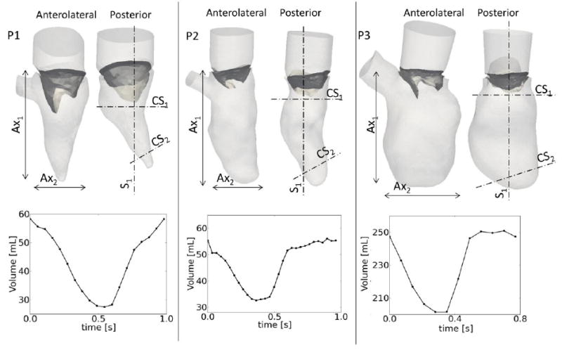
Methods: In this study, we applied a patient-specific computational fluid dynamics model of the left ventricle and mitral valve, with prescribed moving boundaries based on transesophageal ultrasound images for three cardiac pathologies, to verify the abnormal flow patterns in impaired hearts. One case (P1) had normal ejection fraction but low stroke volume and cardiac output, P2 showed low stroke volume and reduced ejection fraction, P3 had a dilated ventricle and reduced ejection fraction.
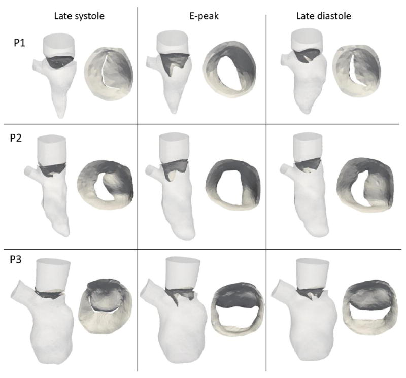
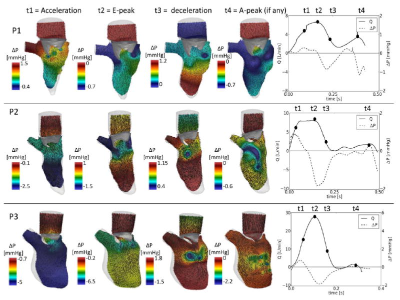
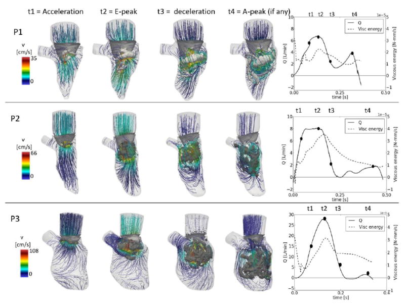
Results: The shape of the ventricle and mitral valve, together with the pathology influence the flow field in the left ventricle, leading to distinct flow features. Of particular interest is the pattern of the vortex formation and evolution, influenced by the valvular orifice and the ventricular shape. The base-to-apex pressure difference of maximum 2mmHg is consistent with reported data.
Conclusion: We used a CFD model with prescribed boundary motion to describe the intraventricular flow field in three patients with impaired diastolic function. The calculated intraventricular flow dynamics are consistent with the diagnostic patient records and highlight the differences between the different cases. The integration of clinical images and computational techniques, therefore, allows for a deeper investigation intraventricular hemodynamics in patho-physiology.
Keywords: CFD with prescribed moving boundaries; Intraventricular flow; Patient-specific models; Ventricular vortex analysis.
Copyright © 2016 Elsevier Ltd. All rights reserved.
Figures
References
-
- Bermejo J, Benito Y, Alhama M, Yotti R, Martinez-Legazpi P, Perez del Villar C, Perez-David E, Gonzalez-Mansilla A, Santa-Maria C, Barrio A, Fernandez-Aviles F, Del Alamo JC. Intraventricular vortex properties in nonischemic dilated cardiomyopahy. American Journal of Physiology - Heart and Circulatory Physiology. 2013;306:H714–H729. - PMC - PubMed
-
- Chnafa C, Mendez S, Nicoud F. Image-based large-eddy simulation in a realistic left heart. Computers and Fluids. 2014;94:173–187.
-
- Greenberg NL, Vandervoort PM, Firstenberg MS, Garcia MJ, Thomas JD. Estimation of diastolic intraventricular pressure gradients by doppler M-mode echocardiography. American Journal of Physiology: Heart and Circulatory Physiology. 2001;280:H2507–H2515. - PubMed
Publication types
MeSH terms
Grants and funding
LinkOut - more resources
Full Text Sources
Other Literature Sources
Research Materials
Miscellaneous
