. 2016 Dec 7;92(5):1049-1062.
doi: 10.1016/j.neuron.2016.10.030. Epub 2016 Nov 17.

Neural Architecture of Hunger-Dependent Multisensory Decision Making in C. elegans

Affiliations

Neural Architecture of Hunger-Dependent Multisensory Decision Making in C. elegans

D Dipon Ghosh et al. Neuron. .

Abstract

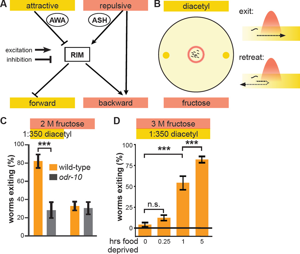
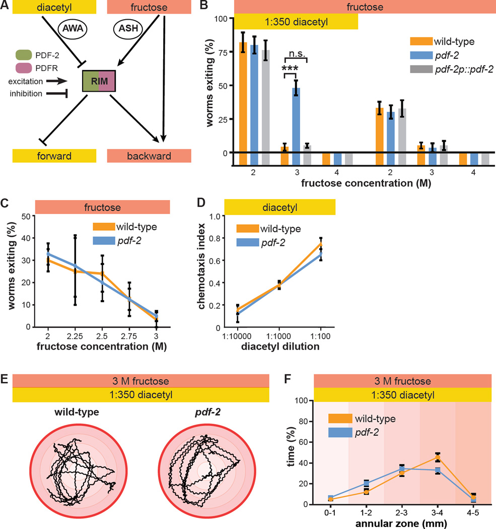
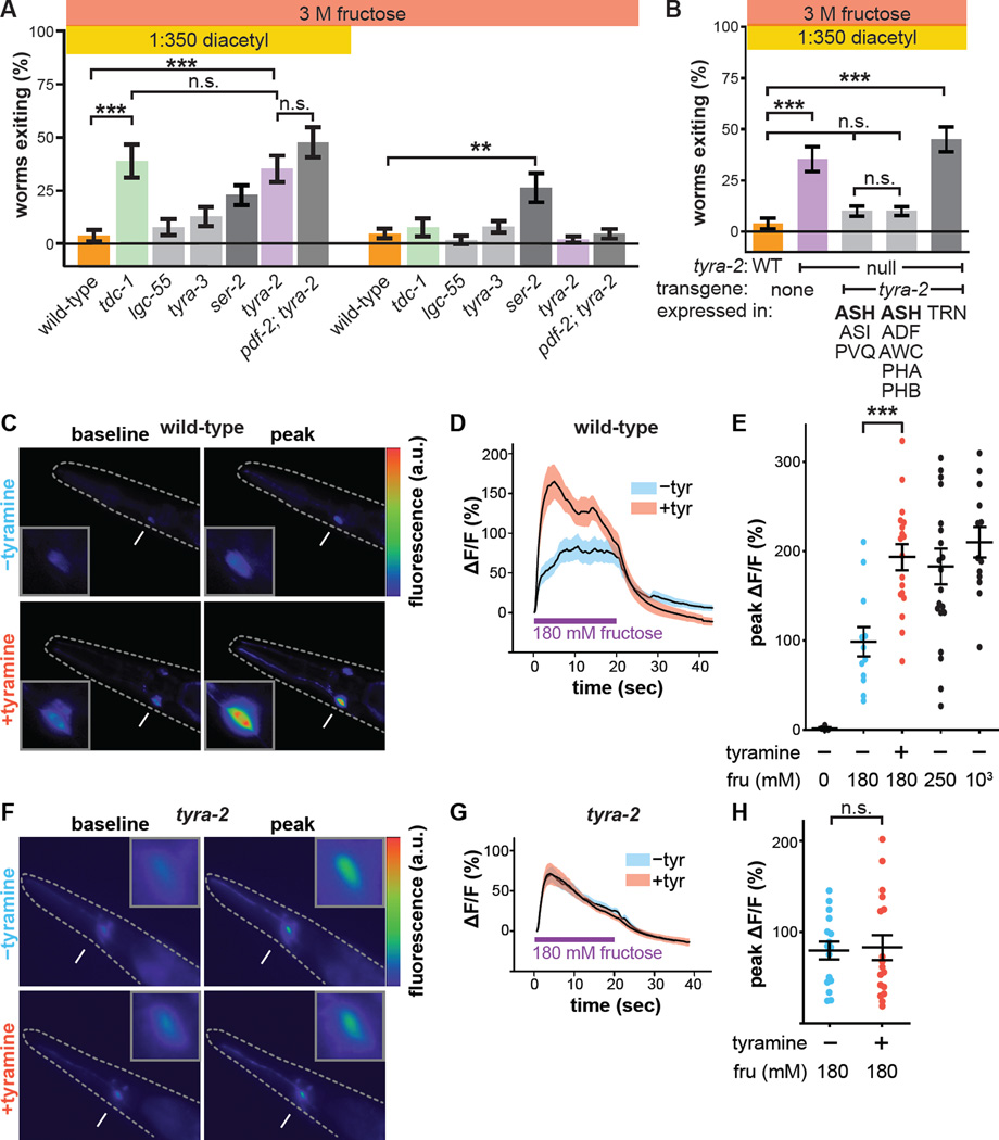
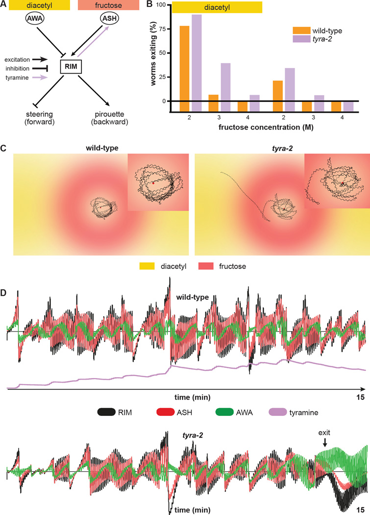
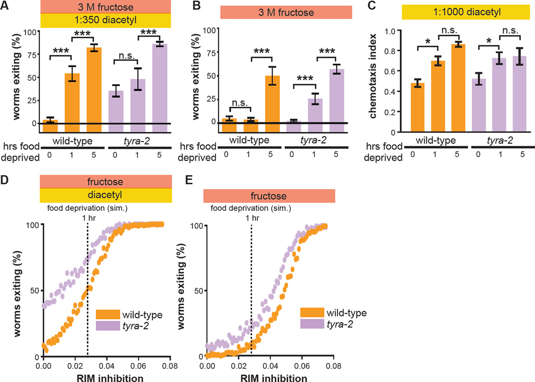
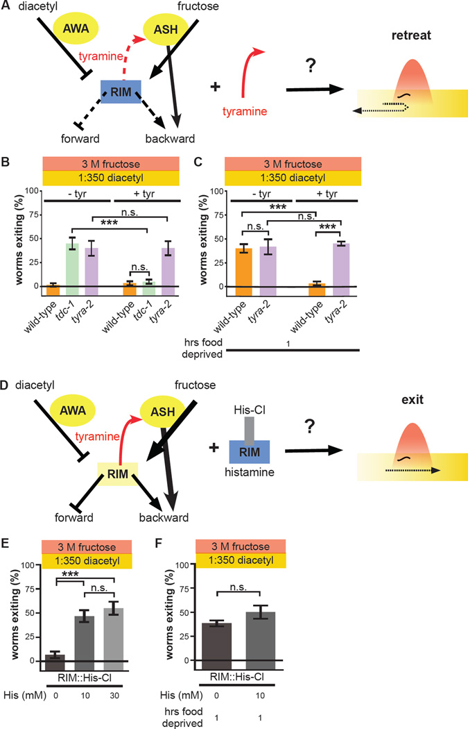
Little is known about how animals integrate multiple sensory inputs in natural environments to balance avoidance of danger with approach to things of value. Furthermore, the mechanistic link between internal physiological state and threat-reward decision making remains poorly understood. Here we confronted C. elegans worms with the decision whether to cross a hyperosmotic barrier presenting the threat of desiccation to reach a source of food odor. We identified a specific interneuron that controls this decision via top-down extrasynaptic aminergic potentiation of the primary osmosensory neurons to increase their sensitivity to the barrier. We also establish that food deprivation increases the worm's willingness to cross the dangerous barrier by suppressing this pathway. These studies reveal a potentially general neural circuit architecture for internal state control of threat-reward decision making.

Keywords: C. elegans; decision making; metabolism; multisensory integration; neural circuits; neuromodulation.


Conflict of interest statement

Author Information The authors declare no conflicts of interest.

Figures

Figure 1
Figure 1. Hunger state modulates a multisensory threat-reward decision
(A) Schematic of information flow in the C. elegans sensorimotor control network. The AWA primary sensory neuron that responds to attractive stimuli like diacetyl food odor indirectly inhibits the RIM interneuron. The ASH primary sensory neuron that responds to aversive stimuli like hyperosmolarity indirectly excites RIM. RIM directly inhibits forward command interneurons and directly excites backward command interneurons to control the balance between forward and backward locomotion. ASH also bypasses RIM to directly excite backward command interneurons. (B) Schematic of the decision making assay. Worms (black squiggles) are placed in the center of a hyperosmotic fructose ring (red circle) with the food odor diacetyl (yellow spots) outside the ring. As each worm ascends the attractive diacetyl gradient, it encounters the aversive hyperosmotic ring, where it either proceeds to exit or retreats to remain in the ring. The decision balance is quantified as the percent of worms that exit the ring during a fifteen minute trial. (C) Wild-type and odr-10 null-mutant worm tolerance of 2 M fructose ring in presence or absence of food odor. odr-10 null-mutant worms lack the olfactory receptor that senses diacetyl. (D) Effect of increasing food deprivation on decision to exit a 3 M fructose ring in the presence of food odor. (Identical data is presented in multiple data sets, as all previously tested genotypes/conditions were not tested every time we extended our analysis to additional genotypes/conditions. Unless otherwise indicated, data in all Figures represent the average of at least five independent assays with ten worms per assay. All statistical comparisons are by one- or two-way ANOVA, as appropriate, with Tukey-Kramer paired-comparison test applied to all pairs of genotypes. Error bars denote s.e.m.; ***, p < 0.001.)
Figure 2
Figure 2. PDF-2 neuropeptide regulates multisensory decision making
(A) RIM is potentially a peptide-modulated locus for internal state control of threat-reward decision making. RIM expresses both the neuropeptide PDF-2 and its G protein-coupled receptor PDFR-1. (B) Decision balance of wild-type and pdf-2 null-mutant worms encountering a 2 M, 3 M, or 4 M fructose ring in the presence or absence of food odor. Rescue of pdf-2 null-mutant phenotype by re-expression of a pdf-2 transgene under the control of its own promoter. (C) Exiting of the osmotic ring in the absence of food odor in wild-type and pdf-2 null-mutant worms. (D) Chemotaxis to various concentrations of diacetyl, measured as the fraction of worms that move to a test spot of diacetyl dilution versus a control spot of water after a fifteen minute trial on a standard assay plate, in wild-type and pdf-2 null-mutant worms. (E) Representative five minute trajectories of wild-type and pdf-2 null-mutant worms inside a 3 M ring with food odor outside. The outer red circle represents the osmotic ring, and the shading indicates 1mm annular zones. (F) Time spent by wild-type and pdf-2 null-mutant worms in each of the radial zones (n=7–10 worms for each genotype, F = 1.688, p = 0.160).
Figure 3
Figure 3. PDF-2 signaling to PDFR-1 in RIM controls the multisensory decision balance by modulating tyramine secretion
(A) Re-expression of PDF-2 in RIM and RIC in pdf-2 null-mutant worms using the tdc-1 promoter. (B) Membrane-tethered peptide (t-peptide) system. Bioactive peptides can be expressed as chimeric fusion proteins with N-terminal secretory signal sequences and C-terminal glycolipid anchor targeting signals. t-peptides are secreted, but remain covalently anchored to the plasma membrane, and thus only activate their cognate receptors cell-autonomously. Adapted from Choi et al., 2009. (C) Expression in pdf-2 null-mutant worms of t-PDF-2 using either of two promoters whose activity overlaps only in RIM; expression of t-PDF-2 in RIC alone; or expression of sequence-scrambled t-SCR in RIM and RIC. The tdc-1, nmr-2, and tbh-1 promoters are used to drive expression in RIM and RIC; RIM, multiple AV neurons, and PVC; and RIC alone, respectively. (D) Tyramine is synthesized from tyrosine by tyrosine decarboxylase (TDC), encoded by the tdc-1 gene expressed in RIM and RIC. Octopamine is synthesized from tyramine by tyramine-β-hydroxylase (TBH), encoded by the tbh-1 gene expressed in RIC. (E) Multisensory and unisensory decision balance of tdc-1 null-mutant worms, which lack both tyramine and octopamine, and tbh-1 null-mutant worms, which lack only octopamine. Decision balance of pdf-2; tdc-1 double-mutant is also shown. (F) Re-expression of tdc-1 in RIM and RIC, but not RIC alone, in tdc-1 null-mutant worms.
Figure 4
Figure 4. Tyramine acts on its receptor TYRA-2 in ASH osmosensory neuron to regulate the multisensory decision balance
(A) Multisensory and unisensory decision balance of tyramine receptor mutants. Tyramine GPCR ser-2 null-mutant worms exhibit increased exiting in both multisensory (p vs wild-type = 0.136) and unisensory contexts, though only the increased exiting in the unisensory context is statistically significant compared to wild-type (**, p = 0.009). Decision balance of the pdf-2; tyra-2 double-mutant is also shown. (B) Re-expression of tyra-2 in tyra-2 null-mutant worms under the control of the sra-6 or gpa-13 promoters, whose activity overlaps solely in ASH. Re-expression of tyra-2 using the mec-17 promoter in touch receptor neurons (TRN), some of which also normally express tyra-2. (C–E) Representative images (C), time course (D), and peak (E) Ca2+ responses of ASH to 180 mM fructose (unless otherwise indicated) in wild-type worms with or without 50 mM tyramine pre-treatment imaged using GCaMP3. (F–H) Representative images (F), time course (G), and peak (H) Ca2+ responses of ASH to 180 mM fructose in tyra-2 null-mutant worms with or without 50 mM tyramine pre-treatment imaged using GCaMP3. (Arrows indicate ASH cell body, magnified in the insets. Dashed lines outline the worm. mean + s.e.m; circles represent peak responses of individual animals; n>12 animals per genotype and treatment condition.)
Figure 5
Figure 5. Computational modeling predicts non-linear slow tyramine signaling by RIM to ASH
(A) Schematic of the simplified nervous system used for computational modeling. AWA and ASH provide direct inhibitory and excitatory inputs onto RIM, respectively. RIM integrates these sensory inputs and directionally biases forward locomotion via inhibition of steering and pirouette modulation. Tyraminergic positive feedback from RIM to ASH increases ASH sensitivity to osmotic stimuli. Simulated tyra-2 null-mutant worms lack the tyraminergic RIM-ASH signal. (B) Decision balance of simulated wild-type and tyra-2 null-mutant worms encountering a 2 M, 3 M, or 4 M fructose ring in the presence or absence of food odor. n=1000 single worm simulations per genotype and condition. (C) Sample fifteen-minute trajectories of simulated wild-type and tyra-2 null-mutant worms inside a 3 M ring with food odor outside. Trajectories are magnified in the insets. (D) Neural activity profiles of AWA, ASH, and RIM during the simulated trajectories in (C). Activity of individual neurons and of tyramine signals are expressed in arbitrary units, plotted to the same scale for the simulated wild-type and tyra-2 null-mutant worms.
Figure 6
Figure 6. One-hour food deprivation fails to increase threat tolerance of tyra-2 null-mutant worms
(A–C) Effect of one hour or five hours of food deprivation on wild-type and tyra-2 null-mutant worm multisensory decision balance (A), exiting of a 3 M fructose ring in the absence of food odor (B), and chemotaxis to 1:1000 diacetyl (C). (*, p<0.05.) (D and E) Effect of increasing RIM inhibition on simulated wild-type and tyra-2 null-mutant worm multisensory (D) or unisensory (E) decision balance. Vertical dashed lines indicate the degree of RIM inhibition that results in exiting rates for simulated wild-type worms that match those of real worms deprived of food for the indicated durations (see panel A). n=250 simulated worms for each genotype and strength of RIM inhibition.
Figure 7
Figure 7. One-hour food deprivation increases threat tolerance by inhibiting RIM activity and suppressing RIM-ASH tyraminergic potentiation
(A) Schematic depicting prediction that exogenous tyramine increases threat sensitivity. Exogenous tyramine is predicted to reverse the effects of suppression of the RIM-ASH positive feedback loop on threat sensitivity. Yellow represents strong activity, while blue represents weak activity. Thickness of solid lines represents strength of signals, and dashed lines represent inactive signals. (B) Effect of exogenous tyramine on multisensory decisions of wild-type, tdc-1 null-mutant, and tyra-2 null-mutant worms. (C) Effect of exogenous tyramine on multisensory decisions of one-hour food-deprived wild-type and tyra-2 null-mutant worms. (D) Schematic depicting prediction that inhibition of RIM increases threat tolerance. Inhibition of RIM expressing His-Cl with exogenous histamine is predicted to increase threat tolerance and mimic one hour of food deprivation. (E) Effect of 0, 10 mM, and 30 mM histamine on multisensory decision balance of worms expressing RIM::His-Cl. (F) Effect of 0 and 10 mM histamine on multisensory decision balance of one-hour food-deprived worms expressing RIM::His-Cl.

References

    1. Alkema MJ, Hunter-Ensor M, Ringstad N, Horvitz HR. Tyramine Functions independently of octopamine in the Caenorhabditis elegans nervous system. Neuron. 2005;46:247–260. - PubMed
    1. Barrios A, Ghosh R, Fang C, Emmons SW, Barr MM. PDF-1 neuropeptide signaling modulates a neural circuit for mate-searching behavior in C. elegans. Nature neuroscience. 2012;15:1675–1682. - PMC - PubMed
    1. Berthoud HR. Metabolic and hedonic drives in the neural control of appetite: who is the boss? Curr Opin Neurobiol. 2011;21:888–896. - PMC - PubMed
    1. Choi C, Cao G, Tanenhaus AK, McCarthy EV, Jung M, Schleyer W, Shang Y, Rosbash M, Yin JC, Nitabach MN. Autoreceptor control of peptide/neurotransmitter corelease from PDF neurons determines allocation of circadian activity in drosophila. Cell Rep. 2012;2:332–344. - PMC - PubMed
    1. Choi C, Fortin JP, McCarthy E, Oksman L, Kopin AS, Nitabach MN. Cellular dissection of circadian peptide signals with genetically encoded membrane-tethered ligands. Current biology : CB. 2009;19:1167–1175. - PMC - PubMed