. 2016 Dec 14;20(6):770-784.
doi: 10.1016/j.chom.2016.10.011. Epub 2016 Nov 17.

A Viral Deamidase Targets the Helicase Domain of RIG-I to Block RNA-Induced Activation

Affiliations

A Viral Deamidase Targets the Helicase Domain of RIG-I to Block RNA-Induced Activation

Jun Zhao et al. Cell Host Microbe. .

Abstract

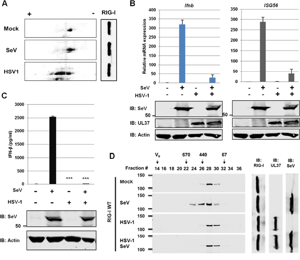
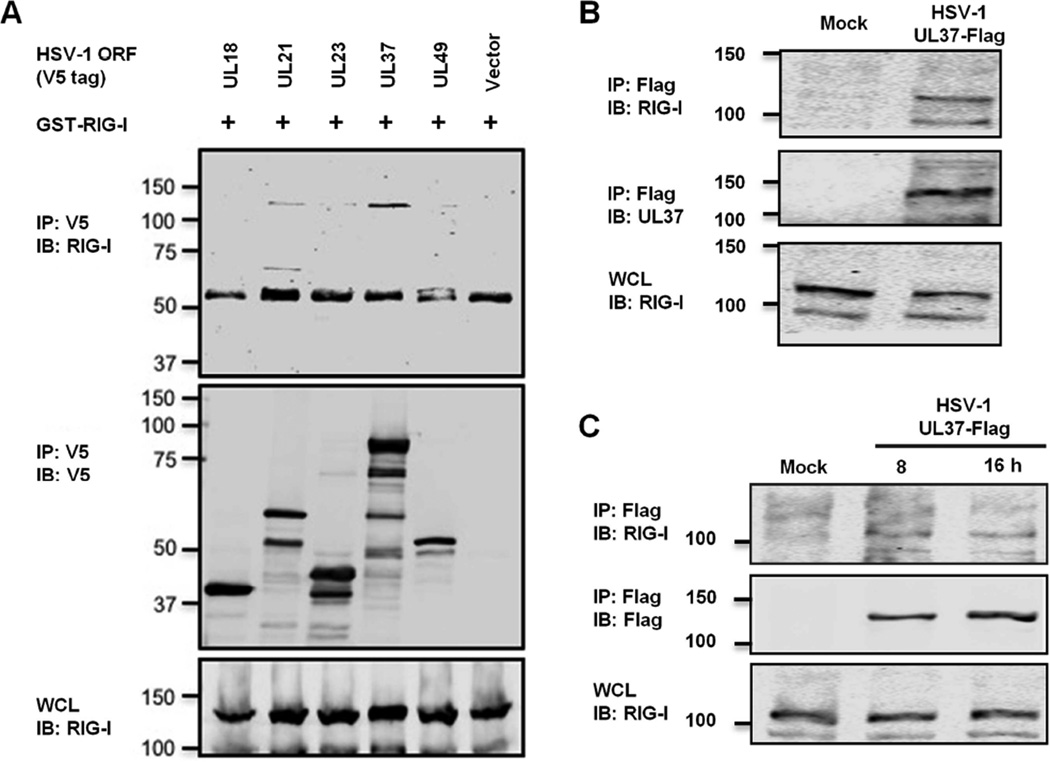
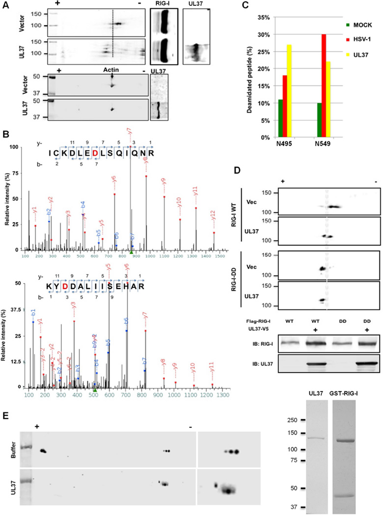
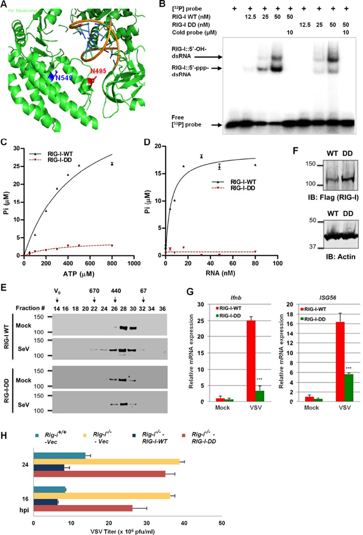
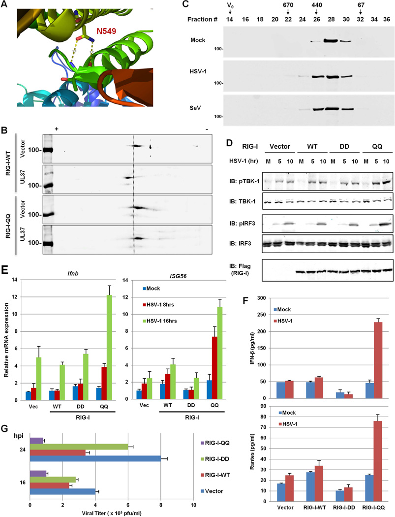
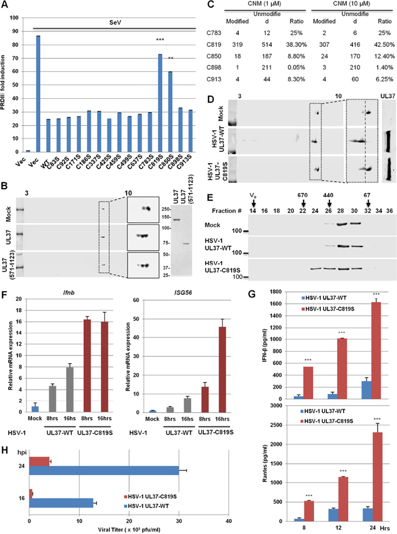
RIG-I detects double-stranded RNA (dsRNA) to trigger antiviral cytokine production. Protein deamidation is emerging as a post-translational modification that chiefly regulates protein function. We report here that UL37 of herpes simplex virus 1 (HSV-1) is a protein deamidase that targets RIG-I to block RNA-induced activation. Mass spectrometry analysis identified two asparagine residues in the helicase 2i domain of RIG-I that were deamidated upon UL37 expression or HSV-1 infection. Deamidation rendered RIG-I unable to sense viral dsRNA, thus blocking its ability to trigger antiviral immune responses and restrict viral replication. Purified full-length UL37 and its carboxyl-terminal fragment were sufficient to deamidate RIG-I in vitro. Uncoupling RIG-I deamidation from HSV-1 infection, by engineering deamidation-resistant RIG-I or introducing deamidase-deficient UL37 into the HSV-1 genome, restored RIG-I activation and antiviral immune signaling. Our work identifies a viral deamidase and extends the paradigm of deamidation-mediated suppression of innate immunity by microbial pathogens.

Keywords: ATPase/helicase; RIG-I; RNA-sensing; UL37; deamidation; herpesvirus; immune evasion.


Figures

Figure 1
Figure 1. HSV-1 evades RNA-induced activation of RIG-I
(A) HEK293/Flag-RIG-I cells were mock-infected or infected with HSV-1 (MOI=2) or Sendai virus (SeV, 100 HAU/ml) for 4 hours. Whole cell lysates (WCLs) were analyzed by two-dimensional gel electrophoresis. (B and C) HEK293 cells were mock-infected or infected with HSV-1 (MOI=2) for 1 h and super-infected with SeV (100 HAU/ml) for 8 (B) or 16 h (C). The expression of the indicated antiviral genes was analyzed by real-time PCR using total RNA (B). Supernatant was collected to determine IFN-β by ELISA (C). (D) HEK293/Flag-RIG-I were mock-infected or infected with HSV-1 for 1 h, followed by SeV infection (100 HAU/ml) for 4 h. RIG-I was purified and analyzed by gel filtration and immunoblotting. Numbers indicate the size of RIG-I in kDa and V0 denotes void volume. For B-D, WCLs were analyzed by immunoblotting with antibodies against SeV, HSV-1 UL37 and β-actin. ***, p<0.001 was calculated in reference to cells infected with SeV. For B and C, data are presented as mean ± SD. See also Figure S1.
Figure 2
Figure 2. HSV-1 UL37 interacts with RIG-I
(A) 293T cells were transfected with plasmids containing GST-RIG-I and the indicated open reading frames of HSV-1. WCLs were precipitated with the indicated antibody. WCLs and precipitated proteins were analyzed by immunoblotting. (B and C) 293T cells were infected with recombinant HSV-1 UL37-Flag at MOI of 30 for 1 h (B) or MOI of 1 for 8 and 16 h (C). WCLs were precipitated with anti-Flag (M2) antibody. RIG-I and WCLs were analyzed by immunoblotting with indicated antibodies. See also Figure S2.
Figure 3
Figure 3. UL37 inhibits RIG-I activation
(A–F) Whole cell lysates (WCLs) of 293T cells stably expressing UL37 were analyzed by immunoblotting with anti-V5 (UL37) and anti-β-actin antibodies (A). Cells were infected with Sendai virus (SeV) (100 HAU, 8 h) and total RNA was analyzed by real-time PCR with primers specific for the indicated genes (B). WCLs were analyzed by immunoblotting with antibodies against V5 (UL37), SeV and β-actin (C). Supernatant was harvested for cytokines determined by ELISA at 16 hpi, and WCLs were analyzed by immunoblotting with antibodies against SeV and β-actin (D). Cells were transfected with poly[I:C] and the PRDIII-luc reporter. Activation of the PRDIII promoter was determined by luciferase reporter assay (E). 293T stable cells were infected with SeV (100 HAU/ml) for 1 and 3 h, and WCLs were analyzed for the phosphorylation of TBK-1 and IRF3 by immunoblotting (F). (G and H) 293T cells stably expressing Flag-RIG-I and RIG-I-V5 were transfected with an empty or UL37-containing plasmid. At 30 h post-transfection, cells were infected with SeV (100 HAU/ml) for 4 h. WCLs were precipitated with anti-Flag. Precipitated proteins and WCLs were analyzed by immunoblotting with the indicated antibodies (G). 293T/Flag-RIG-I cells, without or with UL37-V5 expression (by lentivirus), were mock-infected or infected with SeV (100 HAU/ml) for 4 h. Purified RIG-I was analyzed by gel filtration and immunoblotting. Numbers at the top indicate the size of RIG-I in kDa and V0 denotes void volume (H). (I) Diagram of key components of the RIG-I-mediated IFN induction pathway. (J) 293T cells were transfected with plasmids containing MAVS, TBK1 and the constitutively active IRF3-5D, along with the IFN-β reporter plasmid and a UL37-containing plasmid. Activation of the IFN-β promoter was determined by luciferase assay. WCLs were analyzed by immunoblotting with anti-Flag (M2) (MAVS, TBK-1 and IRF3-5D) and anti-V5 (UL37) antibodies (right panels). ***, p<0.001. For B, D, E and J, data are presented as mean ± SD. See also Figure S3.
Figure 4
Figure 4. UL37 deamidates RIG-I in cells and in vitro
(A) HEK293/Flag-RIG-I cells were transfected with an empty or UL37-containing plasmid. Whole cell lysates (WCLs) were analyzed by two-dimensional gel electrophoresis and immunoblotting with the indicated antibodies. (B and C) HEK293/Flag-RIG-I cells were transfected with a UL37-expressing plasmid or infected with HSV-1 (MOI=1) for 12 h. RIG-I was purified and analyzed by tandem mass spectrometry. Two peptides containing deamidated asparagines were identified. D495 and D549 (in red) were shown (B). Deamidated peptides were quantitatively determined by tandem mass spectrometry analysis and data represents one of two independent experiments (C). (D) HEK293/Flag-RIG-I or HEK293/Flag-RIG-I-DD cells were transfected with an empty or UL37-containing plasmid. WCLs were analyzed by two-dimensional gel electrophoresis and immunoblotting. (E) GST-RIG-I and UL37 were purified from transfected 293T cells and E.coli, respectively, and analyzed by silver staining (right panels). Deamidation reaction was analyzed by two-dimensional gel electrophoresis and immunoblotting with anti-RIG-I antibody. See also Figure S4.
Figure 5
Figure 5. The deamidated RIG-I-DD mutant fails to sense viral dsRNA
(A) N495 and N549 are located in the helicase 2i domain of the RIG-I structure (PDB ID: 3TMI). dsRNA is shown as helices in dark yellow. (B) Purified RIG-I and RIG-I-DD were incubated with [32P]-labeled 5’-triphosphate 19mer dsRNA and analyzed by electrophoresis mobility shift assay. (C and D) Purified RIG-I and RIG-I-DD were used for in vitro ATP hydrolysis with increasing amount of ATP (C) or 5’-triphosphate 19 mer dsRNA (D). (E) 293T cells stably expressing RIG-I-WT and RIG-I-DD were mock-infected or infected with Sendai virus (SeV, 100 HAU/ml) for 4 h. Purified RIG-I was analyzed by gel filtration and immunoblotting. Numbers at the top indicate the size of RIG-I in kDa and V0 denotes void volume. (F–H) Rig-i−/− MEFs “reconstituted” with RIG-I-WT or RIG-I-DD were analyzed by immunoblotting with the indicated antibodies (F). Cells were infected with recombinant eGFP VSV (Indiana Strain, MOI=20) for 8 h, and total RNA was analyzed by real-time PCR (G). Cells were infected with VSV (MOI=0.05) and viral titer in the supernatant was determined by plaque assay (H). hpi, hours post-infection. For C, D, G and H, data are presented as mean ± SD. See also Figure S5.
Figure 6
Figure 6. The deamidation-resistant RIG-I-QQ restores antiviral cytokine production in response to HSV-1 infection
(A) N549 forms two hydrogen bonds with the backbone of T504 within the helicase 2i domain of the RIG-I structure (PDB ID: 4A36). (B) HEK293/Flag-RIG-I-WT or HEK293/Flag-RIG-I-QQ cells were transfected with an empty or UL37-containing plasmid. Whole cell lysates (WCLs) were analyzed by two-dimensional gel electrophoresis. (C) HEK293/Flag-RIG-I-QQ cells were mock-infected or infected with HSV-1 (MOI=5) or Sendai virus (SeV, 100 HAU/ml) for 4 h. Purified RIG-I was resolved by gel filtration and analyzed by immunoblotting. (D–F) Control HEK293 (Vec) or HEK293 cells stably expressing RIG-I-WT, RIG-I-DD or RIG-I-QQ were were infected with HSV-1 (MOI=5) for 4h and WCLs were analyzed by immunoblotting with the indicated antibodies (D). Total RNA was analyzed by real-time PCR (E). Supernatant was collected at 16 hpi and cytokines (IFN-β and RANTES) were quantified by ELISA (F). M, mock-infected; numbers on the top indicate hours post-infection in (D). (G) Stable HEK293 cell lines as above were infected with HSV-1 at MOI=0.5 and viral titer was determined by plaque assay. For E, F and G, data are presented as mean ± SD.
Figure 7
Figure 7. A cysteine is required for UL37 deamidase activity
(A) 293T cells were transfected with plasmids containing UL37 wild-type or mutants, along with the PRDIII-luciferase reporter. Cells were infected with Sendai virus (SeV, 100 HAU/mL) at 30 h later for 15 hours. Activation of the PRDIII promoter was determined by luciferase reporter assay. (B) UL37 and UL37 (571-1123) were purified from E.coli and analyzed by silver staining (right panel). In vitro RIG-I deamidation reaction was analyzed by two-dimensional gel electrophoresis and immunoblotting. (C) UL37C (571-1123) was reacted with CNM (1 and 10 µM) for 45 minutes and analyzed by tandem mass spectrometry. The ratio of peptide containing the indicated cysteines is shown as the percentage of the CNM-modified peptide to total peptide. Data represents one of two independent experiments. (D and E) HEK293/Flag-RIG-I cells were infected with recombinant HSV-1 UL37-WT or HSV-1 UL37-C819S (MOI=5) for 4 h. WCLs were analyzed by two-dimensional gel electrophoresis and immunoblotting (D). RIG-I was purified and analyzed by gel filtration and immunoblotting (E). Numbers indicate the size of RIG-I in kDa and V0 denotes void volume. (F and G) THP-1 cells were harvested at 8 h after HSV-1 infection (MOI=2) and total RNA was analyzed by real-time PCR (F). Supernatant was collected at 16 hpi to quantify cytokines by ELISA (G). (H) HFF cells were infected with HSV-1 UL37-WT and HSV-1 UL37-C819S at MOI of 0.1. Viral replication was determined by plaque assays. **, p<0.01 and ***, p<0.001. For A, F, G and H, data are presented as mean ± SD. See also Figure S6 and S7.

Similar articles

Cited by

References

    1. Anchisi S, Guerra J, Garcin D. RIG-I ATPase activity and discrimination of self-RNA versus non-self-RNA. MBio. 2015;6:e02349. - PMC - PubMed
    1. Bhatt D, Ghosh S. Regulation of the NF-kappaB-Mediated Transcription of Inflammatory Genes. Front Immunol. 2014;5:71. - PMC - PubMed
    1. Bidinosti M, Ran I, Sanchez-Carbente MR, Martineau Y, Gingras AC, Gkogkas C, Raught B, Bramham CR, Sossin WS, Costa-Mattioli M, et al. Postnatal Deamidation of 4E–BP2 in Brain Enhances Its Association with Raptor and Alters Kinetics of Excitatory Synaptic Transmission. Molecular cell. 2010;37:797–808. - PMC - PubMed
    1. Chan YK, Gack MU. RIG-I-like receptor regulation in virus infection and immunity. Curr Opin Virol. 2015;12:7–14. - PMC - PubMed
    1. Chen ZJ, Parent L, Maniatis T. Site-specific phosphorylation of kappaBalpha by a novel ubiquitination-dependent protein kinase activity. Cell. 1996;84:853–862. - PubMed

MeSH terms