Review
. 2017 Feb;15(2):96-108.
doi: 10.1038/nrmicro.2016.157. Epub 2016 Nov 21.

Candida albicans cell-type switching and functional plasticity in the mammalian host

Affiliations
Review

Candida albicans cell-type switching and functional plasticity in the mammalian host

Suzanne M Noble et al. Nat Rev Microbiol. 2017 Feb.

Abstract

Candida albicans is a ubiquitous commensal of the mammalian microbiome and the most prevalent fungal pathogen of humans. A cell-type transition between yeast and hyphal morphologies in C. albicans was thought to underlie much of the variation in virulence observed in different host tissues. However, novel yeast-like cell morphotypes, including opaque(a/α), grey and gastrointestinally induced transition (GUT) cell types, were recently reported that exhibit marked differences in vitro and in animal models of commensalism and disease. In this Review, we explore the characteristics of the classic cell types - yeast, hyphae, pseudohyphae and chlamydospores - as well as the newly identified yeast-like morphotypes. We highlight emerging knowledge about the associations of these different morphotypes with different host niches and virulence potential, as well as the environmental cues and signalling pathways that are involved in the morphological transitions.


Conflict of interest statement

Competing interest statement

There is NO Competing Interest.

Figures

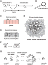
Figure 1
Figure 1. C. albicans Cell Type Transitions
A. C. albicans transitions reversibly among yeast (also known as whitea), hypha, and pseudohypha cell types under different environmental conditions. B. Chlamydospores are generated by terminal (suspensor) cells of mycelia (multicellular hyphae or pseudohyphae) under adverse growth conditions. C and D. In mucocutaneous infection models, such as oropharyngeal candidiasis, yeasts are associated with commensalism (C), whereas the filamentous forms (hyphae and pseudohyphae) are associated with tissue invasion and damage (D). E. Yeasts, hyphae and pseudohyphae all seem to have roles in disseminated disease, for example in abscesses within host internal organs. F. MTLa (“a”) or MTLα (“α”) cells can undergo an epigenetic switch between whitea and opaquea phenotypes. Whitea cells have the same appearance as typical whitea yeasts, while opaquea cells are elongated and have “pimple” structures on their cell surface. G. Mating in C. albicans requires three events: Loss of one allele of MTL (MTLα or MTLa) to generate a whitea phase a or α strain; an epigenetic switch from whitea to opaquea; and pheromone sensing by opaquea cells of opposite mating type, which triggers sexual filament production and mating.
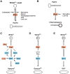
Figure 2
Figure 2. C. albicans Signaling and Morphogenesis
Numerous host signals and fungal signaling pathways have been implicated in the regulation of C. albicans cell shape. Based largely on in vitro analysis of wild-type C. albicans and specific gene deletion mutants, the signals and pathways depicted in this figure have been demonstrated to control the (whitea/α ) yeast-to-hypha transition and, in some cases, the whitea-to-opaquea switch and mating. The PKA pathway (teal blue) incorporates signals via the GTPase Ras1 (gray) and Ras1-independent inputs resulting in the synthesis of cAMP from ATP by the adenylyl cyclase Cyr1 and cAMP-mediated activation of the two catalytic subunits (Tpk1 and Tpk2) of the PKA complex. Once activated, the PKA complex phosphorylates the downstream transcription factor Efg1, eliciting a potent effect on both filamentation and white-to-opaquea switching. The Cek1 Mitogen-Activated Protein Kinase pathway (MAPK, navy blue) initiates a kinase signaling cascade in response to embedded growth (light blue), cell wall damage (navy blue), osmotic damage (beige), and low nitrogen (gray), ultimately phosphorylating the transcription factor Cph1 to induce filamentation. In opaquea cells, mating pheromone (red) signals through the same upstream MAPK signaling cascade but leads to the additional phosphorylation of the MAPK Cek2 and activation of mating genes. The Hog1 MAPK pathway (gray blue) recognizes osmotic and oxidative stress through either the Sln1 two-component protein or the Sho1 adaptor protein and leads to phosphorylation of the MAPK Hog1. Activated Hog1 can inhibit both Cek1- and Brg1-mediated filamentation. The RIM101 pathway (green) senses alkaline pH via two putative receptors (Dfg16 and Rim21) that initiate a proteolytic signaling cascade that results in C-terminal cleavage of the transcription factor Rim101 by the protease Rim13 and activation of Efg1 and filamentation-specific genes. The Ofd1 pathway (pink) and Tor1 pathway (light green) respond to low oxygen and starvation, respectively, to regulate filamentation through the transcription factors, Brg1 and Ume6. References for signalling pathways are provided in the main text, and those for transcription factors (dark rectangles) are listed here: , , , , , , , , , , , , , , , , , , , , , , , , , , , , , . Note that additional transcription factors and some instances of regulation via Ume6 have been omitted for visual clarity.
Figure 3
Figure 3. Opaquea/α, gray, and GUT cells
A) Certain MTLa/α strains switch reversibly between standard whitea/α (round-to-oval) morphology and opaquea/α morphology (elongated, with cell surface pimples). A subset of these strains can also switch to a third, gray (small, elongated, no pimples) morphology. B) Several C. albicans morphotypes exhibit enhanced fitness in specific host niches. MTLa/α hyphae and pseudohyphae exhibit superior virulence in localized oral infection models, whereas whitea/α yeasts, hyphae and pseudohyphae are all required for virulence in disseminated infections. MTL heterozygous opaquea/α and MTL homozygous opaquea cells have both been reported to have superior fitness in colonizing skin, whereas MTLa/α gray cells are the fastest proliferating cell type in an ex vivo tongue infection model. Finally, MTLa/α GUT cells outcompete other cell types in the mammalian gastrointestinal tract, with a relative fitness of GUT≫whitea/α ≫opaquea. C) GUT cells thrive within the digestive tract and rapidly revert to the whitea/α phenotype upon exit from the host, when signals required to maintain the GUT phenotype are removed. Thus, passage of MTLa/α white cells through the mammalian gastrointestinal tract is required for the whitea/α -to-GUT switch.
Figure 4
Figure 4. The Efg1 and Wor1 transcription factors have central roles in C. albicans morphological plasticity
Genetic studies have revealed roles for the transcription factors Efg1 and Wor1 in numerous morphological transitions. In each example below, arrows indicate activation, bars represent inhibition, and dashed lines indicate the direction of Efg1-promoted activity (with the exception of yeast-hypha morphogenesis (A), where Efg1 can support either transition, depending on environmental context). A) Efg1 promotes whitea/α cells to undergo the yeast-to-hypha transition upon exposure to host serum, N-acetylglucosamine, high CO2, nutrient depletion, and/or iron depletion; however, under agar-embedded conditions, Efg1 promotes the reverse transition from hypha-to-yeast. B) Efg1 promotes chlamydospore production by whitea/α hyphal suspensor cells under nutrient poor, oxygen-depleted conditions., C) Efg1 promotes and Wor1 opposes, , the opaquea-to-whitea switch, which is also controlled by additional transcription factors in a complex regulatory circuit. , , , , , Under conditions in which neither cell type is favoured (glucose-containing medium maintained at 25°C), switching may occur in either direction on an infrequent, stochastic basis, depending on changes in the ratio of Efg1 to Wor1 protein in individual cells. , Under conditions that strongly favour whitea (glucose-containing medium at 37°C) or opaquea (N-acetylglucosamine-containing medium in ≥5% CO2) cells, switching occurs in one direction across the entire cell population., , , D) Efg1 promotes the gray-to-whitea/α switch and Wor1 promotes the gray-to-opaquea/α switch., Gray cells are favored under nutrient-rich conditions, whereas opaquea/α cells are favored under nutrient-limited conditions in the presence of N-acetylglucosamine and elevated CO2. E) Efg1 promotes and Wor1 opposes the GUT-to-whitea/α switch. Whitea/α cells are favored under all tested conditions except for within the mammalian gastrointestinal tract.

References

    1. Hoffmann C, et al. Archaea and fungi of the human gut microbiome: correlations with diet and bacterial residents. PLoS One. 2013;8:e66019. - PMC - PubMed
    1. Ghannoum MA, et al. Characterization of the oral fungal microbiome (mycobiome) in healthy individuals. PLoS Pathog. 2010;6:e1000713. - PMC - PubMed
    1. Findley K, et al. Topographic diversity of fungal and bacterial communities in human skin. Nature. 2013;498:367–370. - PMC - PubMed
    1. Oyeka CA, Ugwu LO. Fungal flora of human toe webs. Mycoses. 2002;45:488–491. - PubMed
    1. Drell T, et al. Characterization of the vaginal micro- and mycobiome in asymptomatic reproductive-age Estonian women. PLoS One. 2013;8:e54379. - PMC - PubMed