. 2016 Nov 8:2:16040.
doi: 10.1038/celldisc.2016.40. eCollection 2016.

The HECT domain ubiquitin ligase HUWE1 targets unassembled soluble proteins for degradation

Affiliations

The HECT domain ubiquitin ligase HUWE1 targets unassembled soluble proteins for degradation

Yue Xu et al. Cell Discov. .

Abstract

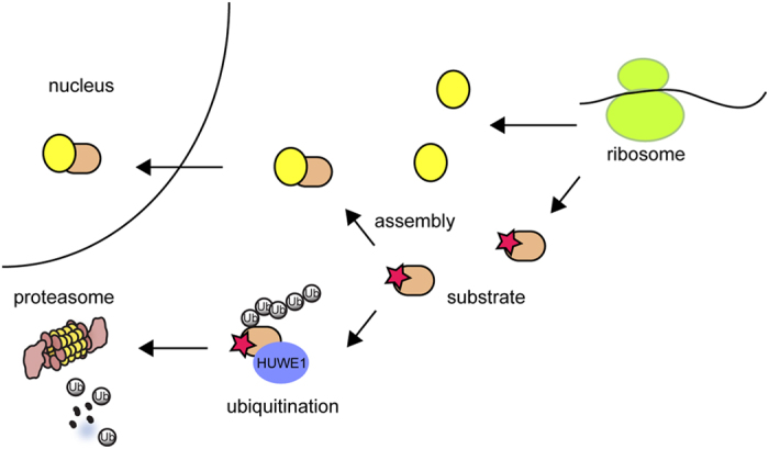
In eukaryotes, many proteins function in multi-subunit complexes that require proper assembly. To maintain complex stoichiometry, cells use the endoplasmic reticulum-associated degradation system to degrade unassembled membrane subunits, but how unassembled soluble proteins are eliminated is undefined. Here we show that degradation of unassembled soluble proteins (referred to as unassembled soluble protein degradation, USPD) requires the ubiquitin selective chaperone p97, its co-factor nuclear protein localization protein 4 (Npl4), and the proteasome. At the ubiquitin ligase level, the previously identified protein quality control ligase UBR1 (ubiquitin protein ligase E3 component n-recognin 1) and the related enzymes only process a subset of unassembled soluble proteins. We identify the homologous to the E6-AP carboxyl terminus (homologous to the E6-AP carboxyl terminus) domain-containing protein HUWE1 as a ubiquitin ligase for substrates bearing unshielded, hydrophobic segments. We used a stable isotope labeling with amino acids-based proteomic approach to identify endogenous HUWE1 substrates. Interestingly, many HUWE1 substrates form multi-protein complexes that function in the nucleus although HUWE1 itself is cytoplasmically localized. Inhibition of nuclear entry enhances HUWE1-mediated ubiquitination and degradation, suggesting that USPD occurs primarily in the cytoplasm. Altogether, these findings establish a new branch of the cytosolic protein quality control network, which removes surplus polypeptides to control protein homeostasis and nuclear complex assembly.

Keywords: HUWE1; p97/Cdc48; protein quality control (PQC); ubiquitin; unassembled soluble protein degradation (USPD).


Figures

Figure 1
Figure 1
Degradation of unassembled soluble proteins by the proteasome. (ac) Unassembled FNTA is degraded by the proteasome. (a) Cells transfected with FNTA-FLAG and FNTB-HA either individually or in combination were treated with dimethyl sulfoxide (DMSO) as a control or MG132 (20 μm, 6 h). Whole-cell extracts were analyzed by immunoblotting (IB). Asterisk, non-specific band. (b) Cycloheximide chase analysis of FNTA degradation in HEK293 T cells transfected with FNTA-FLAG. Where indicated, cells were treated with MG132 (20 μm). (c) Cycloheximide chase analysis of FNTA degradation in HEK293T cells transfected with FNTA-FLAG alone or together with FNTB-HA. (dg) Unassembled Ubl4A is regulated by proteasomal degradation. (d) Whole-cell extracts from control or Bag6 null CRISPR cells were analyzed by immunoblotting. (e) The Ubl4A messenger RNA (mRNA) level in parental 293T cells, control and Bag6 null clones was analyzed by qRT-PCR. Error bars, s.e.m. (n=3). (f) Whole-cell extracts from cells treated as indicated (MG132, 20 μm 6 h; C, chloroquine 50 μM) were analyzed by immunoblotting. (g) Bag6 CRISPR cells transfected as indicated were treated with cycloheximide in the presence of either DMSO as a control or MG132 (20 μm). Cell lysates prepared at the indicated time points were analyzed by immunoblotting. EV, empty vector.
Figure 2
Figure 2
Degradation of unassembled soluble proteins requires p97 and Npl4. (a) A p97 dominant negative mutant inhibits the degradation of unassembled FNTA. HEK293T cells transfected with FNTA-FLAG together with the indicated plasmids were subject to cycloheximide chase analysis. (b) Knockdown of p97 stabilized FNTA. HEK293T cells transfected with FNTA-FLAG together with the indicated siRNA were analyzed by cycloheximide chase. (c) Interaction of p97 with FNTA. Cells transfected with the indicated plasmids and siRNAs were subject to immunoprecipitation and immunoblotting analysis. (d) Knockdown of Npl4 but not Ufd1 stabilizes FNTA. Cells stably expressing FNTA-GFP were transfected with the indicated siRNAs. The level of FNTA-GFP was measured by immunoblotting analysis of immunoprecipitated samples. A fraction of whole-cell extracts were directly analyzed to verify knockdown efficiency.
Figure 3
Figure 3
An exposed hydrophobic residue-containing segment destabilizes unassembled Ubl4A. (a) A schematic illustration of the Ubl4A constructs used in the study. (b) The C-terminus of Ubl4A contains a ‘degron’. HEK293T cells transfected with the indicated plasmids were treated with dimethyl sulfoxide (−) or 10 μm MG132 (+) for 20 h. Whole-cell extracts were analyzed by immunoblotting. (c) Structural analysis of the Ubl4A-Bag6 interactions. Panel 1, The C-terminus of Ubl4A contains 3 helices (H1, H2 and H3). Panel 2, A surface view of the Ubl4A C-terminus. Residues are colored by hydrophobicity. Panel 3, Bag6 shields the hydrophobic residues in H1 and H2. Panel 4 shows a close-up view of the boxed area in panel 2. (d) Cycloheximide chase analysis of cells transfected with GFP appended with either the H1 or H2 segment from Ubl4A.
Figure 4
Figure 4
HUWE1 is required for degradation of unassembled Ubl4A. (a) The domain structure of HUWE1. BH3, BCL-2 homology; UBA, ubiquitin-associated; UIM, ubiquitin-interacting motif. (b, c) siRNA-mediated knockdown of HUWE1 stabilized Ubl4A-GFP. (b) Bag6 CRISPR cells stably expressing Ubl4A-GFP were transfected with two different HUWE1 siRNAs. Cells were imaged 48 h post transfection. (c) Cycloheximide chase analysis of Ubl4A-GFP degradation in Bag6 CRISPR cells stably expressing Ubl4A-GFP. Where indicated, cells were transfected with control or HUWE1 siRNA. (d) HUWE1 knockdown stabilizes endogenous Ubl4A in Bag6 null cells. (e) Generating HUWE1 null HEK293 T CRISPR cells. Two HUWE1 null clones were obtained. The graph indicates the relative rate of proliferation. Error bars, s.e.m. (n=3). (f) Cycloheximide chase analysis of Ubl4A-GFP degradation in control and HUWE1 CRISPR cells. (g) HUWE1 is required for ubiquitination of unassembled Ubl4A. Ubl4A overexpressed in either control or HUWE1 null cells was analyzed by immunoprecipitation and immunoblotting with ubiquitin (top panel) and Ubl4A (bottom panel) antibodies.
Figure 5
Figure 5
Identification of endogenous HUWE1 substrates. (a) Protein fold change comparison between two independent SILAC experiments (exp.). (b) The range of protein fold changes for the 3 433 proteins identified in HUWE1 knockout clone 1. The Venn diagram compared the top 172 proteins increased upon HUWE1 inactivation in the two different CRISPR clones. (c) The biological processes affected by HUWE1 inactivation. (d) Immunoblotting analyses validate the stabilization of proteins involved in DNA damage response pathways using HUWE1 CRISPR cells. The numbers in parentheses indicate fold changes measured by SILAC. The numbers under each gel panel show the band intensity. (e) Immunoblotting validation using HEK293T cells transfected with control or HUWE1 siRNA. (f) Unassembled POLR2G is unstable. Immunoblotting analysis of cells transfected with FLAG-tagged POLR2G (POLR2G-F) either alone or together with HA-tagged POLR2D. (g, h) The ubiquitination state of substrates in control and HUWE1 CRISPR cells. (g) Control or HUWE1 CRISPR cells were transfected with either an empty vector (EV) or a construct expressing FLAG-tagged POLR2G. Cell lysate were subject to immunoprecipitation under denaturing conditions and analyzed by immunoblotting. (h) Cell lysates from control or HUWE1 CRISPR cells transfected with the indicated HUWE1 substrates were subject to immunoprecipitation using FLAG beads. Precipitated proteins were analyzed by immunoblotting.
Figure 6
Figure 6
HUWE1 acts in the cytoplasm in USPD. (a) Localization of HUWE1 by immunostaining of cells transfected with either control or HUWE1 siRNA. (b) Biochemical fractionation analysis of HUWE1 localization in HEK293T cells. (c) Removal of NLS alters the localization of SF3B6. HeLa cells transfected with SF3B6-GFP or SF3B6∆NLS-GFP were stained with DAPI and imaged. (d) Deletion of the NLS increases SF3B6 ubiquitination by HUWE1. Lysates from control or HUWE1 CRISPR cells transfected as indicated were subject to immunoprecipitation under denaturing conditions with anti-FLAG beads. Immunoprecipitated proteins were analyzed by immunoblotting. (e) Expression of RAN Q69L inhibits degradation of unassembled Ubl4A. 293T Cells transfected as indicated were treated with cycloheximide in the presence of dimethyl sulfoxide as a control or MG132 (20 μM). Cell lysates prepared at the indicated time points were analyzed by immunoblotting. The graph shows the quantification of three experiments. Error bars, s.e.m. (n=3).
Figure 7
Figure 7
A model of HUWE1-mediated cytosolic PQC. On translation, proteins are assembled into multi-subunit protein complexes. The assembly process usually shields hydrophobic surfaces that would otherwise be exposed on unassembled subunit. Although assembled protein complexes can be transported to their final destination (for example, nucleus), unassembled protein subunits bearing an exposed hydrophobic segment (marked by the star) is subject to HUWE1-mediated ubiquitination, which targets them for degradation by the proteasome in the cytosol.

Similar articles

Cited by

References

    1. Scazzari M, Amm I, Wolf DH. Quality control of a cytoplasmic protein complex: chaperone motors and the ubiquitin-proteasome system govern the fate of orphan fatty acid synthase subunit Fas2 of yeast. J Biol Chem 2015; 290: 4677–4687. - PMC - PubMed
    1. Shemorry A, Hwang CS, Varshavsky A. Control of protein quality and stoichiometries by N-terminal acetylation and the N-end rule pathway. Mol Cell 2013; 50: 540–551. - PMC - PubMed
    1. Deshaies RJ. Proteotoxic crisis, the ubiquitin-proteasome system, and cancer therapy. BMC Biol 2014; 12: 94. - PMC - PubMed
    1. Hegde RS, Lingappa VR. Membrane protein biogenesis: regulated complexity at the endoplasmic reticulum. Cell 1997; 91: 575–582. - PubMed
    1. Ruggiano A, Foresti O, Carvalho P. Quality control: ER-associated degradation: protein quality control and beyond. J Cell Biol 2014; 204: 869–879. - PMC - PubMed