Review
. 2016 Oct;5(4):186-204.
doi: 10.21037/tp.2016.10.07.

Instrumentation for minimally invasive surgery in pediatric urology

Affiliations
Review

Instrumentation for minimally invasive surgery in pediatric urology

Dalia Gobbi et al. Transl Pediatr. 2016 Oct.

Abstract

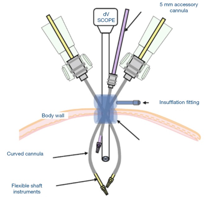
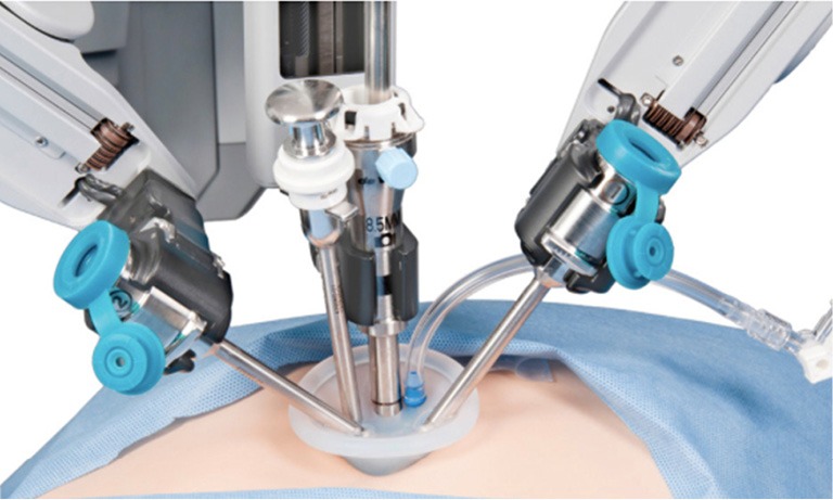
The success of modern surgery is dependent on the availability of good equipment and instruments. This dependence increases along with the degree of sophistication of the surgery performed. Paediatric minimally invasive and endoscopic surgery are sophisticated techniques where imaging is obtained through a video-circuit. Endoscopic surgery has opened the field of virtual reality in surgery, and in minimally invasive surgery the actual operation is done through a limited number of small holes. Robot-assisted urologic surgery is an emerging and safe technology for many urologic paediatric operations, although further documentation, including long-term functional outcome, is deemed necessary before definite conclusions can be drawn regarding the superiority or not of robotic assistance compared to conventional laparoscopic approaches.

Keywords: Paediatric minimally invasive surgery; endourology; laparoscopy; robotics; urology.


Conflict of interest statement

The authors have no conflicts of interest to declare.

Figures

Figure 1
Figure 1
Basic set: 19 or 26 “HD Flat screen”, Image 1 camera control, Light fountain (Xenon), fiber optic light cable, electronic CO2 thermoinflator neutral electrode, connecting cable for neutral electrode, unipolar high frequency cord, bipolar high frequency cord, two-pedal footswitch, double pedal holder, single pedal holder, AIDA compact HD (or alternative: Storz AIDA compact II system): for data management and documentation.
Figure 2
Figure 2
Operative paediatric cystoscope. (A) Removable instrument ports offer a choice between single or dual working channels; (B) the reduced outer diameter ensures minimal patient discomfort.
Figure 3
Figure 3
Right angles irrigation/suction ports prevent positioning instruments too close to the housing, enabling optimal handling.
Figure 4
Figure 4
A wide variety of accessories (forceps, electrodes and other instruments) 8/9.5 Fr.
Figure 5
Figure 5
Grasping and biopsy forceps, flexible, 3 and 5 Fr.
Figure 6
Figure 6
Hook electrode, unipolar, 3 Fr (for both 8 and 9.5 cystoscopes).
Figure 7
Figure 7
Knife, triangular tip, 3 Fr (for 8 and 9.5 cystoscopes) for single use (package of 6).
Figure 8
Figure 8
Grasping forceps, 5 Fr, only for 9.5 cystoscope, for larger stone fragments.
Figure 9
Figure 9
Pediatric operating cysto-urethroscopes. (A) 8 Fr cystoscope for use with reflux needles, 4 Fr working channel; (B) straight forward telescope 0° forward/oblique 30°, 19 mm diameter, for neonate urethrocystoscope 7–9 Fr.
Figure 10
Figure 10
Kit for paediatric urethrotomies. (A) Resectoscope (9 Fr): cutting by means of a spring (in the rest position, the electrode tip is inside the sheath); (B) accessory instruments for the 8 Fr urethrotome; (C) accessory instruments for the 9 Fr resectoscope.
Figure 11
Figure 11
The flexible system to access some of the most challenging areas (the bladder neck) and the entire intrarenal collecting system. (A) Cystourethrofiberscope; (B) tip deflection.
Figure 12
Figure 12
Ultrathin uretero-renoscope 7 Fr sheath, 43 cm L, distal tip 6.5 Fr.
Figure 13
Figure 13
Flexible uretero-renoscope (Storz Flex-X2).
Figure 14
Figure 14
Hopkins telescopes for laparoscopy. (A) Telescope; (B) 2 mm 0°–30°(trocar 2.5), 2.4 mm 0° (trocar 3.5), 3 mm 0°–30° (trocar 3.5), 3.3 mm 0°–30°–45°(trocar 3.9 mm), 5 mm 0°–30° (trocar 6).
Figure 15
Figure 15
Trocars for laparoscopy. (A) Trocar (sizes: 2.5–3.5–3.9–6 mm); (B) pyramidal tip; (C) blunt tip (and conical for the 6 mm).
Figure 16
Figure 16
Minitrocars.
Figure 17
Figure 17
Self-retained minitrocars.
Figure 18
Figure 18
Available instruments. (A) Click-line; (B) Scissors serrated, curved; (C) dissecting and grasping forceps (fenestrated jaws); (D) dissecting and grasping forceps (right angle jaws); (E) grasping forceps fenestrated with atraumatic serration; (F) dissecting and grasping forceps (Kelly); (G) bowel grasper.
Figure 19
Figure 19
Electrodes. Size 3 mm, for use with trocar 3.5 mm. L-shaped, insulated, length 20–30 mm.
Figure 20
Figure 20
Suction and irrigation instruments. (A) Size 3 mm, length 20 and 30 cm, trocar size 3.5 mm for use with two-way stopcock; (B) two-way stopcock device for irrigation and suction.
Figure 21
Figure 21
For extracorporeal knotting, size 3 mm, length 20 cm (or 30 cm), Trocar size 3.5 mm. Atraumatic.
Figure 22
Figure 22
Endobag.
Figure 23
Figure 23
Endoloop.
Figure 24
Figure 24
Endoclip.
Figure 25
Figure 25
2 (ultra micro) and 3 mm, for use with trocar size 2.5 and 3.5 mm, 20 cm length, for use with suture material 7.0-7.0.
Figure 26
Figure 26
Hasson trocar (Ethicon).
Figure 27
Figure 27
Hasson balloon trocar (Covidien).
Figure 28
Figure 28
Cohen procedure.
Figure 29
Figure 29
Landmarks and boundaries of the renal space and port position for retroperitoneoscopic nephrectomy.
Figure 30
Figure 30
Laparoscope 5.5 mm for varicocelectomy.
Figure 31
Figure 31
Bipolar forceps for varicocelectomy.
Figure 32
Figure 32
Endoeye for laparoscopic single-site surgery (LESS).
Figure 33
Figure 33
Platform for laparoscopic single-site surgery (LESS).
Figure 34
Figure 34
Surgeon’s position using platforms.
Figure 35
Figure 35
TRIPORT.
Figure 36
Figure 36
Gelpoint.
Figure 37
Figure 37
SILS.
Figure 38
Figure 38
Platform instrumentation.
Figure 39
Figure 39
da Vinci operating room (OR).
Figure 40
Figure 40
Needle driver.
Figure 41
Figure 41
Round tip scissors.
Figure 42
Figure 42
Curved scissors.
Figure 43
Figure 43
Monopolar cautery hook tip.
Figure 44
Figure 44
Monopolar cautery spatule tip.
Figure 45
Figure 45
Harmonic ACE curved scissors.
Figure 46
Figure 46
Endowrist instruments. (A) Maryland; (B) DeBakey; (C) Schertel; (D) bowel.
Figure 47
Figure 47
Robotic set-up for pyeloplasty.
Figure 48
Figure 48
Bipolar cautery PK.
Figure 49
Figure 49
Mega suture cut.
Figure 50
Figure 50
Prograsp forceps.
Figure 51
Figure 51
DeBakey forceps.
Figure 52
Figure 52
Round tip scissors.
Figure 53
Figure 53
Single site port has a target anatomy arrow indicator and five lumens (one the insufflation adapter and four to hold cannulae).
Figure 54
Figure 54
Single site for robotics.

References

    1. Kim C, Docimo SG. Use of laparoscopy in pediatric urology. Rev Urol 2005;7:215-23. - PMC - PubMed
    1. Godbole P,Koyle MA, Wilcox DT. editors. Pediatric Endourology Techniques. London: Springer-Verlag London, 2014.
    1. Smith AD, Badlani G, Bagley D, et al. editors. Smith's Textbook of Endourology. PMPH-USA, 2007.
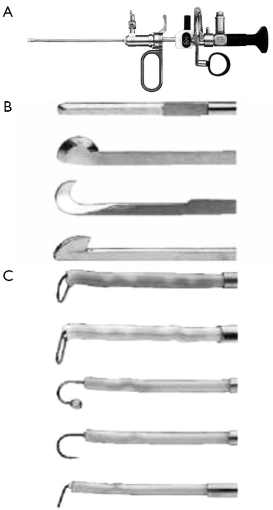
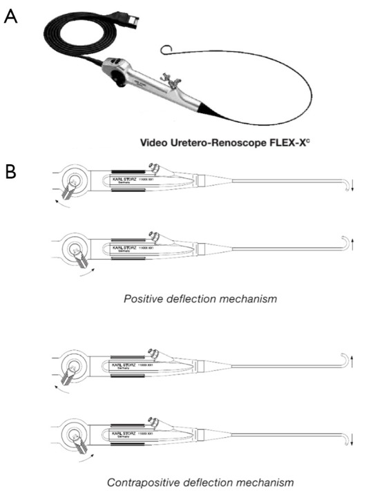
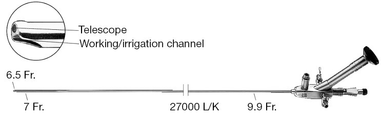
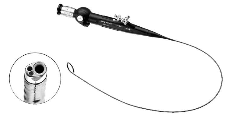
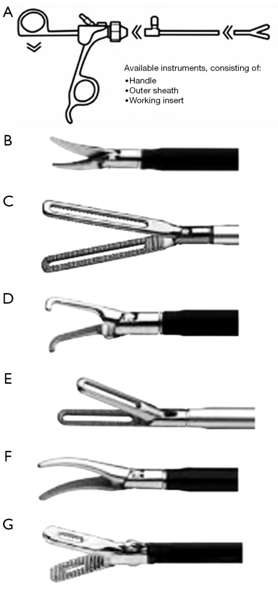
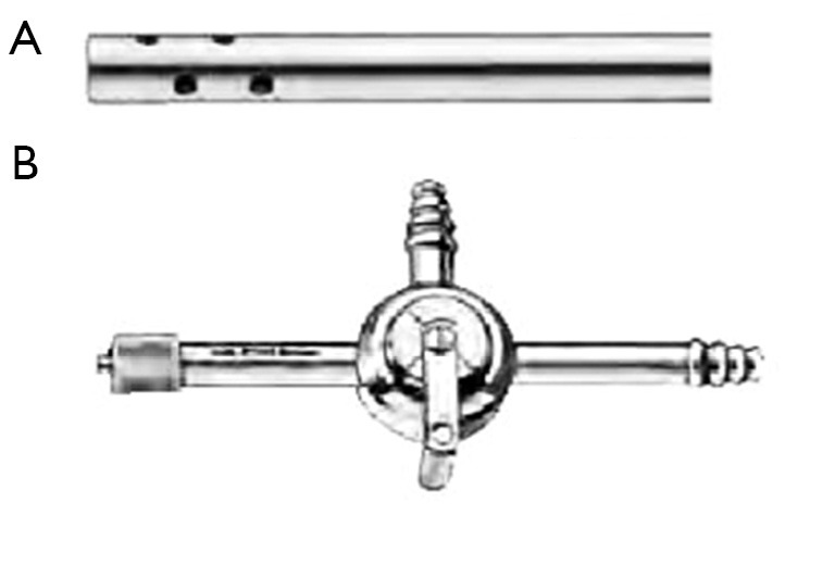
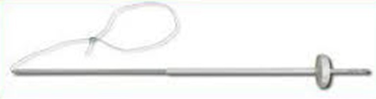
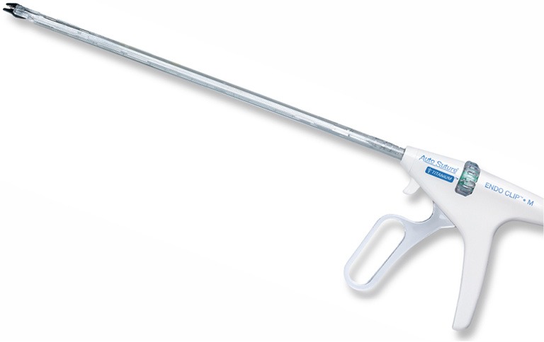
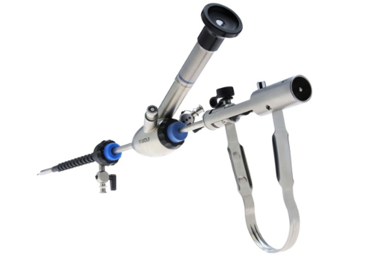
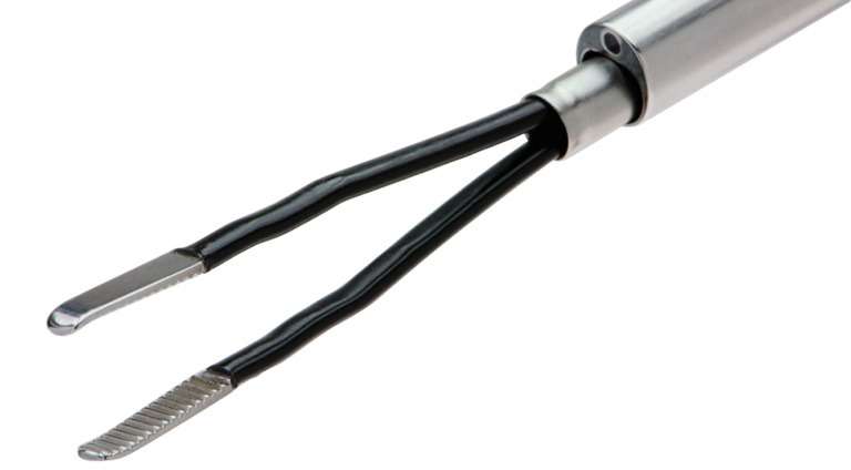
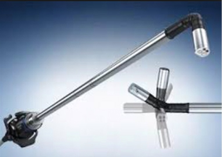
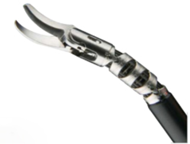
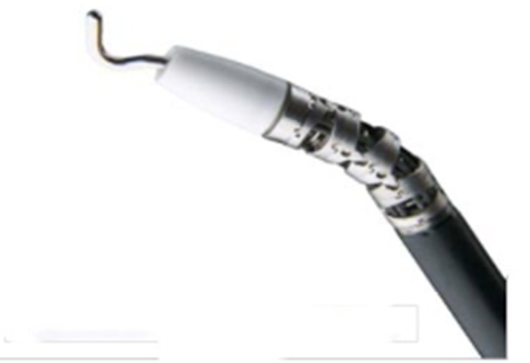
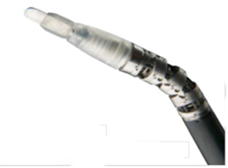
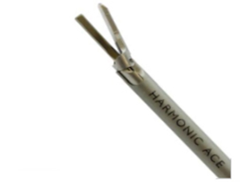
    1. Instrument set for laparoscopic and endourologic pediatric surgery-Karl Storz Endoskope Catalogue-Endoscopes, Instruments, accessories, Units and Imaging Systems. Available online: http://www.karlstorz.com/bt/en/online-catalog.htm
    1. Hidas G, Watts B, Khoury AE. The evolving role of laparoscopic surgery in paediatric urology. Arab J Urol 2012;10:74-80. 10.1016/j.aju.2011.12.010 - DOI - PMC - PubMed

LinkOut - more resources