Central Neurocytoma: A Review of Clinical Management and Histopathologic Features
- PMID: 27867912
- PMCID: PMC5114192
- DOI: 10.14791/btrt.2016.4.2.49
Central Neurocytoma: A Review of Clinical Management and Histopathologic Features
Abstract
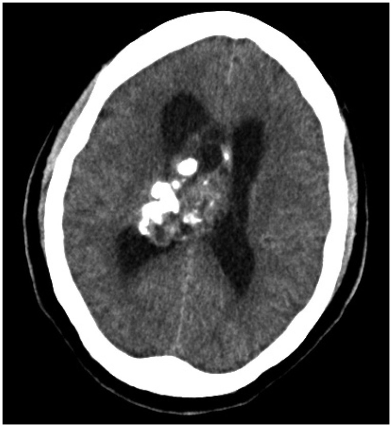
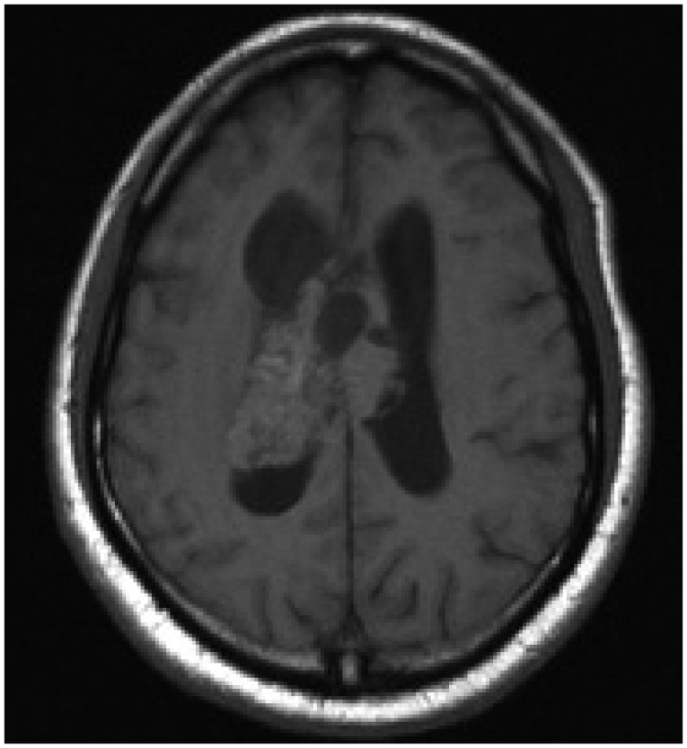
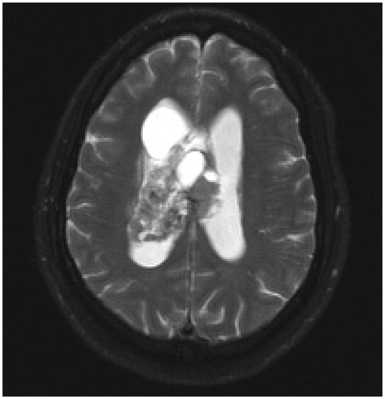
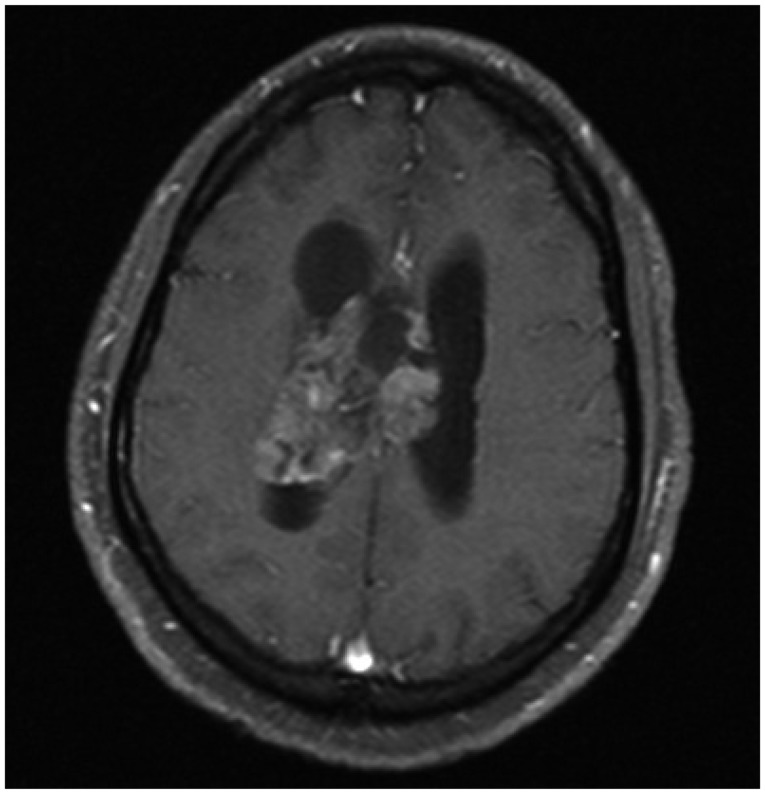
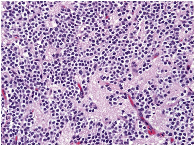
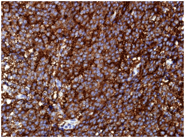
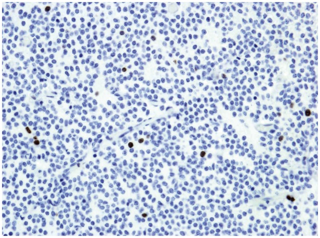
Central neurocytoma (CN) is a rare, benign brain tumor often located in the lateral ventricles. CN may cause obstructive hydrocephalus and manifest as signs of increased intracranial pressure. The goal of treatment for CN is a gross total resection (GTR), which often yields excellent prognosis with a very high rate of tumor control and survival. Adjuvant radiosurgery and radiotherapy may be considered to improve tumor control when GTR cannot be achieved. Chemotherapy is also not considered a primary treatment, but has been used as a salvage therapy. The radiological features of CN are indistinguishable from those of other brain tumors; therefore, many histological markers, such as synaptophysin, can be very useful for diagnosing CNs. Furthermore, the MIB-1 Labeling Index seems to be correlated with the prognosis of CN. We also discuss oncogenes associated with these elusive tumors. Further studies may improve our ability to accurately diagnose CNs and to design the optimal treatment regimens for patients with CNs.
Keywords: Central neurocytoma; Histopathology; Management.
Conflict of interest statement
The authors have no financial conflicts of interest.
Figures
References
-
- Hassoun J, Gambarelli D, Grisoli F, et al. Central neurocytoma. An electron-microscopic study of two cases. Acta Neuropathol. 1982;56:151–156. - PubMed
-
- Yang I, Ung N, Chung LK, et al. Clinical manifestations of central neurocytoma. Neurosurg Clin N Am. 2015;26:5–10. - PubMed
-
- Choudhari KA, Kaliaperumal C, Jain A, et al. Central neurocytoma: a multi-disciplinary review. Br J Neurosurg. 2009;23:585–595. - PubMed
-
- Coca S, Moreno M, Martos JA, Rodriguez J, Barcena A, Vaquero J. Neurocytoma of spinal cord. Acta Neuropathol. 1994;87:537–540. - PubMed
Publication types
LinkOut - more resources
Full Text Sources
Other Literature Sources