. 2016:2016:5214561.
doi: 10.1155/2016/5214561. Epub 2016 Oct 27.

Shrimp Protein Hydrolysate Modulates the Timing of Proinflammatory Macrophages in Bupivacaine-Injured Skeletal Muscles in Rats

Affiliations

Shrimp Protein Hydrolysate Modulates the Timing of Proinflammatory Macrophages in Bupivacaine-Injured Skeletal Muscles in Rats

Junio Dort et al. Biomed Res Int. 2016.

Abstract

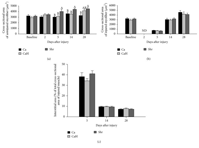
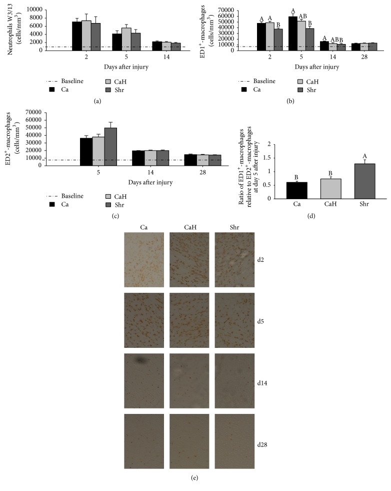
This study was designed to determine whether marine-derived proteins other than cod could have beneficial effects on inflammation following muscle injury. Macrophage and neutrophil densities were measured from bupivacaine-injured tibialis anterior muscle of rats fed isoenergetic diets containing either shrimp hydrolysate (Shr), casein hydrolysate (CaH), or whole casein (Ca). In this study, Shr reduced ED1+-macrophages at day 2 (p = 0.013), day 5 (p = 0.006), and day 14 after injury (p = 0.038) compared with Ca, indicating faster resolution of inflammation in Shr. Except for day 2 after injury where Shr led to lower ED1+-macrophages compared with CaH (p = 0.006), both Shr and CaH responded similarly at days 5, 14, and 28 after injury. This findings suggest that beneficial effects of Shr on ED1+-cells might be related to generation of anti-inflammatory peptides through the hydrolysis process, in addition to its high content of anti-inflammatory amino acids. However, while increasing myofiber cross-sectional area in noninjured muscles compared with both Ca and CaH, Shr failed to have a positive effect in corresponding injured muscles. These data indicate that shrimp hydrolysate can facilitate resolution of inflammation after muscle injury mainly through modulating proinflammatory macrophage accumulation but have less effect on optimal recovery in terms of muscle mass and fiber size.


Figures

Figure 1
Figure 1
Effect of dietary proteins on zootechnical parameters of rats after muscle injury. Rats were fed diets for 56 days (including 28 days before injury and 28 days after injury), containing either Ca, CaH, or Shr. Timeline of sacrifice and tibialis anterior muscle collection after bupivacaine injection (BPV) is shown in (a). Initial body weight (b), mean food intake (c), final body weight (d), and body weight gain (e) are given for each studied time point. Values are mean ± SEM (n = 7-8 rats per group/time point). Ca, casein; CaH, casein hydrolysate; Shr, shrimp protein hydrolysate.
Figure 2
Figure 2
Representative H/E stained cross-sections from injured tibialis anterior muscle.
Figure 3
Figure 3
Muscle mass recovery of regenerating tibialis anterior muscle in rats. Noninjured and bupivacaine-injured muscles were collected at days 0, 2, 5, 14, and 28 after injury, weighed, and normalized to body weight. Absolute muscle weights are presented for noninjured (a) and injured (b) muscles. A recovery index, as the percentage of injured values relative to noninjured values, was calculated and presented for muscle mass (c). Muscle mass is also presented as a percentage of body weight for injured (d) and noninjured (e) muscles. BCA Assay Kit was used to determine protein content in injured muscles (f). Values are mean ± SEM (n = 7-8 rats per group/time point). Groups bearing different letters for a given time point are significantly different (p ≤ 0.05). Ca, casein; CaH, casein hydrolysate; Shr, shrimp protein hydrolysate.
Figure 4
Figure 4
Muscle fiber cross-sectional area and interstitial area (%TCSA) of regenerating tibialis anterior muscle in rat. Myofiber cross-sectional area (MCSA) of three H/E-stained cryosections was quantified at days 0, 5, 14, and 28 after injury. Graphs are presented for noninjured (a) and injured (b) muscles. Two images were captured at 200x magnification from each cryosection and MCSA was traced using Image J analysis software. Total cross-sectional area (TCSA) was then traced in the injured muscle, and the interstitial area (IA) was quantified by subtracting the sum of individual MCSA from the TCSA at days 5, 14, and 28 after injury (c). Values are expressed as mean ± SEM (n = 7-8 rats per group/time point). Groups bearing different letters for a given time point are significantly different (p ≤ 0.05). ND, nondetermined because of massive destruction of the myofibers in the necrotic area. Ca, casein; CaH, casein hydrolysate; Shr, shrimp protein hydrolysate.
Figure 5
Figure 5
Time course of inflammatory cell accumulation in rat tibialis anterior muscle following bupivacaine injection. Transverse sections (10 μm) were immunoassayed with specific antibodies against neutrophils (W3/13) (a), ED1+-macrophages (b), or ED2+-macrophages (c). (d) shows the ratio of ED2+ relative to ED1+-cells at day 5 after injury. Labelled cells were counted at 400x magnification and expressed as a number of cells/mm3. Immunostained sections for ED1+-macrophages are also presented in (e); labelled ED1+-macrophages are identified by brown dots (e). Values are mean ± SEM (n = 7-8 rats per group/time point). Groups bearing different letters for a given time point are significantly different (p ≤ 0.05). Ca, casein; CaH, casein hydrolysate; Shr, shrimp protein hydrolysate.
Figure 6
Figure 6
MyoD and myogenin proteins in regenerating muscles. Expression of MyoD (a) and myogenin (b) (both are usually upregulated during muscle regeneration) was measured by immunoblotting in regenerating muscles at day 2 and day 5, respectively. A representative blot is displayed in the upper panel for each. All values were corrected for GAPDH as a protein loading control. Results are expressed as a percentage of values obtained at day 0 (mean optical density ± SEM, n = 7-8/group). Ca, casein; CaH, casein hydrolysate; Shr, shrimp protein hydrolysate.

Similar articles

Cited by

References

    1. Shireman P. K., Contreras-Shannon V., Ochoa O., Karia B. P., Michalek J. E., McManus L. M. MCP-1 deficiency causes altered inflammation with impaired skeletal muscle regeneration. Journal of Leukocyte Biology. 2007;81(3):775–785. doi: 10.1189/jlb.0506356. - DOI - PubMed
    1. Lapointe B. M., Frenette J., Côté C. H. Lengthening contraction-induced inflammation is linked to secondary damage but devoid of neutrophil invasion. Journal of Applied Physiology. 2002;92(5):1995–2004. - PubMed
    1. Dort J., Leblanc N., Maltais-Giguère J., Liaset B., Côté C. H., Jacques H. Beneficial effects of cod protein on inflammatory cell accumulation in rat skeletal muscle after injury are driven by its high levels of arginine, glycine, taurine and lysine. PLoS ONE. 2013;8(10, article e77274) doi: 10.1371/journal.pone.0077274. - DOI - PMC - PubMed
    1. Tidball J. G. Mechanisms of muscle injury, repair, and regeneration. American Journal of Physiology. Regulatory, Integrative and Comparative Physiology. 2029;1(4):2029–2062. - PubMed
    1. Tidball J. G., Villalta S. A. Regulatory interactions between muscle and the immune system during muscle regeneration. American Journal of Physiology—Regulatory Integrative and Comparative Physiology. 2010;298(5):R1173–R1187. doi: 10.1152/ajpregu.00735.2009. - DOI - PMC - PubMed

MeSH terms

LinkOut - more resources