. 2016 Nov 21:6:37403.
doi: 10.1038/srep37403.

Irradiance-dependent UVB Photocarcinogenesis

Affiliations

Irradiance-dependent UVB Photocarcinogenesis

Cheng-Che E Lan et al. Sci Rep. .

Abstract

Ultraviolet B (UVB) radiation from the sun may lead to photocarcinogenesis of the skin. Sunscreens were used to protect the skin by reducing UVB irradiance, but sunscreen use did not reduce sunburn episodes. It was shown that UVB-induced erythema depends on surface exposure but not irradiance of UVB. We previously showed that irradiance plays a critical role in UVB-induced cell differentiation. This study investigated the impact of irradiance on UVB-induced photocarcinogenesis. For hairless mice receiving equivalent exposure of UVB radiation, the low irradiance (LI) UVB treated mice showed more rapid tumor development, larger tumor burden, and more keratinocytes harboring mutant p53 in the epidermis as compared to their high irradiance (HI) UVB treated counterpart. Mechanistically, using cell models, we demonstrated that LI UVB radiation allowed more keratinocytes harboring DNA damages to enter cell cycle via ERK-related signaling as compared to its HI UVB counterpart. These results indicated that at equivalent exposure, UVB radiation at LI has higher photocarcinogenic potential as compared to its HI counterpart. Since erythema is the observed sunburn at moderate doses and use of sunscreen was not found to associate with reduced sunburn episodes, the biological significance of sunburn with or without sunscreen use warrants further investigation.


Figures

Figure 1
Figure 1. Effect of irradiance on UVB-induced skin tumor.
The hairless mice were treated with high or LI UVB radiation at equivalent surface exposure (200 mJ/cm2) three times per week. (A) The number of skin tumors per mouse after indicated UVB treatment. (B) The representative presentation of dorsal mouse skin after 18 weeks of indicated UVB treatment. Left: control mouse; middle: high irradiance (HI) UVB treated mouse; right: low irradiance (LI) UVB treated mouse. (C) The tumor burden per mouse after 18 weeks of indicated UVB treatment. (D) The expression of mutant p53 in the normal-appearing skin of hairless mice after indicated UVB treatment. Representative presentation of immunohistochemical staining of p53 (PAb240 antibody) on dorsal skin of control (left), HI UVB treated (middle), and LI UVB (right) mice after 8 wks (top panel) and 18 wks (bottom panel) of indicated UVB treatment. (E) The quantitative analyses of mutant p53 expression on the dorsal skin of control, HI, and LI UVB treated mice after 8 wks and 18 wks of indicated UVB treatment. Scale bar indicates 100 μm. *Indicates p < 0.05.
Figure 2
Figure 2. CPD and Brdu expression in cultured keratinocytes after indicated UVB irradiation.
(A) The CPD lesions of normal keratinocytes at 4 and 6 hr after high irradiance (HI) and low irradiance (LI) UVB (23 mJ/cm2) treatment as determined by ELISA specific for CPD. (B) Double labeling of HaCaT keratinocytes 3 hr after HI and LI UVB (23 mJ/cm2) treatment with anti-CPD antibody (green) and anti-BrdU (red). (C) Double labeling of ERK inhibitor (U0126; 5 μM) pretreated HaCaT keratinocytes 3 hr after HI and LI UVB (23 mJ/cm2) treatment with anti-CPD antibody (green) and anti-BrdU (red). The numbers show percentage (mean ± SD) of BrdU + CPD keratinocytes.
Figure 3
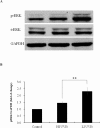
Figure 3. The expression of ERK pathway after indicated UVB treatment.
(A) Cultured HaCaT keratinocytes were treated with high irradiance (HI) and low irradiance (LI) UVB (23 mJ/cm2) treatment. The protein extracts were analyzed with western blotting using antibodies against phosphorylated ERK (p-ERK), total ERK (t-ERK), and GAPDH. (B) Densitometric analyses of (A) n = 3. **Indicates p < 0.05.
Figure 4
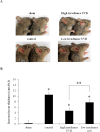
Figure 4. Contact hypersensitivity (CHS) was performed to compare the levels of immunosuppression after indicated UVB treatment.
(A) The representative presentation of ear swelling response after indicated treatment. The increase in ear thickness was defined as the amount of swelling of the 2,4,-dinitrofluorobenzene (DNFB)-challenged ear as compared to the thickness of vehicle treated ear. Upper Left: control mouse with no prior sensitization; Lower Left: Positive control mouse with prior sensitization; Upper Right: high irradiance (HI) UVB treated mouse before sensitization; Lower Right: low irradiance (LI) UVB treated mouse before sensitization. (B) The quantitative analyses of increase in ear thickness after indicated treatment. Sham: control mice with no prior sensitization; Control: mice with prior sensitization; HI UVB: mice treated with HI UVB prior to sensitization; LI UVB: mice treated with LI UVB treated mice prior to sensitization. *Indicates p < 0.05 as compared to sham treated group; **Indicates p < 0.05.

References

    1. Matsumura Y. & Ananthaswamy H. N. Toxic effects of ultraviolet radiation on the skin. Toxicol Appl Pharmacol 195, 298–308 (2004). - PubMed
    1. IARC Working Group. IARC Handbooks of cancer prevention. Vol. 5 (eds International Agency for Research on Cancer) Ch.5, Cancer-preventive effects of sunscreens. Pp69–124, (Lyon, France, 2001).
    1. Marrot L. & Meunier J. R. Skin DNA photodamage and its biological consequences. J Am Acad Dermatol 58, S139–148 (2008). - PubMed
    1. Christensen D. Data still cloudy on association between sunscreen use and melanoma risk. J Natl Cancer Inst 95, 932–933 (2003). - PubMed
    1. Ghiasvand R., Lund E., Edvardsen K., Weiderpass E. & Veierod M. B. Prevalence and trends of sunscreen use and sunburn among Norwegian women. Br J Dermatol 172, 475–483 (2015). - PubMed

Publication types

MeSH terms