. 2017 Jan 3;127(1):321-334.
doi: 10.1172/JCI87532. Epub 2016 Nov 21.

Pericyte MyD88 and IRAK4 control inflammatory and fibrotic responses to tissue injury

Pericyte MyD88 and IRAK4 control inflammatory and fibrotic responses to tissue injury

Irina A Leaf et al. J Clin Invest. .

Abstract

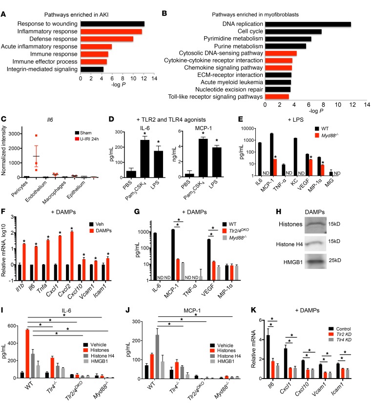
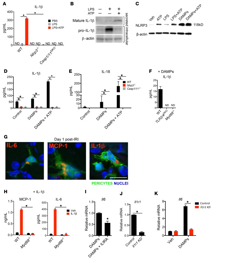
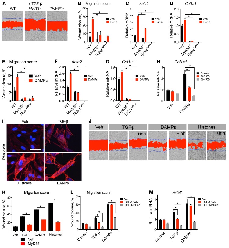
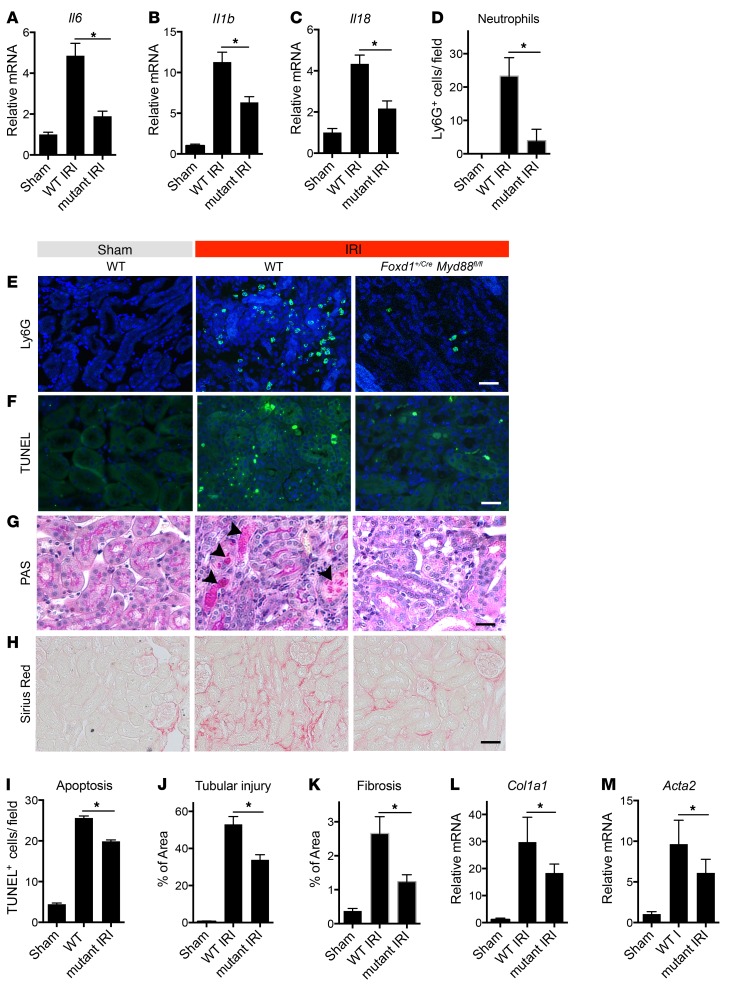
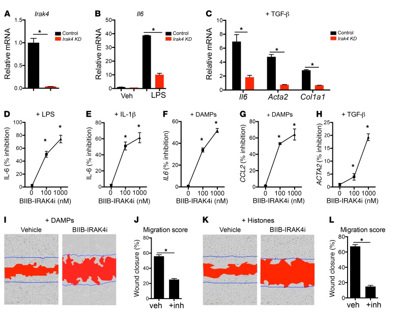
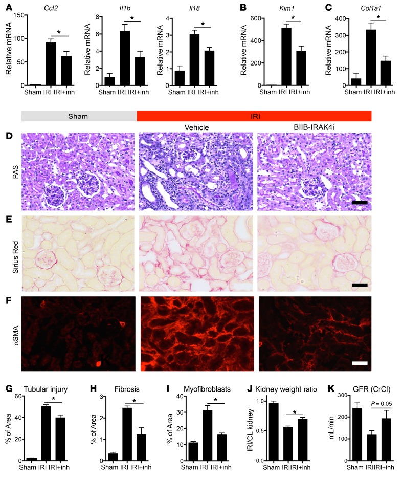
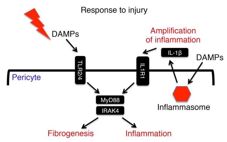
Fibrotic disease is associated with matrix deposition that results in the loss of organ function. Pericytes, the precursors of myofibroblasts, are a source of pathological matrix collagens and may be promising targets for treating fibrogenesis. Here, we have shown that pericytes activate a TLR2/4- and MyD88-dependent proinflammatory program in response to tissue injury. Similarly to classic immune cells, pericytes activate the NLRP3 inflammasome, leading to IL-1β and IL-18 secretion. Released IL-1β signals through pericyte MyD88 to amplify this response. Unexpectedly, we found that MyD88 and its downstream effector kinase IRAK4 intrinsically control pericyte migration and conversion to myofibroblasts. Specific ablation of MyD88 in pericytes or pharmacological inhibition of MyD88 signaling by an IRAK4 inhibitor in vivo protected against kidney injury by profoundly attenuating tissue injury, activation, and differentiation of myofibroblasts. Our data show that in pericytes, MyD88 and IRAK4 are key regulators of 2 major injury responses: inflammatory and fibrogenic. Moreover, these findings suggest that disruption of this MyD88-dependent pathway in pericytes might be a potential therapeutic approach to inhibit fibrogenesis and promote regeneration.


Conflict of interest statement

I.A. Leaf, B.G. Johnson, K.M. Guckian, I.G. Gomez, and J.S. Duffield own Biogen stock. K.M. Guckian, I.A. Leaf, and J.S. Duffield have filed a patent for the use of IRAK4 inhibition in the treatment of fibrosis (WO 2016011390 A1).

Figures

Figure 1
Figure 1. TLR2/4– and MyD88-dependent injury responses in pericytes.
(A and B) Enrichment analysis of biological process ontology in human biopsies from acute kidney injury (AKI) patients compared with healthy controls (A) and myofibroblasts from control and kidneys with acute injury (B). Innate immune–related pathways are highlighted in red. The x axis of the graph shows −log P values calculated using t test for the enrichment of a specific pathway. ECM, extracellular matrix. (C) RNA expression levels of Il6 in pericytes, macrophages, and epithelial and endothelial cells. Translated RNA was isolated from unilateral IRI (U-IRI) and sham control kidneys 24 hours after injury. The y axis shows normalized intensity for Il6 transcript in microarrays; n = 3 per group. (D) Cytokine concentration in supernatants from cultured pericytes 24 hours after stimulation with TLR ligands. (E) Secretion in WT or Myd88–/– pericytes 24 hours after treatment with LPS. (F) Transcriptional response of pericytes to diseased kidney DAMPs at 6 hours. (G) Cytokine concentration in supernatants of WT, Myd88–/–, or Tlr2/4DKO pericytes in response to diseased kidney DAMPs at 24 hours. (H) Western blot of kidney DAMPs. (I and J) IL-6 and MCP-1 concentration in supernatants of WT, Tlr4–/–, TLR2/4DKO, and Myd88–/– pericytes treated with either total histones, histone H4, or HMGB1 for 24 hours. (K) Effect of silencing of Tlr4 or Tlr2 on pericyte response to kidney DAMPs. (n = 3–6 per group; *P < 0.05, 1-way and 2-way ANOVA, Bonferroni’s multiple comparisons test.)
Figure 2
Figure 2. Activation of NLRP3 inflammasome by pericytes in response to DAMPs.
(A) Concentration of secreted IL-1β in WT and Nlrp3–/– and Casp1/11DKO pericytes 24 hours after treatment with LPS and ATP. (B) Western blot of intracellular pro–IL-1β and secreted mature IL-1β from pericytes 24 hours after treatment with LPS and ATP. (C) Western blot of pericyte cell lysates 24 hours after treatment with LPS, ATP, and DAMPs. (D and E) IL-1β and IL-18 concentration in WT and mutant pericyte supernatants after treatment with DAMPs and ATP. (F) IL-1β concentration in supernatants of WT, TLR2/4DKO, and Myd88–/– pericytes 24 hours after treatment with DAMPs. (G) Immunofluorescence staining for IL-1β, IL-6, and MCP-1 on sections from kidneys on day 1 after IRI. (H) IL-6 and MCP-1 cytokine response to IL-1β treatment in the supernatant of WT and Myd88–/– pericytes. (I) The effect of recombinant IL-1 receptor antagonist (IL1RA) on transcriptional response to DAMPs. (J and K) The effect of siRNA against Il1r1 on Il1r1 expression (J) and Il6 response to DAMPs (K). ND, not detected. KD, knockdown. (Scale bar: 25 μm; n = 3–6 per group; *P < 0.05, 2-tailed Student’s t test or 2-way ANOVA, Bonferroni’s multiple comparisons test.)
Figure 3
Figure 3. TLR2/4 and MyD88 control fibrogenic responses of mouse and human pericytes.
(A) Representative images showing migratory response of WT, Myd88–/–, and Tlr2/4DKO pericytes to 24-hour TGF-β treatment. Blue lines mark the boundaries of the scratch at 0 hours; red area designates scratch boundaries at 24 hours. (B) Quantification of TGF-β migration assays. (C and D) Transcription of Acta2 and Col1a1 induced by TGF-β. (EG) Responses of WT, Myd88–/–, and Tlr2/4DKO pericytes at 24 hours to kidney DAMP treatment. (E) Quantification of migration. (F and G) Transcription of Acta2 and Col1a1. (H) Effect of Tlr2 or Tlr4 silencing with siRNA on upregulation of Col1a1 stimulated by DAMPs. (I) Stress fiber formation shown by phalloidin staining in human kidney pericytes treated with TGF-β, histones, or kidney DAMPs. (J and K) Representative images (J) and quantification (K) of migratory response of human pericytes to TGF-β, histones, or DAMPs in the presence of a MyD88 inhibitor or vehicle. (L) Quantification of migration induced by TGF-β or DAMPs in the presence or absence of TGF-β1,2,3 neutralizing antibody (NAb) or TGFRβI/II inhibitor LY2109761, 24 hours after treatment. (M) Transcription of myofibroblast marker Acta2 in response to TGF-β or DAMPs in the presence or absence of the TGFRβI/II inhibitor LY2109761, in mouse pericytes after 24-hour treatment. (Scale bar: 25 μm; n = 3–6 per group; *P < 0.05, 2-tailed Student’s t test or 2-way ANOVA, Bonferroni’s multiple comparisons test.)
Figure 4
Figure 4. Myd88 deletion specifically in pericytes attenuates injury and fibrotic responses following IRI.
(AC) Transcription of inflammatory genes in kidneys from control and day 5 post-IRI kidneys from WT and Foxd1+/Cre Myd88fl/fl (referred to as mutant) mice. (D) Quantification of Ly6G+ neutrophils on kidney sections. (EH) Representative images of kidney sections stained with Ly6G antibody, TUNEL, PAS, or Sirius red. (F) TUNEL+ cells are labeled green. (G) Necrotic tubules in PAS-stained sections are labeled with arrowheads. (H) Fibrosis is detected as red stain. (IK) Graphs quantifying apoptotic (TUNEL+) cells (I), tubular injury score based on PAS-stained sections (J), and fibrosis score based on Sirius red staining (K). (L and M) Transcription of Col1a1 and Acta2. (Scale bars: 50 μm; n = 6 per group; *P < 0.05, 2-tailed Student’s t test or 2-way ANOVA, Bonferroni’s multiple comparisons test.)
Figure 5
Figure 5. Role of IRAK4 in pericytes and its inhibition in vitro by BIIB-IRAK4i.
(A) Irak4 transcript levels in pericytes after treatment with either scramble or Irak4-specific siRNA. (B) Il6 transcript levels in control and Irak4 knockdown (KD) pericytes 24 hours after treatment with LPS. (C) Il6, Col1a1, and Acta2 expression in control and Irak4 KD pericytes 24 hours after TGF-β treatment. (DH) Inhibition of injury responses by IRAK4 inhibitor BIIB-IRAK4i in kidney pericytes in vitro. (D and E) Inhibition of IL-6 secretion by mouse kidney pericytes stimulated with LPS (D) or IL-1β (E) in the presence of BIIB-IRAK4i. (F and G) Inhibition of IL6 and CCL2 transcription by human kidney pericytes in response to kidney DAMPs. (H) Inhibition of ACTA2 transcription by human kidney pericytes following TGF-β treatment. (IL) Inhibition of pericyte migration by IRAK4 inhibitors. (I and K) Representative images of human pericytes stimulated with kidney DAMPs (I) or histones (K) and either vehicle or IRAK4 inhibitors in scratch-wound assays. Blue lines mark the scratch boundaries at 0 hours, and red area designates the scratch at 16 hours. (J and L) Migration scores of DAMP-induced (J) or histone-induced (L) migration. (n = 3–6 per group; *P < 0.05, 2-tailed Student’s t test, 1-way or 2-way ANOVA, Bonferroni’s multiple comparisons test.)
Figure 6
Figure 6. IRAK4 inhibition attenuates inflammatory and fibrotic responses in vivo and improves organ function.
(AC) Effect of kidney IRI and BIIB-IRAK4i (inh) on whole-kidney transcriptional responses at 7 days after injury showing inflammatory genes (A), epithelial injury marker (B), and Col1a1 (C). (DI) Representative histological and immunofluorescence images (DF) and quantitative analysis of kidneys from IRI at day 7 after injury treated with BIIB-IRAK4i compared with vehicle or sham control kidneys. (GI) Quantification of tubular injury, interstitial fibrosis, and extent of myofibroblasts detected by αSMA. (J) Quantification of kidney weight changes associated with unilateral IRI. Ratios of ischemic kidneys’ to contralateral (CL) kidneys’ weights 14 days after injury are shown. (K) Measurement of glomerular filtration rate (GFR) by creatinine clearance (CrCl) in post-IRI kidney at 16 days, following contralateral nephrectomy at 14 days. (Scale bars: 50 μm; n = 3–11 per group; *P < 0.05, 2-tailed Student’s t test or 2-way ANOVA, Bonferroni’s multiple comparisons test.)
Figure 7
Figure 7. Schematic of injury sensing by MyD88 in pericytes.
Pericytes sense tissue injury through TLR2 and TLR4. Both MyD88 and IRAK4 are important in transducing this signal and activation of inflammatory responses. Injurious molecules also activate inflammasome and release of additional proinflammatory cytokines including IL-1β. Pericytes respond to IL-1β, and this generates an autocrine loop that amplifies the inflammatory response. In addition to inflammatory responses, activation of the TLR/MyD88/IRAK4 axis by tissue injury induces myofibroblast differentiation.

Comment in

References

    1. Duffield JS. Cellular and molecular mechanisms in kidney fibrosis. J Clin Invest. 2014;124(6):2299–2306. doi: 10.1172/JCI72267. - DOI - PMC - PubMed
    1. Zeisberg M, Kalluri R. Fibroblasts emerge via epithelial-mesenchymal transition in chronic kidney fibrosis. Front Biosci. 2008;13:6991–6998. - PubMed
    1. LeBleu VS, et al. Origin and function of myofibroblasts in kidney fibrosis. Nat Med. 2013;19(8):1047–1053. doi: 10.1038/nm.3218. - DOI - PMC - PubMed
    1. Cieslik KA, Trial J, Entman ML. Mesenchymal stem cell-derived inflammatory fibroblasts promote monocyte transition into myeloid fibroblasts via an IL-6-dependent mechanism in the aging mouse heart. FASEB J. 2015;29(8):3160–3170. doi: 10.1096/fj.14-268136. - DOI - PMC - PubMed
    1. Asada N, et al. Dysfunction of fibroblasts of extrarenal origin underlies renal fibrosis and renal anemia in mice. J Clin Invest. 2011;121(10):3981–3990. doi: 10.1172/JCI57301. - DOI - PMC - PubMed

Publication types

MeSH terms