. 2016 Dec;28(12):10.1111/jne.12444.
doi: 10.1111/jne.12444.

Timing of Maternal Exposure and Foetal Sex Determine the Effects of Low-level Chemical Mixture Exposure on the Foetal Neuroendocrine System in Sheep

Affiliations

Timing of Maternal Exposure and Foetal Sex Determine the Effects of Low-level Chemical Mixture Exposure on the Foetal Neuroendocrine System in Sheep

M Bellingham et al. J Neuroendocrinol. 2016 Dec.

Abstract

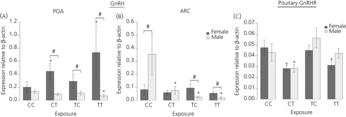
We have shown that continuous maternal exposure to the complex mixture of environmental chemicals (ECs) found in human biosolids (sewage sludge), disrupts mRNA expression of genes crucial for development and long-term regulation of hypothalamic-pituitary gonadal (HPG) function in sheep. The present study investigated whether exposure to ECs only during preconceptional period or only during pregnancy perturbed key regulatory genes within the hypothalamus and pituitary gland and whether these effects were different from chronic (life-long) exposure to biosolid ECs. The findings demonstrate that the timing and duration of maternal EC exposure influences the subsequent effects on the foetal neuroendocrine system in a sex-specific manner. Maternal exposure prior to conception, or during pregnancy only, altered the expression of key foetal neuroendocrine regulatory systems such as gonadotrophin-releasing hormone and kisspeptin to a greater extent than when maternal exposure was 'life-long'. Furthermore, hypothalamic gene expression was affected to a greater extent in males than in females and, following EC exposure, male foetuses expressed more 'female-like' mRNA levels for some key neuroendocrine genes. This is the first study to show that 'real-life' maternal exposure to low levels of a complex cocktail of chemicals prior to conception can subsequently affect the developing foetal neuroendocrine system. These findings demonstrate that the developing neuroendocrine system is sensitive to EC mixtures in a sex-dimorphic manner likely to predispose to reproductive dysfunction in later life.

Keywords: GnRH; endocrine disruptors; foetal; hypothalamus; kisspeptin; oestrogen receptor.


Figures

Figure 1
Figure 1
Maternal exposure to biosolids chemicals before conception only (TC), during pregnancy only (CT) or life‐long (TT) has sex‐ and region‐ specific effects on foetal male and female gonadotrophin‐releasing hormone (GnRH) mRNA expression in the hypothalamus. In the preoptic area (POA) (a), a divergent effect on expression can be seen between males (suppression) and females (increase) in the TT group. Similarly in the arcuate nucleus (ARC) (b), exposure is associated with suppression in males but no effect in females. In the pituitary gland (c), the effect of exposure on GnRH receptor (GnRHR) expression is also sexually dimorphic, with a greater effect in females compared to males relative to unexposed foetuses (CC); n = 12 (CC and TT); n = 11 (CT); n = 10 (TC). *Significant difference compared to respective control group (P < 0.05). #Significant sex difference within a particular group (P < 0.05).
Figure 2
Figure 2
Maternal exposure to biosolids chemicals before conception only (TC), during pregnancy only (CT) or life‐long (TT) has sex‐ and region‐ specific effects on foetal male and female KiSS1 and KiSS1R mRNA expression. For KiSS1 (a, b) and KiSSR expression (c, d), a greater number of effects are seen in males compared to females. Interestingly, for KiSS1 expression in both the preoptic area (POA) (a) and arcuate nucleus (ARC) (b) regions of hypothalamus, life‐long exposure reduced expression whereas pregnancy only exposure (CT) caused an increase in expression relative to unexposed foetuses (CC). For both KISS1 and KISSR, life‐long exposure resulted in sexually dimorphic expression levels that were not seen when comparing control males and females; n = 12 (CC and TT); n = 11 (CT); n = 10 (TC). *Significant difference compared to respective control group (P < 0.05). #Significant sex difference within a particular group (P < 0.05).
Figure 3
Figure 3
Maternal exposure to biosolids chemicals before conception only (TC), during pregnancy only (CT) or life‐long (TT) has sex‐ and region‐ specific effects on foetal male and female oestrogen receptor (ER)α mRNA expression in the (a) preoptic area (POA) and (b) arcuate nucleus (ARC) regions of hypothalamus and (c) in the pituitary gland relative to each other and to unexposed foetuses (CC). In both the POA and ARC, there is sexually dimorphic expression between CC males and females that is subsequently lost in the TT group. Interestingly, pituitary gland ERα expression is only affected in females exposed to biosolids chemicals; n = 12 (CC and TT); n = 11 (CT); n = 10 (TC). *Significant difference compared to respective control group (P < 0.05). #Significant sex difference within a particular group (P < 0.05).
Figure 4
Figure 4
Aryl hydrocarbon receptor (AhR) mRNA expression in the (a) preoptic area (POA) and (b) arcuate nucleus (ARC) regions of hypothalamus. An effect of exposure was only seen in pregnancy only (CT) exposed males in the ARC relative to controls (CC) with no effect of exposure in the preconception (TC) or life‐long (TT) exposure groups. Interestingly, female foetal hypothalamic AhR expression was not affected by treatement, which contrasts with the effects on AhR expression in the pituitary gland (c) where expression was reduced in all exposed groups relative to controls, whereas male AhR expression was unaffected; n = 12 (CC and TT); n = 11 (CT); n = 10 (TC). *Significant difference compared to respective control group (P < 0.05). #Significant sex difference within a particular group (P < 0.05).

References

    1. Barker D, Eriksson J, Forsén T, Osmond C. Fetal origins of adult disease: strength of effects and biological basis. Int J Epidemiol 2002; 31: 1235–1239. - PubMed
    1. Seckl JR. Prenatal glucocorticoids and long‐term programming. Eur J Endocrinol. 2004; 151(Suppl 3): U49–U62. - PubMed
    1. Grandjean P, Barouki R, Bellinger DC, Casteleyn L, Chadwick LH, Cordier S, Etzel RA, Gray KA, Ha EH, Junien C, Karagas M, Kawamoto T, Paige Lawrence B, Perera FP, Prins GS, Puga A, Rosenfeld CS, Sherr DH, Sly PD, Suk W, Sun Q, Toppari J, van den Hazel P, Walker CL, Heindel JJ. Life‐long implications of developmental exposure to environmental stressors: new perspectives. Endocrinology 2015; 156: 3408–3415. - PMC - PubMed
    1. Rogers JM. Tobacco and pregnancy: overview of exposures and effects. Birth Defects Res C Embryo Today 2008; 84: 1–15. - PubMed
    1. Bruin JE, Gerstein HC, Holloway AC. Long‐term consequences of fetal and neonatal nicotine exposure: a critical review. Toxicol Sci 2010; 116: 364–374. - PMC - PubMed

Publication types

MeSH terms