Review
. 2017 Jan 2;95(1-2):24-39.
doi: 10.1002/jnr.23809.

Understanding the broad influence of sex hormones and sex differences in the brain

Affiliations
Review

Understanding the broad influence of sex hormones and sex differences in the brain

Bruce S McEwen et al. J Neurosci Res. .

Abstract

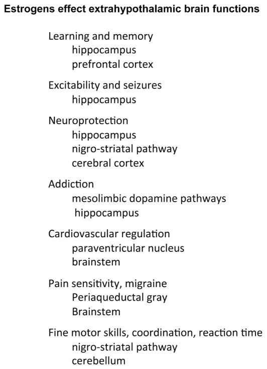
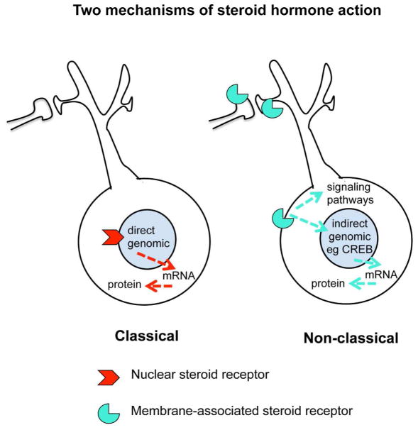
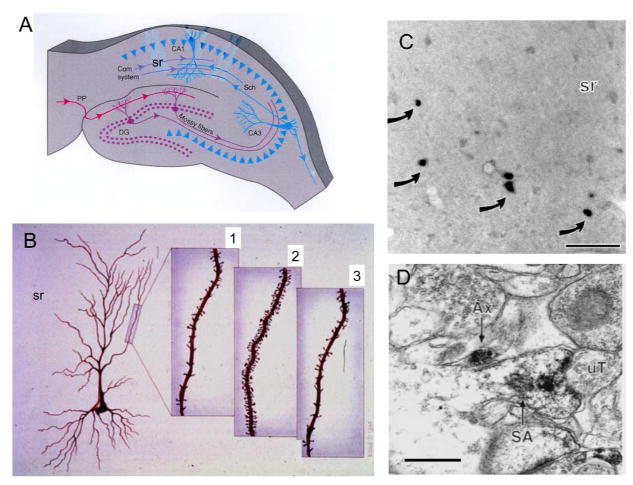
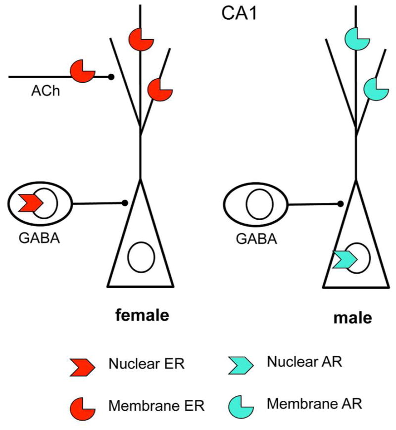
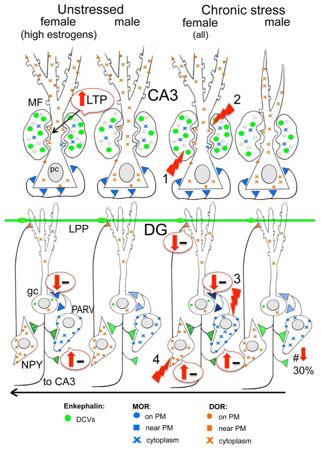
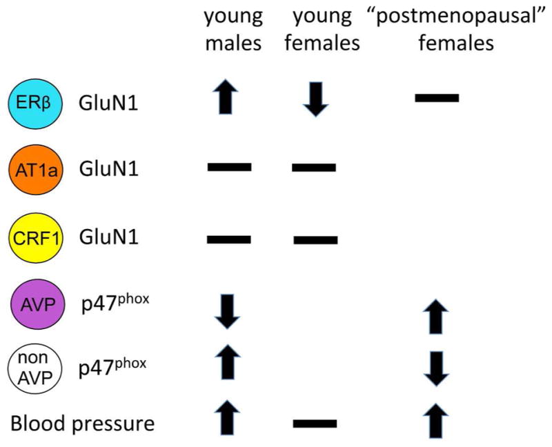
Sex hormones act throughout the entire brain of both males and females via both genomic and nongenomic receptors. Sex hormones can act through many cellular and molecular processes that alter structure and function of neural systems and influence behavior as well as providing neuroprotection. Within neurons, sex hormone receptors are found in nuclei and are also located near membranes, where they are associated with presynaptic terminals, mitochondria, spine apparatus, and postsynaptic densities. Sex hormone receptors also are found in glial cells. Hormonal regulation of a variety of signaling pathways as well as direct and indirect effects on gene expression induce spine synapses, up- or downregulate and alter the distribution of neurotransmitter receptors, and regulate neuropeptide expression and cholinergic and GABAergic activity as well as calcium sequestration and oxidative stress. Many neural and behavioral functions are affected, including mood, cognitive function, blood pressure regulation, motor coordination, pain, and opioid sensitivity. Subtle sex differences exist for many of these functions that are developmentally programmed by hormones and by not yet precisely defined genetic factors, including the mitochondrial genome. These sex differences and responses to sex hormones in brain regions, which influence functions not previously regarded as subject to such differences, indicate that we are entering a new era of our ability to understand and appreciate the diversity of gender-related behaviors and brain functions. © 2016 Wiley Periodicals, Inc.

Keywords: cardiovascular; cerebellum; estrogens; hippocampus; prefrontal cortex; stress.


Conflict of interest statement

The authors have no conflicts to declare.

Figures

Figure 1
Figure 1
Estrogens have many effects throughout the brain.
Figure 2
Figure 2
Steroid hormones can act via classical (genomic) and non-classical (non-genomic) receptors. In many cases, the same receptor molecule has different functions in the nucleus and non-nuclear sites in the cell.
Figure 3
Figure 3
The discovery of estrogen actions on synapse formation in hippocampus via both genomic and non-genomic mechanisms has opened the way to understanding actions of estrogens and other steroid hormones throughout the brain where nuclear steroid hormone receptors are not evident. A. Schematic of coronal section through the dorsal rat hippocampus. Com, commissural; DG, dentate gyrus; PP, perforant path; Sch, Schaffer collaterals; sr, stratum radiatum. B. Representation of CA1 pyramidal neurons in the female rat hippocampus during the 4–5 day estrous cycle. 1. Diestrus, when estradiol levels are lowest. 2. Proestrus, when estradiol levels are decreasing. 3. Estrous, when estradiol levels are decreasing (A and B from McEwen and Schmeck. The Hostage Brain. Rockefeller Univ. Press, 1994. Drawings by Lidia Kibiuk.) C. Light micrograph shows nuclei with ERα-ir (arrows) in the stratum radiatum (sr) of CA1. D. Electron micrograph shows ERα-ir in a dendritic spine identified by a spine apparatus (SA) that is contacted by an unlabeled terminal (uT). An axon with ERα-ir is nearby. Bar C 40 μm; D, 500nm (C and D modified from Milner et al. (Milner et al 2001).
Fig. 4
Fig. 4
Sex differences in CA1 steroid hormone receptor distribution. Nuclear and nonnuclear steroid receptors are found on different cell types and/or cellular locations in females and males. Both females and males have membrane receptors on dendrites and dendritic spines. However, in females nuclear ERs are in GABAergic neurons whereas in males ARs are in pyramidal cell neurons. Moreover, membrane estrogen receptors are found on cholinergic (ACh) afferents.
Fig. 5
Fig. 5
Schematic shows sex differences in the hippocampal opioid system in unstressed and CIS rats. Arrows indicate predicted effects of MOR/DOR trafficking changes on inhibition (minus signs). In unstressed conditions, low frequency (1 Hz) stimulation of the granule cells elicits a DOR-dependent LTP in CA3 of proestrus females that is not seen in diestrus females or males (Harte-Hargrove et al 2015). The opioid system in all females after CIS resembles that of females in elevated estrogen states: (1) the pool of available enkephalin is elevated in mossy fibers; (2) DORs are decreased in the dendritic shafts but increased in the spines of CA3 pyramidal cells; and (3) MORs are increased in the dendrites and terminals of PARV GABA interneurons in the dentate gyrus (DG). Moreover, (4) after CIS in females, DORs have mobilized to the near-plasmalemma of the dendrites of GABAergic NPY/SOM interneurons known to project to granule cell dendrites where they converge with entorhinal afferents. Abbreviations: δ opioid receptors (DORs), dense-core vesicles (DCVs), lateral perforant path (LPP), long-term potentiation (LTP), μ opioid receptors (MORs), neuropeptide Y (NPY), plasma membrane (PM), somatostatin (SOM)
Fig. 6
Fig. 6
Sex differences in PVN neuron responses to slow pressor AngII. Fourteen days following slow pressor Angiotensin II delivered through osmotic minipumps, blood pressor increases in young males and in accelerated ovarian failure females from a timepoint that corresponds to “postmenopause” in humans. The trafficking of GluN1 and p47phox within PVN neurons varies depending on sex, ovarian hormone status and cell type. Arrows indicate direction of movement towards the plasma membrane. Receptors on the plasma membrane are available for ligand binding. Thus, an increase of GluN1 would indicate potential for greater excitability.

References

    1. Akama KT, McEwen BS. Estrogen stimulates postsynaptic density-95 rapid protein synthesis via the Akt/protein kinase B pathway. J Neurosci. 2003;23:2333–39. - PMC - PubMed
    1. Almey A, Cannell E, Bertram K, Filardo E, Milner TA, Brake WG. Medial prefrontal cortical estradiol rapidly alters memory system bias in female rats: ultrastructural analysis reveals membrane-associated estrogen receptors as potential mediators. Endocrinology. 2014;155:4422–32. - PMC - PubMed
    1. Almey A, Filardo EJ, Milner TA, Brake WG. Estrogen receptors are found in glia and at extranuclear neuronal sites in the dorsal striatum of female rats: evidence for cholinergic but not dopaminergic colocalization. Endocrinology. 2012;153:5373–83. - PMC - PubMed
    1. Almey A, Milner TA, Brake WG. Estrogen receptors in the central nervous system and their implication for dopamine-dependent cognition in females. Horm Behav. 2015;74:125–38. - PMC - PubMed
    1. Altman J, Das GD. Autoradiographic and histological evidence of postnatal hippocampal neurogenesis in rats. J Comp Neurol. 1965;124:319–36. - PubMed

Substances