. 2016 Nov 21;11(1):21.
doi: 10.1186/s13064-016-0076-8.

Akirin2 is essential for the formation of the cerebral cortex

Affiliations

Akirin2 is essential for the formation of the cerebral cortex

Peter J Bosch et al. Neural Dev. .

Abstract

Background: The proper spatial and temporal regulation of dorsal telencephalic progenitor behavior is a prerequisite for the formation of the highly-organized, six-layered cerebral cortex. Premature differentiation of cells, disruption of cell cycle timing, excessive apoptosis, and/or incorrect neuronal migration signals can have devastating effects, resulting in a number of neurodevelopmental disorders involving microcephaly and/or lissencephaly. Though genes encoding many key players in cortical development have been identified, our understanding remains incomplete. We show that the gene encoding Akirin2, a small nuclear protein, is expressed in the embryonic telencephalon. Converging evidence indicates that Akirin2 acts as a bridge between transcription factors (including Twist and NF-κB proteins) and the BAF (SWI/SNF) chromatin remodeling machinery to regulate patterns of gene expression. Constitutive knockout of Akirin2 is early embryonic lethal in mice, while restricted loss in B cells led to disrupted proliferation and cell survival.

Methods: We generated cortex-restricted Akirin2 knockouts by crossing mice harboring a floxed Akirin2 allele with the Emx1-Cre transgenic line and assessed the resulting embryos using in situ hybridization, EdU labeling, and immunohistochemistry.

Results: The vast majority of Akirin2 mutants do not survive past birth, and exhibit extreme microcephaly, with little dorsal telencephalic tissue and no recognizable cortex. This is primarily due to massive cell death of early cortical progenitors, which begins at embryonic day (E)10, shortly after Emx1-Cre is active. Immunostaining and cell cycle analysis using EdU labeling indicate that Akirin2-null progenitors fail to proliferate normally, produce fewer neurons, and undergo extensive apoptosis. All of the neurons that are generated in Akirin2 mutants also undergo apoptosis by E12. In situ hybridization for Wnt3a and Wnt-responsive genes suggest defective formation and/or function of the cortical hem in Akirin2 null mice. Furthermore, the apical ventricular surface becomes disrupted, and Sox2-positive progenitors are found to "spill" into the lateral ventricle.

Conclusions: Our data demonstrate a previously-unsuspected role for Akirin2 in early cortical development and, given its known nuclear roles, suggest that it may act to regulate gene expression patterns critical for early progenitor cell behavior and cortical neuron production.

Keywords: Apoptosis; Cortical development; Dorsal telencephalon; Microcephaly; Neural progenitor; Neuronal differentiation.


Figures
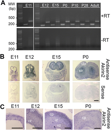
Fig. 1
Fig. 1
The Akirin2 gene is expressed throughout cortical development. a RT-PCR using two different primer-sets that generate amplicons across multiple exons. Akirin2 is expressed in telencephalic/cortical tissue at E11, E12, E15, P0, P10, P28 and in the adult mouse. The -RT control confirms that none of the bands are due to genomic DNA contamination. b, c Low magnification (b) and higher magnification (c) images of in situ hybridization experiments utilizing an antisense riboprobe for Akirin2 indicate that it is expressed throughout the developing telencephalon, with high expression in the early ventricular zone (vz) and in post-mitotic neurons situated in the preplate (pp; E12), intermediate zone (iz) or cortical plate (cp). No appreciable signal was observed using an Akirin2 sense probe in these or any other experiments. ctx, cortex; str, striatum. Scale bar: 1 mm in (b), 200 μm in (c)
Fig. 2
Fig. 2
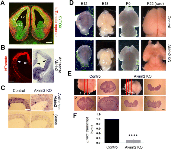
Disruption of the Akirin2 gene results in loss of the cerebral cortex. a A coronal section of the telencephalon of an Emx1-Cre; Ai14-tdTomato E12 embryo confirms the restriction of floxed allele excision to the dorsal (dCx) and lateral (lCx) cortex; Sytox Green is used as a cellular counterstain. b, c Low (b) and higher (c) magnification views of in situ hybridization utilizing Akirin2 probes on E11 telencephalon. Loss of Akirin2 begins in the dorsal cortex (arrows mark similar positions on two different E11 sections showing tdTomato Cre reporter fluorescence and antisense Akirin2 staining); some Akirin2 expression is still detected in the lateral cortex at this stage, but is not observed later as Cre activity expands (b). In knockout cortex at E11, Akirin2 antisense signal is reduced compared to controls, and is nearing background sense probe levels (c). d Akirin2 knockout causes severe microcephaly, which is already apparent by E12 and which leads to little, if any, remaining cortex in perinatal animals and in the rare animals that survive past P0 (one such knockout that survived to P22 is shown). e Cresyl violet staining of a rare surviving Akirin2 knockout and its littermate control at P16. There is no identifiable cortex or hippocampus in the knockout brain, though ventral structures appear to be present (note, for example, the prominent mammillary bodies in the lower right image). f Quantitative RT-PCR of control and Akirin2 knockout E15 forebrain RNA using a Taqman probe set for Emx1. Transcripts are nearly absent in the knockout tissue, consistent with loss of the cortex. ****p < 0.0001. LGE, lateral ganglionic eminence; LV, lateral ventricle. Scale bar: 200 μm in (a); 200 μm in (b); 100 μm in (c); 1 mm in (e)
Fig. 3
Fig. 3
Severe loss of cortical tissue in Akirin2 knockout embryos. a Hematoxylin and eosin staining of coronal sections of control and Akirin2 knockout cortex (ctx) at E12, shown at two locations on the rostro-caudal axis. The knockout contains only a thin epithelium dorsomedially and exhibits cells falling/spilling into the lateral ventricle (LV). b Closeup images of boxed areas in (a); arrows show cells spilling into the LV. c, d Comparative low (c) and higher (d) magnification images of tdTomato reporter and Sytox Green fluorescence in coronal sections of control and Akirin2 knockout cortex at 4 locations on the rostro-caudal axis. In the knockout, there are still tdTomato cells present, although these are drastically fewer in number and found only laterally, or situated in a thin epithelium dorsally. e Control and knockout sagittal sections at E11 showing tdTomato-positive cells. In the knockout at this timepoint, cells are already observed spilling in the LV (arrows) and cell loss is more pronounced rostrally than caudally. f Higher magnification of boxed area in (e); arrows show individual cells that are double positive for tdTomato and Sytox Green within the LV. di, diencephalon; LGE, lateral ganglionic eminence; MGE, medial ganglionic eminence; SA, septal area. Scale bar: 200 μm in (b, c, d, e); 90 μm in (f)
Fig. 4
Fig. 4
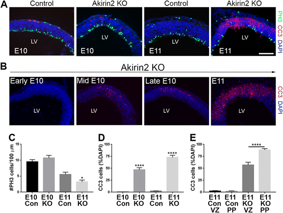
Apoptosis of Akirin2 knockout cortical progenitors and neurons. a Sections from mid-E10 and E11 (a) control and knockout cortex were stained for the mitotic marker phospho-histone H3 (PH3; green), apoptotic cell marker CC3 (red), and DAPI (blue). At mid-E10, controls and knockouts exhibit a similar number of PH3+ profiles at the ventricular surface (c); a significantly higher number of CC3+ dying cells are observed close to the emerging preplate (a, d). The cell death progresses between E10 and E11 (b), with no apoptotic cells apparent at early E10; CC3+ cells appear at mid-E10, and their number increases at late E10 and peaks at E11. At E11, Akirin2 knockouts exhibit significantly fewer PH3-positive mitotic cells (a, c), CC3 staining is greatly increased (d, e), and the tissue is already thinner (a). Further analysis of the CC3+ cells indicates that a greater percentage of cell death occurs in cells close to the pial surface at the developing preplate (PP) compared with cells closer to the apical venticular surface (VZ; e). *p < 0.05,****p < 0.0001. LV, lateral ventricle. Scale bar: 200 μm
Fig. 5
Fig. 5
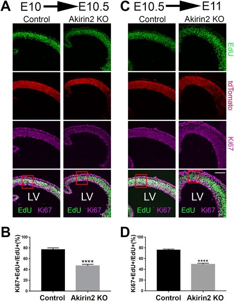
Cell cycle analysis of cortical progenitors in Akirin2 knockout embryos. Pregnant dams were injected with EdU at E10 and embryos collected 12 h later at E10.5 (a, b), or injected at E10.5 and embryos collected 12 h later at E11 (c, d). Coronal sections were double stained for EdU and Ki67 and cells from the medial dorsal telencephalon were assessed for expression of these two markers. The ratio of Ki67 + EdU+/EdU+ cells was significantly reduced in Akirin2 knockout embryos at both E10.5 (b) and E11 (d). Red boxes in (a and c) demarcate the regions quantified in (b and d); all analyzed cells were positive for the Cre reporter, tdTomato. ****p < 0.0001. Scale bar: 100 μm
Fig. 6
Fig. 6
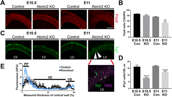
Disrupted production of cortical neurons in Akirin2 knockout embryos. a, b The number of Pax6+ progenitors in the Akirin2 knockout is comparable to controls at E10.5, but significantly reduced by E11 (a, b). c, d In contrast, there are already significantly fewer Tuj1+ neurons in knockouts at E10.5; this difference is maintained at E11. Unlike in controls, where all neurons migrate superficially to form a tight preplate (PP) band, at E11 some knockout neurons that double-stain for Tuj1 and TBR2 appear “trapped” at the apical ventricular surface (arrowheads, and inset, from c). e Neurons that do successfully migrate to the PP in the Akirin2 knockout form a less tightly-defined layer (black lines) compared with control (blue lines), indicated by a line-scan of Tuj1 intensity across the thickness of the cortical wall (e). Subsequently, Tuj1 fluorescence intensity is spread more broadly at the PP and is also more apparent at the apical ventricular surface (VS) and throughout the ventricular zone (VZ) in the Akirin2 knockout. **p < 0.01, ****p < 0.0001. LV, lateral ventricle. Scale bar: 100 μm in (a, c); 50 μm in (c) inset
Fig. 7
Fig. 7
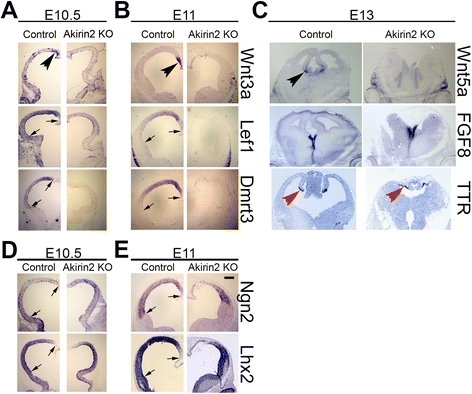
Disruption of the cortical hem in the absence of Akirin2. In situ hybridization for a number of cortical patterning genes at E10.5 (a, d), E11 (b, e) and E13 (c) is shown. The cortical hem, a medial structure that releases Wnt signaling molecules to pattern the cortex, is disrupted in Akirin2 knockouts, as few if any Wnt3a or Wnt5a transcripts are detected (a, b). Consistent with this, the Wnt-responsive genes Lef1 and Dmrt3 are reduced or absent in the knockout cortex (a, b). The transthyretin (TTR)-positive choroid plexus, and the ventromedial domain of Fgf8 expression, both of which are wholly or partially spared by Emx1-Cre, remain present in the knockout (c), though the location of the choroid plexus is shifted dorsally due to the loss of cortical tissue. Expression patterns of the proneural transcription factors Ngn2 and Lhx2 appear fairly normal in the dorsal cortical region, although the loss of tissue documented previously is observed at E11 (d, e). Arrows represent the region of Emx1-Cre activity at E10.5 and E11, identified by tdTomato reporter boundaries in other sections not shown. Black arrowheads show the Wnt3a and Wnt5a-positive cortical hem and red arrowheads show the TTR-positive choroid plexus. Scale bar: 200 μm in (a, b, d, e); 300 μm in (c)
Fig. 8
Fig. 8
Disruption of the ventricular zone apical surface in Akirin2 knockout embryos. a, b High magnification images of the control (a) and Akirin2 knockout (b) ventricular zone at E11 stained for N-cadherin and connexin-43. In controls, these proteins are very prominent at apical cell-cell junctions at the ventricular surface, and connexin-43 is also observed at gap junctions through which the cell bodies of progenitors are coupled (a). Both proteins are patchy and severely reduced in Akirin2 knockouts, and are absent at sites where cells are “spilling” into the lateral ventricle (b). Red arrowheads show chromatin condensation in pyknotic DAPI-stained nuclei observed in the Akirin2 knockout cortex (b). Akirin2 knockout embryos at E11 also display “rosette-like” circular cell clusters within the VZ that contain some Sox2-positive cells but do not stain for Pax6 (white arrows; c). The cells that spill into the lateral ventricle in Akirin2 knockout cortex appear to be Sox2+ but Pax6– (d, white arrowheads). The dashed line indicates the apical ventricular surface. LV, lateral ventricle; VS, apical ventricular surface. Scale bar: 50 μm

Similar articles

Cited by

References

    1. Florio M, Huttner WB. Neural progenitors, neurogenesis and the evolution of the neocortex. Development. 2014;141(11):2182–2194. doi: 10.1242/dev.090571. - DOI - PubMed
    1. Hu WF, Chahrour MH, Walsh CA. The diverse genetic landscape of neurodevelopmental disorders. Annu Rev Genomics Hum Genet. 2014;15:195–213. doi: 10.1146/annurev-genom-090413-025600. - DOI - PMC - PubMed
    1. Chenn A, Walsh CA. Regulation of cerebral cortical size by control of cell cycle exit in neural precursors. Science. 2002;297(5580):365–369. doi: 10.1126/science.1074192. - DOI - PubMed
    1. Kuida K, Zheng TS, Na S, Kuan C, Yang D, Karasuyama H, et al. Decreased apoptosis in the brain and premature lethality in CPP32-deficient mice. Nature. 1996;384(6607):368–372. doi: 10.1038/384368a0. - DOI - PubMed
    1. Ross ME, Walsh CA. Human brain malformations and their lessons for neuronal migration. Annu Rev Neurosci. 2001;24:1041–1070. doi: 10.1146/annurev.neuro.24.1.1041. - DOI - PubMed

Publication types