. 2016 Nov 17;64(4):826-834.
doi: 10.1016/j.molcel.2016.09.033. Epub 2016 Oct 27.

Conformational Control of Cascade Interference and Priming Activities in CRISPR Immunity

Affiliations

Conformational Control of Cascade Interference and Priming Activities in CRISPR Immunity

Chaoyou Xue et al. Mol Cell. .

Abstract

During type I-E CRISPR-Cas immunity, the Cascade surveillance complex utilizes CRISPR-derived RNAs to target complementary invasive DNA for destruction. When invader mutation blocks this interference activity, Cascade instead triggers rapid primed adaptation against the invader. The molecular basis for this dual Cascade activity is poorly understood. Here we show that the conformation of the Cse1 subunit controls Cascade activity. Using FRET, we find that Cse1 exists in a dynamic equilibrium between "open" and "closed" conformations, and the extent to which the open conformation is favored directly correlates with the attenuation of interference and relative increase in priming activity upon target mutation. Additionally, the Cse1 L1 motif modulates Cascade activity by stabilizing the closed conformation. L1 mutations promote the open conformation and switch immune response from interference to priming. Our results demonstrate that Cascade conformation controls the functional outcome of target recognition, enabling tunable CRISPR immune response to combat invader evolution.


Figures

Figure 1
Figure 1
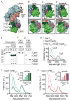
Direct detection of Cse1 conformational changes by FRET. (A) Full view of apo Cascade structure (PDB 4TVX). Cse1 NTD: dark green, Cse1 CTD: light green, Cas5: purple, Cse2 subunits: light orange, Cas7 subunits: light blue, Cas6: light red. (B–D) Close-up of Cse1 DNA-binding face (top) and solvent-exposed face (bottom) in (B) apo, (C) ssDNA-bound and (D) dsDNA-bound Cascade. PDB IDs: (B) 4TVX; (C) 4QYZ with modeled chains from 4TVX (see Supplemental Experimental Procedures); (D) 5H9F. The L1 motif is shown in red, and the PAM is shown in yellow in the dsDNA bound structure. Cy3 (green stars) and Cy5 (red stars) labeling sites are indicated, with corresponding distances between sites. See also Table S1 and Movies S1–2. (E–G) SDS-PAGE analysis of Cy3 and Cy5 labeling. The gel was analyzed by (E) Coomassie blue staining, (F) Cy3 scan at 560–580 nm or (G) Cy5 scan at ≥665 nm. Gel lanes: (1) Wild-type (WT) Cse1 or Cse2-Cas6, containing all native Cys residues. (2) Minimal-Cys Cse1 or Cse2-Cas6. (3) Minimal-Cys N73C Cse1 or Minimal-Cys Cse2-Cas6 containing K169C Cas5. See also Figure S1. (H) Fluorescence emission spectra for individually labeled Cse1 and Cse2-Cas6, and reconstituted FRET-enabled Cascade. Excitation wavelength = 530 nm. (I–J) Fluorescence emission spectra for FRET-enabled Cascade containing (I) Cse1-NTDCy3 or (J) Cse1-CTDCy3 binding to various concentrations of (I) ssDNA or (J) dsDNA. The insets show EFRET at various molar equivalents of the DNA substrates, error represents mean (n = 3) ± SD. Dashed lines are spectra for Cy3-labeled Cse1 + DNA in the absence of acceptor, solid lines are spectra for FRET-enabled Cascade + DNA.
Figure 2
Figure 2
Target mutations inhibit interference through alteration of Cse1 conformation. (A) Flow cytometry spectra for E. coli cells harboring empty GFP-reporter plasmid (–), or GFP-reporter plasmid with targets containing a canonical PAM and fully-matching (FM) or seed mismatch (MM, position 1 rG-dG mismatch) protospacer after one growth cycle (8 h). (B) Flow cytometry spectra for E. coli cells harboring GFP-reporter plasmids containing fully-matching protospacer with canonical PAM (AAG) or mutant PAMs (AAA, AGA) after two growth cycles (12 h each). For (A–B) high GFP fluorescence (green) indicates full retention of plasmid, low GFP fluorescence (blue) indicates partial plasmid loss, and GFP– cells (red) indicate complete plasmid loss. (C–D) Primed spacer acquisition against GFP-reporter targets after (C) one 8 h or (D) two 12 h growth cycles. CRISPR arrays from genomic DNA were PCR amplified and visualized by gel electrophoresis to detect the acquisition of new spacers (+1 band) relative to original (O) product. (E–F) Quantified interference and priming efficiencies. Interference efficiencies are the percentage of GFP– cells in flow cytometry experiments. Priming efficiencies are quantified from CRISPR amplicons (see Supplemental Experimental Procedures). Error represents mean (n = 3) ± SD. (F–G) Changes in EFRET for Cascade containing (F) Cse1-NTDCy3 or (G) Cse1-CTDCy3 upon binding to DNA targets. For each domain, FRET was measured with Cy3 located at two different positions. For (F–G), error represents mean (n = 3) ± propagated SD (see Supplemental Experimental Procedures). See also Figures S2A–C and S2E–N. (H) In vitro Cascade-dependent Cas3 cleavage of pUC19 plasmid DNA containing either a FM or MM protospacer (with AAG PAM), analyzed by gel electrophoresis. Cascade was bound to plasmid prior to initiation of Cas3 cleavage, and aliquots were quenched at indicated time points. See also Figure S2D. DNA is labeled as follows: OC – Open Circle; L – Linear; nSC – Negatively supercoiled; D – Degraded.
Figure 3
Figure 3
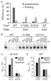
Cse1 L1 mutants switch immune response by promoting the open conformation. (A) Plasmid loss and spacer acquisition rates for strains expressing WT, F129A and N131A Cse1 against a GFP-reporter plasmid containing a bona fide target after 8 h growth. For comparison, WT activity against an AGA target after 8 h growth is also plotted. WT plasmid loss against the canonical target are reproduced from Figures 2B–C. Error represents mean (n = 3) ± SD. See also Figures S4A–B. (B) In vitro interference assay against plasmid target. Cascade bearing WT, F129A or N131A Cse1 were bound to DNA prior to initiation of Cas3 cleavage. See also Figures S4C–D. (C) Change in EFRET for Cascade containing N131A Cse1-NTDCy3 or N131A Cse1-CTDCy3 upon binding to a canonical dsDNA target with fully-matching protospacer and AAG PAM, or ssDNA with fully-matching protospacer. Cy3 labeling positions: NTD – N73C; CTD – N376C. See also Figures S4E–I. (D) Change in EFRET for Cascade containing WT (minimal-Cys) Cse1-NTDCy3 or Cse1-CTDCy3 upon binding to a canonical dsDNA target with fully-matching protospacer and AAA PAM, or ssDNA with fully-matching protospacer. Values are reproduced from experiments shown in Figure 2G–H for comparison with (C). For (C–D), error represents mean (n = 3) ± SD propagated for subtraction (see Experimental Procedures).
Figure 4
Figure 4
Model for conformational control of Cascade function. (A) When Cse1 adopts the closed conformation, CTD locking leads to recruitment of the Cas3 endonuclease and interference. (B) Alternatively, Cse1 adopts the open conformation, which may promote priming through Cas1–Cas2-dependent recruitment of Cas3 (Redding et al., 2015). The two conformations in (A) and (B) exist in a dynamic equilibrium. Favorable interactions between the PAM-NTD and L1-crRNA promote the closed and locked conformation, while disruption of these interactions through mutation of the PAM, seed or L1 motif promote the open conformation.

Comment in

References

    1. Barrangou R, Fremaux C, Deveau H, Richards M, Boyaval P, Moineau S, Romero DA, Horvath P. CRISPR provides acquired resistance against viruses in prokaryotes. Science (80– ) 2007;315:1709–1712. - PubMed
    1. Blosser TR, Loeff L, Westra ER, Vlot M, Kunne T, Sobota M, Dekker C, Brouns SJJ, Joo C. Two distinct DNA binding modes guide dual roles of a CRISPR-Cas protein complex. Mol Cell. 2015;58:60–70. - PMC - PubMed
    1. Brouns SJJ, Jore MM, Lundgren M, Westra ER, Slijkhuis RJH, Snijders APL, Dickman MJ, Makarova KS, Koonin EV, van der Oost J. Small CRISPR RNAs guide antiviral defense in prokaryotes. Science (80– ) 2008;321:960–964. - PMC - PubMed
    1. Datsenko KA, Pougach K, Tikhonov A, Wanner BL, Severinov K, Semenova E. Molecular memory of prior infections activates the CRISPR/Cas adaptive bacterial immunity system. Nat Commun. 2012;3:945. - PubMed
    1. Davis JH, Baker TA, Sauer RT. Small-molecule control of protein degradation using split adaptors. ACS Chem Biol. 2011;6:1205–1213. - PMC - PubMed

Publication types

MeSH terms

LinkOut - more resources