

Experimental, Systems, and Computational Approaches to Understanding the MicroRNA-Mediated Reparative Potential of Cardiac Progenitor Cell-Derived Exosomes From Pediatric Patients
- PMID: 27872050
- PMCID: PMC5315680
- DOI: 10.1161/CIRCRESAHA.116.309935
Experimental, Systems, and Computational Approaches to Understanding the MicroRNA-Mediated Reparative Potential of Cardiac Progenitor Cell-Derived Exosomes From Pediatric Patients
Erratum in
-
Correction to: Experimental, Systems, and Computational Approaches to Understanding the MicroRNA-Mediated Reparative Potential of Cardiac Progenitor Cell-Derived Exosomes From Pediatric Patients.Circ Res. 2017 May 12;120(10):e45. doi: 10.1161/RES.0000000000000151. Circ Res. 2017. PMID: 28495998 No abstract available.
Abstract
Rationale: Studies have demonstrated that exosomes can repair cardiac tissue post-myocardial infarction and recapitulate the benefits of cellular therapy.
Objective: We evaluated the role of donor age and hypoxia of human pediatric cardiac progenitor cell (CPC)-derived exosomes in a rat model of ischemia-reperfusion injury.
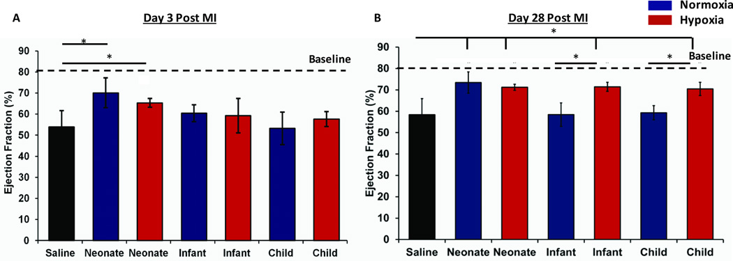
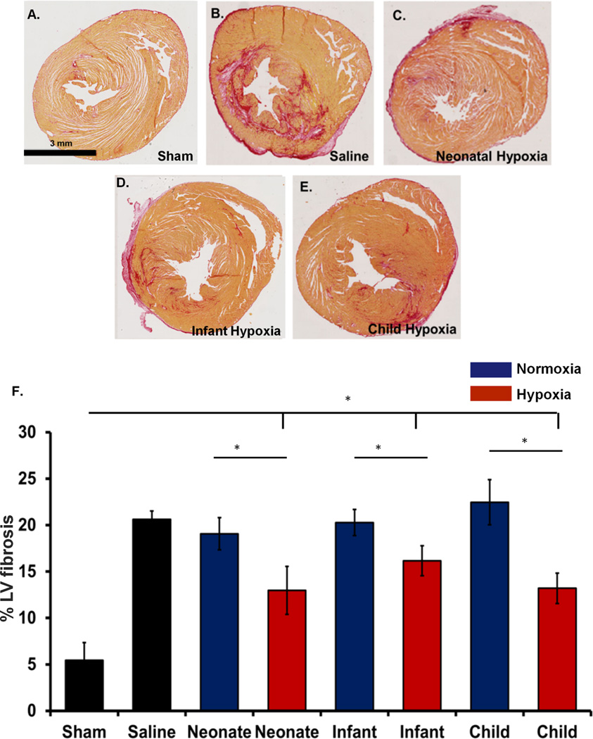
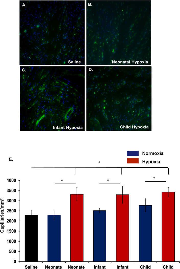
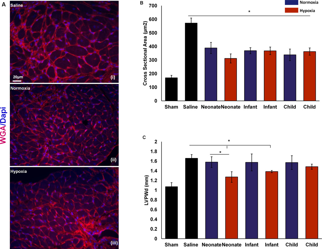
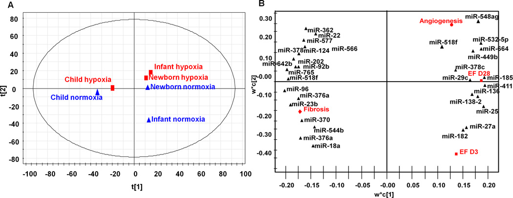
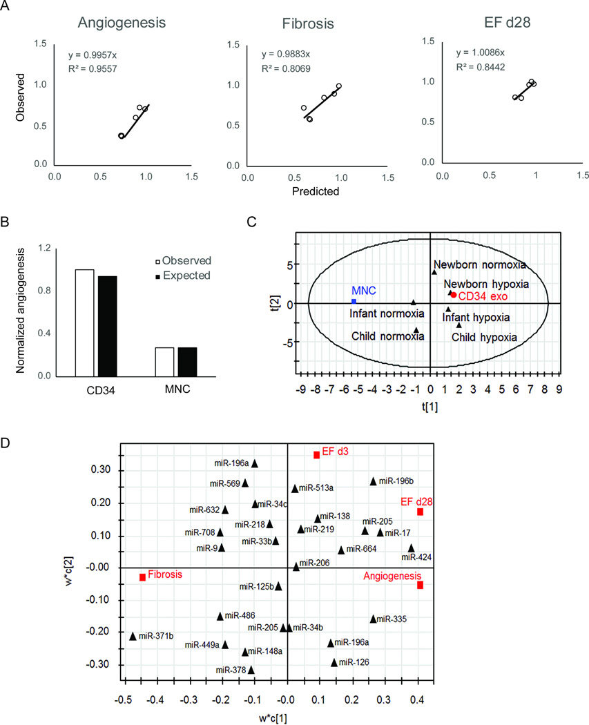
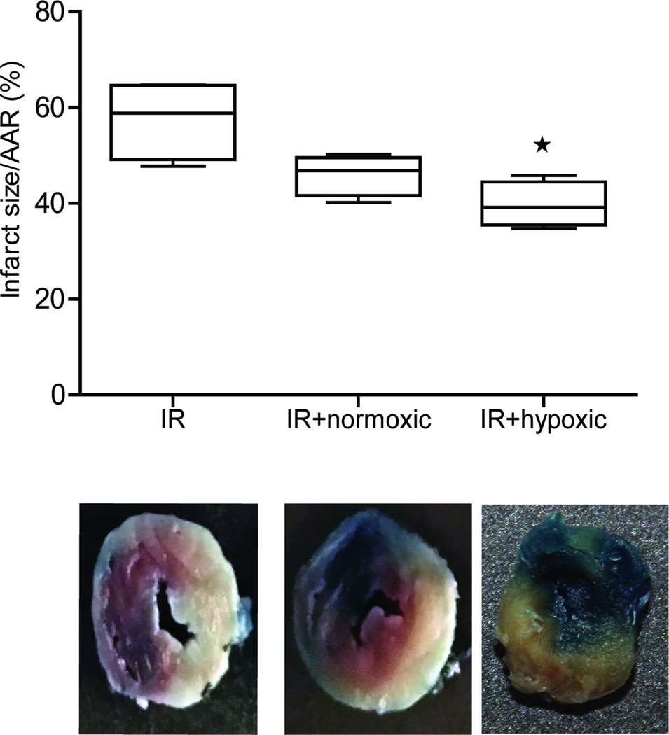
Methods and results: Human CPCs from the right atrial appendages from children of different ages undergoing cardiac surgery for congenital heart defects were isolated and cultured under hypoxic or normoxic conditions. Exosomes were isolated from the culture-conditioned media and delivered to athymic rats after ischemia-reperfusion injury. Echocardiography at day 3 post-myocardial infarction suggested statistically improved function in neonatal hypoxic and neonatal normoxic groups compared with saline-treated controls. At 28 days post-myocardial infarction, exosomes derived from neonatal normoxia, neonatal hypoxia, infant hypoxia, and child hypoxia significantly improved cardiac function compared with those from saline-treated controls. Staining showed decreased fibrosis and improved angiogenesis in hypoxic groups compared with controls. Finally, using sequencing data, a computational model was generated to link microRNA levels to specific outcomes.
Conclusions: CPC exosomes derived from neonates improved cardiac function independent of culture oxygen levels, whereas CPC exosomes from older children were not reparative unless subjected to hypoxic conditions. Cardiac functional improvements were associated with increased angiogenesis, reduced fibrosis, and improved hypertrophy, resulting in improved cardiac function; however, mechanisms for normoxic neonatal CPC exosomes improved function independent of those mechanisms. This is the first study of its kind demonstrating that donor age and oxygen content in the microenvironment significantly alter the efficacy of human CPC-derived exosomes.
Keywords: cardiac progenitor cells; exosome; microRNA; modeling; systems biology.
© 2016 American Heart Association, Inc.
Figures
Comment in
-
Exosomal MicroRNAs Released by Pediatric Cardiac Progenitor Cells.Circ Res. 2017 Feb 17;120(4):607-609. doi: 10.1161/CIRCRESAHA.117.310443. Circ Res. 2017. PMID: 28209789 No abstract available.
References
-
- Go AS, Mozaffarian D, Roger VL, Benjamin EJ, Berry JD, Blaha MJ, Dai S, Ford ES, Fox CS, Franco S, Fullerton HJ, Gillespie C, Hailpern SM, Heit JA, Howard VJ, Huffman MD, Judd SE, Kissela BM, Kittner SJ, Lackland DT, Lichtman JH, Lisabeth LD, Mackey RH, Magid DJ, Marcus GM, Marelli A, Matchar DB, McGuire DK, Mohler ER, 3rd, Moy CS, Mussolino ME, Neumar RW, Nichol G, Pandey DK, Paynter NP, Reeves MJ, Sorlie PD, Stein J, Towfighi A, Turan TN, Virani SS, Wong ND, Woo D, Turner MB. Heart disease and stroke statistics--2014 update: A report from the american heart association. Circulation. 2014;129:e28–e292. - PMC - PubMed
-
- Beltrami AP, Barlucchi L, Torella D, Baker M, Limana F, Chimenti S, Kasahara H, Rota M, Musso E, Urbanek K, Leri A, Kajstura J, Nadal-Ginard B, Anversa P. Adult cardiac stem cells are multipotent and support myocardial regeneration. Cell. 2003;114:763–776. - PubMed
-
- Maxeiner H, Krehbiehl N, Muller A, Woitasky N, Akinturk H, Muller M, Weigand MA, Abdallah Y, Kasseckert S, Schreckenberg R, Schluter KD, Wenzel S. New insights into paracrine mechanisms of human cardiac progenitor cells. Eur J Heart Fail. 2010;12:730–737. - PubMed
-
- Chugh AR, Beache GM, Loughran JH, Mewton N, Elmore JB, Kajstura J, Pappas P, Tatooles A, Stoddard MF, Lima JA, Slaughter MS, Anversa P, Bolli R. Administration of cardiac stem cells in patients with ischemic cardiomyopathy: The scipio trial: Surgical aspects and interim analysis of myocardial function and viability by magnetic resonance. Circulation. 2012;126:S54–S64. - PMC - PubMed
-
- Makkar RR, Smith RR, Cheng K, Malliaras K, Thomson LE, Berman D, Czer LS, Marban L, Mendizabal A, Johnston PV, Russell SD, Schuleri KH, Lardo AC, Gerstenblith G, Marban E. Intracoronary cardiosphere-derived cells for heart regeneration after myocardial infarction (caduceus): A prospective, randomised phase 1 trial. Lancet. 2012;379:895–904. - PMC - PubMed
MeSH terms
Substances
Grants and funding
LinkOut - more resources
Full Text Sources
Other Literature Sources
Medical
