. 2017 Jan 15;77(2):390-400.
doi: 10.1158/0008-5472.CAN-16-1303. Epub 2016 Nov 21.

Mutational Landscape of Pediatric Acute Lymphoblastic Leukemia

Affiliations

Mutational Landscape of Pediatric Acute Lymphoblastic Leukemia

Ling-Wen Ding et al. Cancer Res. .

Erratum in

Abstract

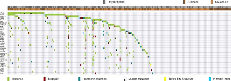
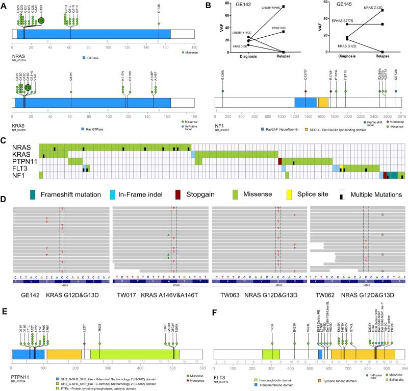
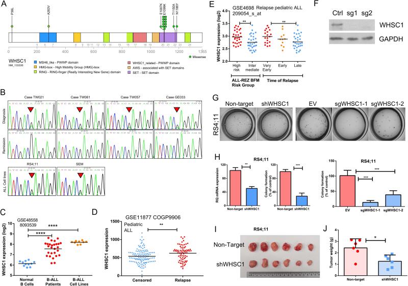
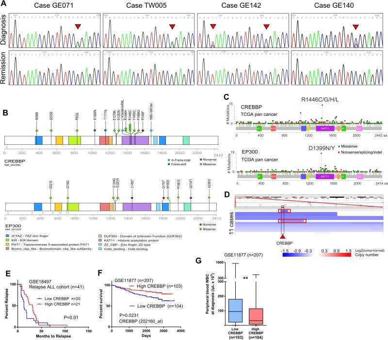
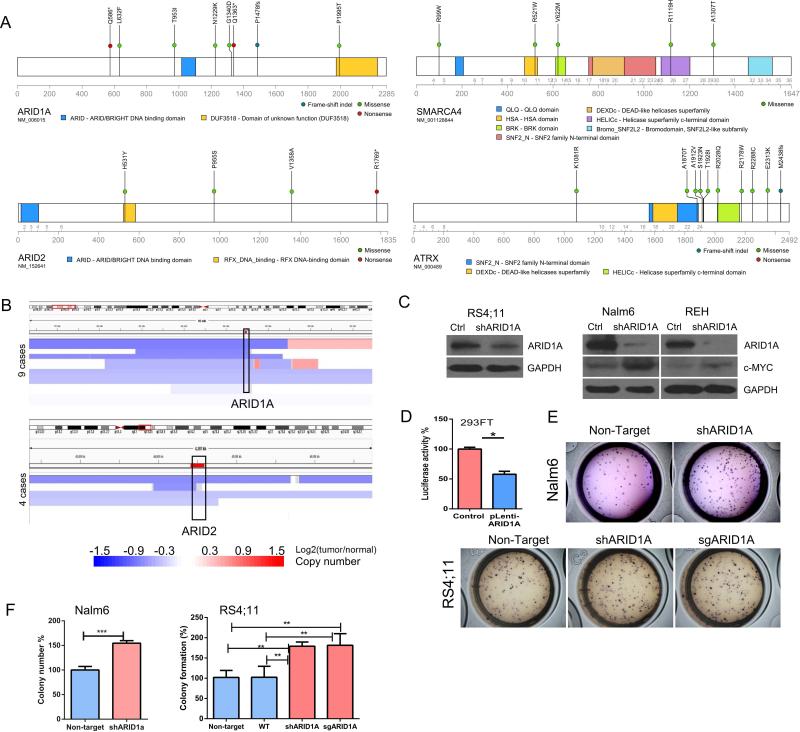
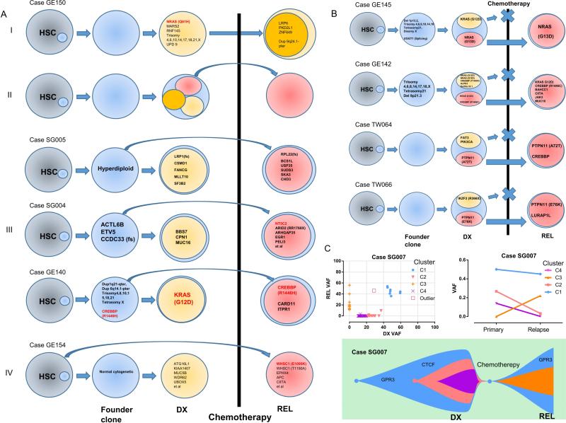
Current standard of care for patients with pediatric acute lymphoblastic leukemia (ALL) is mainly effective, with high remission rates after treatment. However, the genetic perturbations that give rise to this disease remain largely undefined, limiting the ability to address resistant tumors or develop less toxic targeted therapies. Here, we report the use of next-generation sequencing to interrogate the genetic and pathogenic mechanisms of 240 pediatric ALL cases with their matched remission samples. Commonly mutated genes fell into several categories, including RAS/receptor tyrosine kinases, epigenetic regulators, transcription factors involved in lineage commitment, and the p53/cell-cycle pathway. Unique recurrent mutational hotspots were observed in epigenetic regulators CREBBP (R1446C/H), WHSC1 (E1099K), and the tyrosine kinase FLT3 (K663R, N676K). The mutant WHSC1 was established as a gain-of-function oncogene, while the epigenetic regulator ARID1A and transcription factor CTCF were functionally identified as potential tumor suppressors. Analysis of 28 diagnosis/relapse trio patients plus 10 relapse cases revealed four evolutionary paths and uncovered the ordering of acquisition of mutations in these patients. This study provides a detailed mutational portrait of pediatric ALL and gives insights into the molecular pathogenesis of this disease. Cancer Res; 77(2); 390-400. ©2016 AACR.


Figures

Figure 1
Figure 1. Mutational landscapes of 240 ALL patients
Heatmap diagram showing the mutational landscape of ALL patients. Patients are labeled on the horizontal axis and mutant genes on the vertical axis.
Figure 2
Figure 2. Recurrent mutations in RAS-RTK pathway
A, schematic of the mutations in NRAS, KRAS and NF1. B, comparison of mutational allele frequency (VAF) of two cases harboring different RAS mutations at diagnosis and relapse. C, mutational heatmap of the RAS-RTK pathway genes. D, mutation phasing analysis depicting the mutually-exclusive pattern of RAS mutations in the leukemic cells of the same patient, four representative cases are shown. Sequencing reads are displayed at each locus as a gray bar. Mutations found in the reads (nucleotide differs from the reference sequence) are indicated and highlighted. E, F, schematic diagrams showing mutations of PTPN11 and FLT3, respectively.
Figure 3
Figure 3. WHSC1 is a unique oncogene in B-ALL
A, mutational diagram illustrating the mutations of WHSC1. B, representative Sanger sequence tracings that show somatic E1099K mutation in 4 patients (upper and middle panel, diagnosis and the matched remission sample, respectively) and 2 ALL cell lines (RS4;11, SEM, lower panel). C, elevation of WHSC1 mRNA in B-ALL samples or B-ALL cell lines compared with normal B-cells (data were retrieved from GSE48558). ****, p<0.0001. D, patients whose ALL cells at diagnosis had higher levels of WHSC1, also had a higher likelihood to undergo relapse (GSE11877, 207 Pediatric ALL. Censored: Patients in clinical remission, or with a second malignancy, or with a toxic death as a first event were censored at the date of last contact). **, p<0.01. E, aberrant WHSC1 expression was associated with high risk leukemia and early occurrence of relapse: very early relapse (within 18 months after initial diagnosis); early relapse (>18 months after initial diagnosis but <6 months after cessation of frontline treatment); late relapse (>6 months after cessation of frontline treatment). Data were retrieved from GSE4698 (60 childhood ALL (28)). **, p<0.01. F, western blot shows the silencing effect of CRISPR-Cas9 sgRNA targeting WHSC1. G, H, silencing of WHSC1 by either shRNA or CRISPR-Cas9 guide RNAs markedly reduced the clonogenic growth of B-ALL cells RS4;11 (cell line carrying WHSC1 E1099K mutation) in methylcellulose assay. Non-target, Non-target shRNA Control. EV, empty vector control. **, p<0.01, ***, p<0.001. I, J, silencing of WHSC1 impairs in vivo cell growth of RS4;11. *, p<0.05.
Figure 4
Figure 4. CREBBP and EP300 are frequently mutated in ALL
A, Sanger sequencing chromatograms showing the hotspot R1446 mutation of CREBBP (arrowhead). B, Diagram showing the sites of mutations in CREBBP and EP300. C, TCGA pan cancer sequencing analysis identified mutational hotspot in the HAT domain of CREBBP and EP300. D, CREBBP loci is frequently deleted in ALL patients. Each blue color bar indicates a deletion event in one individual. Two cases with focal deletion of CREBBP were highlighted with a red rectangle. Data were retrieved from Tumorscape ALL patients SNP-Array database. E, The lower CREBBP expression in the leukemic cells at diagnosis is associated with early relapse in the relapse ALL cohort GSE18497. The patients are separated into “high” (upper 50%, n=20) and “low” (lower 50%, n=21) groups based on the CREBBP expression of their leukemic cells at diagnosis. F, Kaplan-Meier plot of overall survival: comparison of cases with high versus low expression of CREBBP in 207 ALL patients (GSE11877). “High” and “low” ALL cohorts are defined as upper and lower 50% of expression of CREBBP in their diagnosis leukemic cells. P value was calculated by log-rank test. G, Box plots for peripheral white blood cell counts (WBC) in individuals whose leukemic cells at diagnosis expressed either higher (upper 50%) or lower (lower 50%) level of CREBBP. **, p<0.01.
Figure 5
Figure 5. Mutation of chromatin remodeling (SWI/SNF complex) genes
A, diagrams depict the mutations of ARID1A, ARID2, SMARAC4 and ATRX identified in this study. B, deletion of ARID1A (upper panel) or ARID2 (lower panel) in ALL samples. Each blue bar indicates a deletional event in an ALL patient. Data were retrieved from Tumorscape SNP-array database. C, western blots show shRNA silencing of ARID1A in RS4;11, Nalm6 and REH. Ctrl, Non-target shRNA control. D, luciferase assay stimulated by c-MYC activity in 293FT cells (control) versus forced expression of ARID1A (pLenti-ARID1A, see Supplementary Materials and Methods). Mean ± SD, n=3. *, p<0.05. E, F, silencing ARID1A enhances in vitro clonogenic growth of Nalm6 and RS4;11 in methylcellulose assays. Mean ± SD, n=3. **, p<0.01, ***, p<0.001.
Figure 6
Figure 6. Clonal architecture and clonal evolution of ALL
A, different clonal evolutionary paths inferred from the sequencing data. HSC, Hematopoietic stem cells. B, four representative cases showing the evolution of relapse (REL) ALL from a subclone at diagnosis (DX). C, clonal architecture and evolution of ALL patient SG007. Variant allele frequency (VAF) of mutations of ALL at diagnosis and relapse (upper left panel). Mean VAF mutational clusters (C1, C2, C3, C4) in the primary and relapse ALL samples (upper right panel). Lower panel: schematic diagram illustrating the clonal evolution of ALL cells of patient SG007.

References

    1. Mullighan CG. Molecular genetics of B-precursor acute lymphoblastic leukemia. J Clin Invest. 2012;122:3407–15. - PMC - PubMed
    1. Mar BG, Bullinger LB, McLean KM, Grauman PV, Harris MH, Stevenson K, et al. Mutations in epigenetic regulators including SETD2 are gained during relapse in paediatric acute lymphoblastic leukaemia. Nat Commun. 2014;5:3469. - PMC - PubMed
    1. Ma X, Edmonson M, Yergeau D, Muzny DM, Hampton OA, Rusch M, et al. Rise and fall of subclones from diagnosis to relapse in pediatric B-acute lymphoblastic leukaemia. Nat Commun. 2015;6:6604. - PMC - PubMed
    1. Paulsson K, Lilljebjorn H, Biloglav A, Olsson L, Rissler M, Castor A, et al. The genomic landscape of high hyperdiploid childhood acute lymphoblastic leukemia. Nat Genet. 2015;47:672–6. - PubMed
    1. Papaemmanuil E, Rapado I, Li Y, Potter NE, Wedge DC, Tubio J, et al. RAG-mediated recombination is the predominant driver of oncogenic rearrangement in ETV6-RUNX1 acute lymphoblastic leukemia. Nat Genet. 2014;46:116–25. - PMC - PubMed

Publication types