. 2017 Jan 15;77(2):279-290.
doi: 10.1158/0008-5472.CAN-16-0442. Epub 2016 Nov 21.

Macrophage-Secreted TNFα and TGFβ1 Influence Migration Speed and Persistence of Cancer Cells in 3D Tissue Culture via Independent Pathways

Affiliations

Macrophage-Secreted TNFα and TGFβ1 Influence Migration Speed and Persistence of Cancer Cells in 3D Tissue Culture via Independent Pathways

Ran Li et al. Cancer Res. .

Abstract

The ability of a cancer cell to migrate through the dense extracellular matrix within and surrounding the solid tumor is a critical determinant of metastasis. Macrophages enhance invasion and metastasis in the tumor microenvironment, but the basis for their effects is not fully understood. Using a microfluidic 3D cell migration assay, we found that the presence of macrophages enhanced the speed and persistence of cancer cell migration through a 3D extracellular matrix in a matrix metalloproteinases (MMP)-dependent fashion. Mechanistic investigations revealed that macrophage-released TNFα and TGFβ1 mediated the observed behaviors by two distinct pathways. These factors synergistically enhanced migration persistence through a synergistic induction of NF-κB-dependent MMP1 expression in cancer cells. In contrast, macrophage-released TGFβ1 enhanced migration speed primarily by inducing MT1-MMP expression. Taken together, our results reveal new insights into how macrophages enhance cancer cell metastasis, and they identify TNFα and TGFβ1 dual blockade as an antimetastatic strategy in solid tumors. Cancer Res; 77(2); 279-90. ©2016 AACR.


Conflict of interest statement

The authors disclose no potential conflict of interest.

Figures

Figure 1
Figure 1. Macrophages enhance cancer cell migration total speed and directedness in 3D ECM
(A) Schematics of the microfluidic device. Cancer cells and macrophages were suspended in 3D collagen I ECM (orange) encased in the device. (B) Representative MDA-MB-231 cancer cell (MDA231) migration trajectories for cancer cell monoculture (left) and cancer cell-Raw 264.7 macrophages (MΦ) co-culture (right). (C and D) Co-culture of Raw macrophages (MΦ) with cancer cells significantly enhanced cancer cell migration total speed (C) and directedness (D) for MDA231 cells, PC3 prostate cancer cells, and MDA-MB-435S melanoma cells (MDA435). (E and F) Co-culture of primary human monocyte-derived macrophages (MDMΦ), as well as murine bone marrow-derived macrophages (BMDM), with MDA231 cells enhanced migration total speed (E) and directedness (F) of MDA231 cells.
Figure 2
Figure 2. Cancer cell migration speed and directedness are MMP-dependent, and macrophages enhance cancer cell MMP expression
(A) Representative confocal image showing MDA231 cells (green) migrating through dense collagen I ECM (magenta) by degrading the matrix, leaving behind a micro-track (arrow). (B and C) Compared to the untreated and DMSO controls, inhibition of MMP activity by GM6001 significantly reduced MDA231 migration total speed (B) and directedness (C). (D and E) Representative western blot images (left) and quantification (right) showing that co-culture of Raw macrophages with MDA231 cells (MDA231 Cocul) in 3D collagen I gels significantly enhanced the expression of MMP1 (D) and MT1-MMP protein (E) in MDA231 relative to monoculture control (MDA231, Ctrl=Control).
Figure 3
Figure 3. Macrophage-released TNFα and TGFβ1 are responsible for the increase in cancer cell migration total speed and directedness
MDA231 cancer cells (CC) co-cultured with Raw cells (MΦ) were treated with neutralizing antibodies against TNFα (a-TNFα) and/or TGFβ1 (a-TGFβ1). (A, B, and C) Neutralizing TNFα released by macrophages (CCMΦ a-TNFα) led to a decrease in MDA231 migration total speed compared to no-treatment control (CCMΦ) (A). However, inhibiting macrophage-released TGFβ1 (CCMΦ a-TGFβ1) led to an almost complete inhibition of macrophage’s effect on MDA231 migration total speed (B), similar to the simultaneous inhibition of both TNFα and TGFβ1 (C). (D, E, and F) Neutralizing macrophage-released TNFα (CCMΦ a-TNFα) or TGFβ1 (CCMΦ a-TGFβ1) alone did not significantly reduce MDA231 migration directedness (D and E). However, simultaneous inhibition of both TNFα and TGFβ1 led to an almost complete abolishment of macrophage-enhanced MDA231 migration directedness (F).
Figure 4
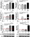
Figure 4. TGFβ1 increases cancer cell migration total speed via the induction of MT1-MMP expression, while TNFα and TGFβ1 synergistically increase cancer cell migration directedness via the induction of MMP1 expression
MDA231 monoculture (CC) was treated with TNFα and/or TGFβ1, and the resulting cell migration dynamics and MMP expressions were analyzed. (A–C) Treatment of MDA231 with TGFβ1 (CC TGFβ1) led to larger increases in MDA231 migration total speed (A), MT1-MMP mRNA (B) and protein (C) expressions than TNFα mono-treatment (CC TNFα). However, co-treatment of both TNFα and TGFβ1 led to no further increase in migration total speed, MT1-MMP mRNA and protein expressions compared to TGFβ1 mono-treatment. Data in (A), (B), and (C) follow a similar trend. (D–F) TNFα and TGFβ1 synergistically increased MDA231 migration directedness (D), MMP1 mRNA expression (E), and MMP1 protein production (F) when compared to mono-treatment conditions. Data in (D), (E), and (F) follow a similar trend. Data in (C) and (F) were obtained from cells cultured in 3D collagen I ECM.
Figure 5
Figure 5. Macrophage-induced cancer cell migration total speed is mediated via MT1-MMP, while directedness is mediated by MMP1
MDA231 cancer cells (CC)-Raw macrophages (MΦ) co-culture was treated with blocking antibodies against MT1-MMP or MMP1. (A and B) Treatment of co-culture with anti-MT1-MMP antibody (CCMΦ a-MT1-MMP) decreased MDA231 migration total speed (A) but not directedness (B). (C and D) Treatment of co-culture with anti-MMP1 antibody (CCMΦ a-MMP1) reduced MDA231 migration directedness (D) while having a minimal effect on total speed (C). (E and F) Treatment of MDA231 cancer cell monoculture with recombinant MMP1 (CC MMP1) enhanced MDA231 migration directedness (F), while having a minimal effect on total speed (E).
Figure 6
Figure 6. TNFα and TGFβ1 synergistically increase NF-κB nuclear localization
(A) Western blot quantification (top) and representative images (bottom) showing 48-hr TNFα and/or TGFβ1 treatments of MDA231 cells did not alter the production of NF-κB (NF-κB total). (B and C) Western blot quantifications (top) and representative images (bottom) showing 2-hr TNFα and TGFβ1 co-treatment of MDA231 synergistically increased the level of NF-κB in the nuclear fraction of MDA231 (NF-κB Nuc, C), but not in the cytoplasmic fraction of the cells (NF-κB Cyto, B).
Figure 7
Figure 7. Proposed mechanism explaining the effects of macrophages (MΦ) on cancer cell (CC) migration speed and persistence
Macrophage-released TNFα and TGFβ1 synergistically enhance NF-κB nuclear localization in cancer cells, leading to synergistic increases in cancer cell MMP1 mRNA expression, protein production, and protein secretion. This increase in MMP1 secretion by cancer cells leads to an increase in cancer cell migration persistence (directedness). Meanwhile, macrophages increase cancer cell migration speed (total speed), mainly through TGFβ1-induced cancer cell expression of MT1-MMP.

References

    1. Balkwill FR, Capasso M, Hagemann T. The tumor microenvironment at a glance. J Cell Sci. 2012;125:5591–5596. - PubMed
    1. Joyce JA, Pollard JW. Microenvironmental regulation of metastasis. Nat Rev Cancer. Nature Publishing Group. 2009;9:239–252. - PMC - PubMed
    1. Lu P, Weaver VM, Werb Z. The extracellular matrix: a dynamic niche in cancer progression. J Cell Biol. 2012;196:395–406. - PMC - PubMed
    1. Friedl P, Wolf K. Tumour-cell invasion and migration: diversity and escape mechanisms. Nat Rev Cancer. 2003;3:362–374. - PubMed
    1. Egeblad M, Werb Z. New functions for the matrix metalloproteinases in cancer progression. Nat Rev Cancer. 2002;2:161–174. - PubMed

Publication types

MeSH terms

Substances