. 2016 Dec 6;113(49):14049-14054.
doi: 10.1073/pnas.1607574113. Epub 2016 Nov 21.

Atomistic insight into lipid translocation by a TMEM16 scramblase

Affiliations

Atomistic insight into lipid translocation by a TMEM16 scramblase

Neville P Bethel et al. Proc Natl Acad Sci U S A. .

Abstract

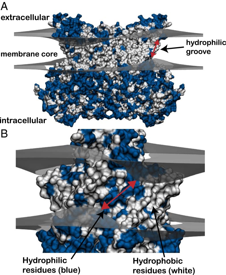
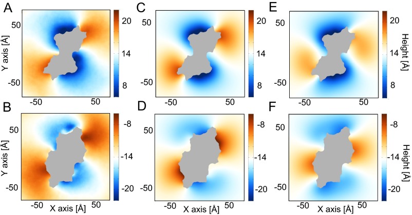
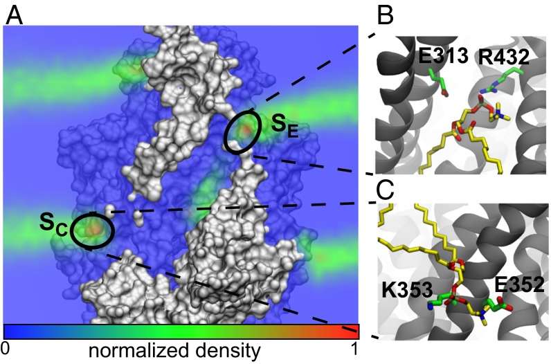
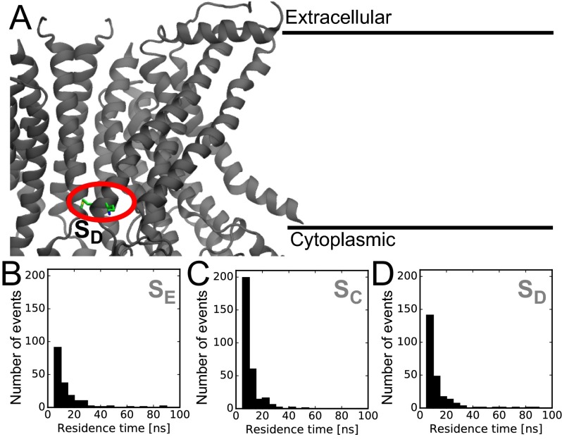
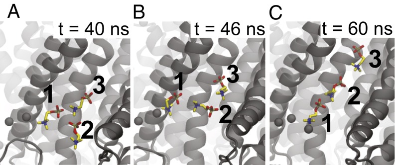
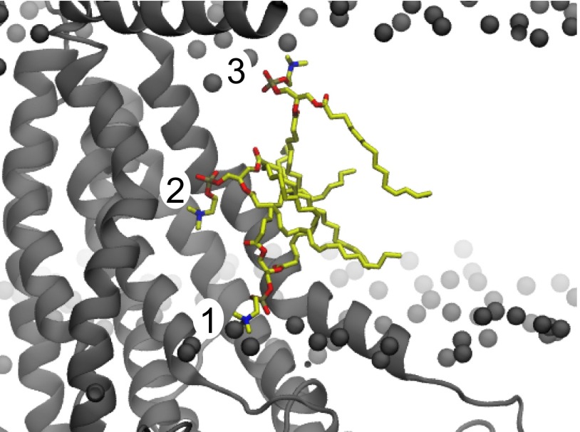
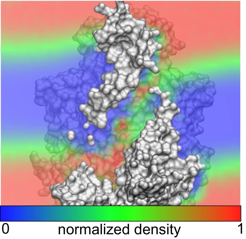
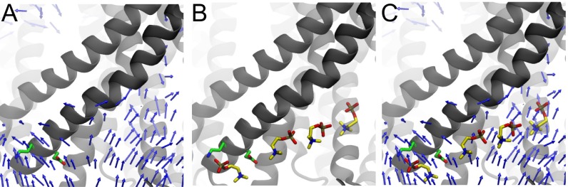
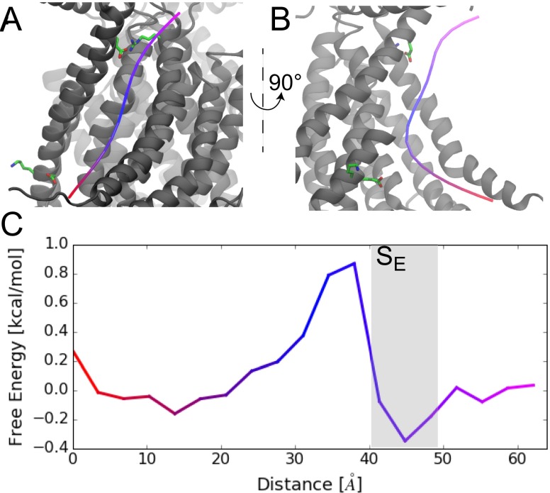
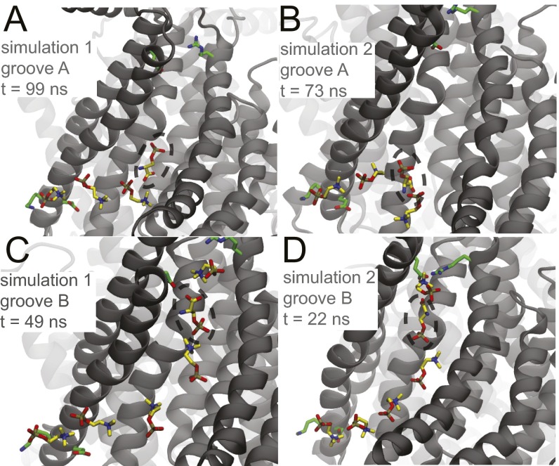
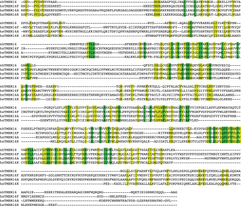
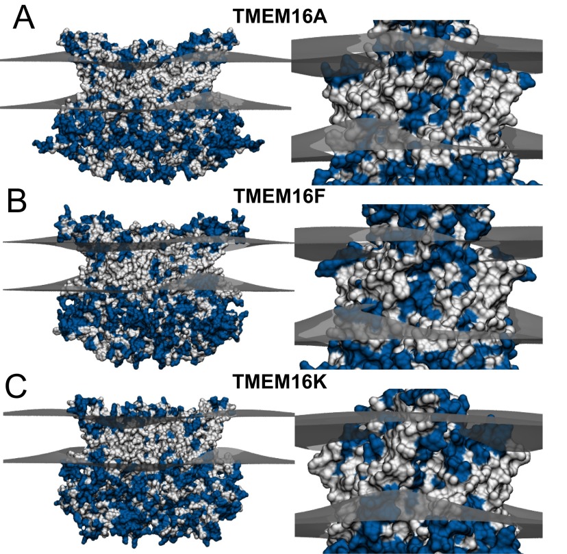
The transmembrane protein 16 (TMEM16) family of membrane proteins includes both lipid scramblases and ion channels involved in olfaction, nociception, and blood coagulation. The crystal structure of the fungal Nectria haematococca TMEM16 (nhTMEM16) scramblase suggested a putative mechanism of lipid transport, whereby polar and charged lipid headgroups move through the low-dielectric environment of the membrane by traversing a hydrophilic groove on the membrane-spanning surface of the protein. Here, we use computational methods to explore the membrane-protein interactions involved in lipid scrambling. Fast, continuum membrane-bending calculations reveal a global pattern of charged and hydrophobic surface residues that bends the membrane in a large-amplitude sinusoidal wave, resulting in bilayer thinning across the hydrophilic groove. Atomic simulations uncover two lipid headgroup-interaction sites flanking the groove. The cytoplasmic site nucleates headgroup-dipole stacking interactions that form a chain of lipid molecules that penetrate into the groove. In two instances, a cytoplasmic lipid interdigitates into this chain, crosses the bilayer, and enters the extracellular leaflet, and the reverse process happens twice as well. Continuum membrane-bending analysis carried out on homology models of mammalian homologs shows that these family members also bend the membrane-even those that lack scramblase activity. Sequence alignments show that the lipid-interaction sites are conserved in many family members but less so in those with reduced scrambling ability. Our analysis provides insight into how large-scale membrane bending and protein chemistry facilitate lipid permeation in the TMEM16 family, and we hypothesize that membrane interactions also affect ion permeation.

Keywords: TMEM16; anoctamin; continuum membrane models; lipid scrambling; simulation.


Conflict of interest statement

The authors declare no conflict of interest.

Figures

Fig. 1.
Fig. 1.
Predicted membrane bending around nhTMEM16 dimer based on continuum modeling. (A) Crystal structure of the fungal scramblase, nhTMEM16 (PDB ID code 4WIS), deforming the upper and lower membrane leaflets (gray surfaces) predicted from our hybrid continuum-atomistic model. Each surface represents the hydrophobic–hydrophilic interface where the polar headgroups meet the hydrophobic core of the acyl chains. The membrane bends in a large amplitude wave that attempts to expose polar and charged residues (blue), while burying hydrophobic residues (white). (B) Ninety-degree rotation of the view in A showing one of the two hydrophilic grooves that span the membrane core. The membrane is maximally distorted at each groove, and the leaflet-to-leaflet distance is 18.3 Å (red arrow).
Fig. S1.
Fig. S1.
Extracellular view of upper and lower leaflet heights predicted from MD and continuum calculations. (A and B) The upper and lower leaflet surfaces, respectively, calculated from averaging results from MD simulations. (C and D) The upper and lower leaflet surfaces, respectively, determined from continuum calculations. (E and F) The upper and lower leaflet surfaces, respectively, determined from continuum calculations when all residues within the hydrophilic groove are neutralized. Surfaces are colored by height, where red is an upward deflection, blue is a downward deflection, and white is no deflection. The area occupied by the protein is shaded gray.
Fig. 2.
Fig. 2.
Phosphate density computed from MD simulations reveals headgroup localization. (A) Cross-section of phosphate density overlaid on the nhTMEM16 structure. Two maxima (red) are highlighted at both sides of the hydrophilic groove. (B) Simulation snapshot showing a lipid (yellow) interacting with two residues, E313 and R423, at the extracellular site (SE). (C) Simulation snapshot showing a lipid (yellow) interacting with two residues, E352 and K353, at the cytoplasmic site (SC).
Fig. S2.
Fig. S2.
Residence times of lipid-binding sites compared with control site. (A) Residues forming the dummy site SD used as a control in the residence time calculations. The methionine and lysine forming this site are circled in red. (BD) Residence times of lipid headgroups at the SE, SC, and a control SD site, respectively. Two outlier residence times of 248 and 317 ns were omitted from B.
Fig. 3.
Fig. 3.
Lipid penetration into the groove occurs via a dipole-stacking mechanism. AC are sequential MD snapshots during the lipid-flopping event. The negative phosphate groups (gold) interact with the positive choline groups (blue) of neighboring lipids. Lipid 2 inserts between lipids 1 and 3 and then stretches out to push lipid 3 far into the groove on the way to the extracellular space over the course of 20 ns. For clarity, only the phosphatidylcholine group is shown. The phosphorus atoms of all other lipids in the cytoplasmic leaflet are shown as black spheres.
Fig. S3.
Fig. S3.
Phospholipid configurations during a flopping event where 1, 2, and 3 are early, middle, and late in the processes, respectively. TM3 and TM4 were removed for clarity.
Fig. S4.
Fig. S4.
Cross-section of normalized water density overlaid on nhTMEM16 structure. Water enters the hydrophilic groove achieving densities close to bulk values (red) at several locations.
Fig. S5.
Fig. S5.
Atomic details of lipid flipping. AC are sequential MD snapshots showing dipole stacking during a lipid-flipping event. Before A, lipid 3 crossed the narrow portion of the groove from the SE site to add itself to the top of the dipole stack already composed of lipids 1 and 2. In B, lipid 2 dissolves from the stack, and then in C, lipid 3 forms a new dipole interaction with lipid 1, resulting in a shorter stack. These interactions closely resemble the time-reversed stacking interactions seen during the lipid-flopping events (Fig. 4).
Fig. 4.
Fig. 4.
Lipid stacking originates at the SC site. (A) Stacking headgroup–dipole interactions from Fig. 3 trace back to the SC. The lipid numbering and the coloring scheme are the same as Fig. 3. (B) Height of the dipole stack in time for the simulation in which a lipid permeates the groove. The permeation event occurs in the gray region. (C) Cartoon model of SC-mediated dipole stacking. One headgroup binds to SC. Lipids randomly insert at different locations on the dipole stack. As the stack grows, the top headgroup is placed deeper into the hydrophilic groove.
Fig. S6.
Fig. S6.
Lipid dipoles align near SC site. (A) Three-dimensional vector field of PC lipid dipoles at the cytoplasmic end of the hydrophilic groove. Arrows point from the positive to the negative charge. (B) Dipole lipid stack from the SC. This snapshot is from a simulation where a lipid did not fully permeate. (C) Dipole stack superimposed on the 3D dipole vector field.
Fig. S7.
Fig. S7.
Permeating lipids experience a small energy barrier. (A) Minimum-energy pathway through the hydrophilic groove determined using a string method. (B) Rotated view of the minimum-energy path. The extracellular half of TM4 is removed for clarity. (C) Free-energy profile along the minimum energy path. The color scheme matches the coloring of the path in A and B.
Fig. S8.
Fig. S8.
POPC dipole stacking stabilizes POPS in the hydrophilic groove. Single PC headgroups were replaced with PS (indicated by dashed circles) and restarted for two simulations (two grooves per simulation; four POPS lipids total). Two POPS molecules were placed in the cytoplasmic side of the groove (A and B), and two were placed in the extracellular end (C and D). Simulations were run for 80 ns, and the snapshots were taken at different times after 20 ns of production. Dipole stacks were observed in all four hydrophilic grooves. For C and D, the dipole stacks observed were not present at the beginning of the simulations. Interestingly in B, when POPS in the middle of the dipole chain, the PC dipoles switch directions, as expected. This result could have an important consequence on lipid flipping through the midplane barrier.
Fig. S9.
Fig. S9.
Sequence alignment of TMEM16A, TMEM16F, TMEM16K, and nhTMEM16. This alignment was carried out with the program Promals3d (47) using all 10 human TMEM16 family members plus nhTMEM16. No hand adjustments were performed. Strictly conserved residues are highlighted green and homologous residues are yellow.
Fig. S10.
Fig. S10.
Predicted membrane deformations around mammalian TMEM16 family members based on continuum modeling: TMEM16A (A), TMEM16F (B), and TMEM16K (C). Ninety-degree rotations of each protein showing the hydrophilic groove are displayed to the right. The predicted membrane surfaces at the upper and lower hydrophobic–hydrophilic interfaces are gray. Hydrophobic and hydrophilic residues on the proteins are white and blue, respectively.
Fig. 5.
Fig. 5.
Sequence alignment of TMEM16 family members at the SC and SE sites. (A) The SC site in nhTMEM16, composed of E352 (red) and K353 (blue), is in TM4. Family members with robust scrambling activity all have an SC-like site with adjacent glutamate and lysine pairs, whereas this positive and negative pairing is not present in the family members with weak or nonexistent scrambling. The minimal scramblase domain [SCRD* (13)] encompasses the SC site. (B) The SE site is composed of residue E313 (red) in TM3 and R432 (blue) in TM6. The charge pairing is conserved in many TMEM16 members, but glutamate is found in TM6 and the arginine in TM3. In both A and B, basic and acidic residues that we suggest influence scrambling activity are highlighted in cyan and red, respectively.

References

    1. Bevers EM, Rosing J, Zwaal RFA. Development of procoagulant binding sites on the platelet surface. Adv Exp Med Biol. 1985;192:359–371. - PubMed
    1. Yang YD, et al. TMEM16A confers receptor-activated calcium-dependent chloride conductance. Nature. 2008;455(7217):1210–1215. - PubMed
    1. Caputo A, et al. TMEM16A, a membrane protein associated with calcium-dependent chloride channel activity. Science. 2008;322(5901):590–594. - PubMed
    1. Schroeder BC, Cheng T, Jan YN, Jan LY. Expression cloning of TMEM16A as a calcium-activated chloride channel subunit. Cell. 2008;134(6):1019–1029. - PMC - PubMed
    1. Yang H, et al. TMEM16F forms a Ca2+-activated cation channel required for lipid scrambling in platelets during blood coagulation. Cell. 2012;151(1):111–122. - PMC - PubMed

Publication types