. 2016:2016:7625947.
doi: 10.1155/2016/7625947. Epub 2016 Oct 30.

Oxygenation of the Intraportally Transplanted Pancreatic Islet

Affiliations

Oxygenation of the Intraportally Transplanted Pancreatic Islet

Thomas M Suszynski et al. J Diabetes Res. 2016.

Abstract

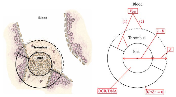
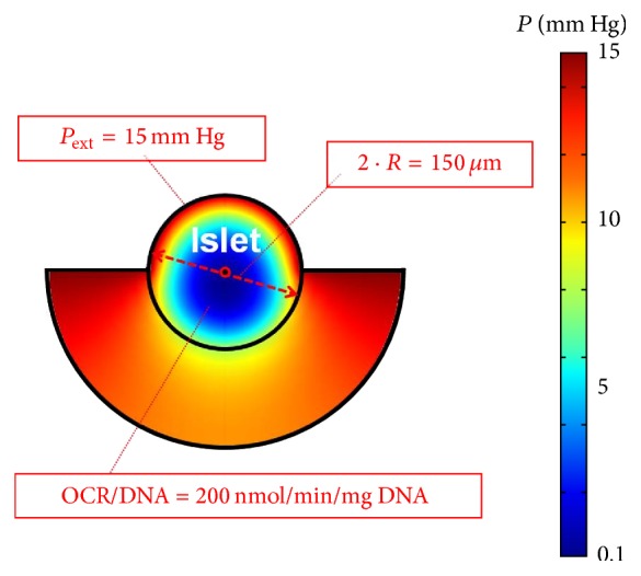
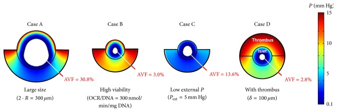
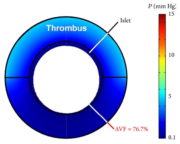
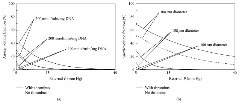
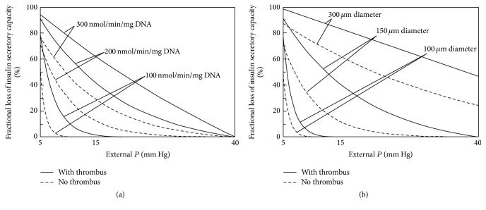
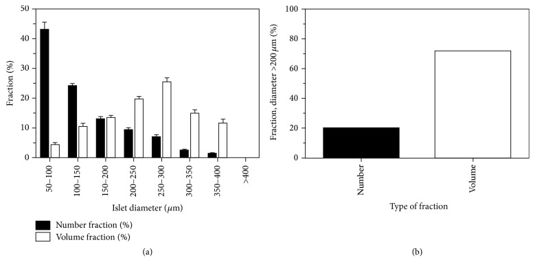
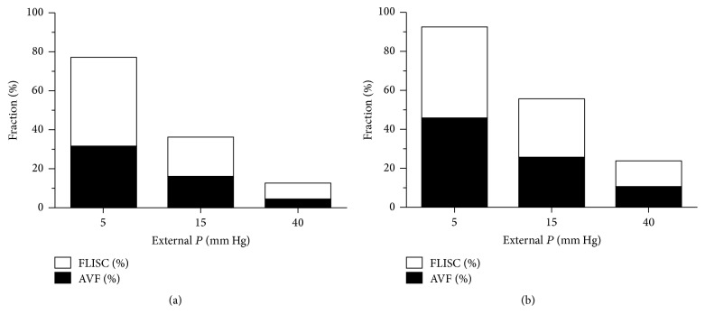
Intraportal islet transplantation (IT) is not widely utilized as a treatment for type 1 diabetes. Oxygenation of the intraportally transplanted islet has not been studied extensively. We present a diffusion-reaction model that predicts the presence of an anoxic core and a larger partly functional core within intraportally transplanted islets. Four variables were studied: islet diameter, islet fractional viability, external oxygen partial pressure (P) (in surrounding portal blood), and presence or absence of a thrombus on the islet surface. Results indicate that an islet with average size and fractional viability exhibits an anoxic volume fraction (AVF) of 14% and a function loss of 72% at a low external P. Thrombus formation increased AVF to 30% and function loss to 92%, suggesting that the effect of thrombosis may be substantial. External P and islet diameter accounted for the greatest overall impact on AVF and loss of function. At our institutions, large human alloislets (>200 μm diameter) account for ~20% of total islet number but ~70% of total islet volume; since most of the total transplanted islet volume is accounted for by large islets, most of the intraportal islet cells are likely to be anoxic and not fully functional.


Figures

Figure 1
Figure 1
Schematic depicting the intraportal islet which is modeled as a spherical body containing viable oxygen-consuming cells. The transplanted islet is lodged at a bifurcation in a distal hepatic sinusoid and has access to portal blood at its proximal half-surface. The distal half of the islet equilibrates with the surrounding environment and is modeled by the presence of a no-flux boundary condition at a specified distance away from its back surface (∂P/∂r = 0). There are 4 parameters that are adjusted in this model, including (1) fractional viability, or oxygen consumption rate normalized to DNA content (OCR/DNA); (2) islet diameter (2 · R); (3) external blood P (P ext); and (4) presence or absence of thrombus with a specified thickness (δ), located only on the proximal half-surface.
Figure 2
Figure 2
Surface plot illustrating model results for a “Baseline Case,” which involves an islet with average diameter (2 · R = 150 μm) and fractional viability (OCR/DNA = 200 nmol/min/mg DNA), exposed to a reasonable oxygen supply (P ext = 15 mm Hg), and no thrombus formation at its proximal half-surface (δ = 0 μm). The colors within and behind the islet depict the calculated spatial P gradients, as indicated by the legend (right). δ, thickness of the thrombus; OCR, oxygen consumption rate; P, oxygen partial pressure; P ext, external blood P; R, islet radius.
Figure 3
Figure 3
Surface plots illustrating model results for four different cases in which the Baseline Case (no thrombus, OCR/DNA = 200 nmol/min/mg DNA, P ext = 15 mm Hg, and 2 · R = 150-μm) is perturbed by adjusting only 1 of the 4 parameters with each case. Case A depicts the Baseline Case with an increase in the islet diameter from 150 to 300 μm. Case B depicts the Baseline Case with an increase in the OCR/DNA from 200 to 300 nmol/min/mg DNA. Case C depicts the Baseline Case with a decrease in P ext from 15 to 5 mm Hg. Case D depicts the Baseline Case with the addition of a 100 μm thrombus on the proximal half-surface of the islet. The anoxic volume fraction (AVF) is depicted by the achromatic core. Note the magnitude of the AVF associated with each perturbation. AVF, anoxic volume fraction; OCR, oxygen consumption rate; P, oxygen partial pressure; P ext, external blood P; R, islet radius.
Figure 4
Figure 4
Surface plot illustrating model results for the worst case scenario (Worst Case) analyzed in this study, which combines the Baseline Case and the 4 individual perturbations of 4 parameters (increase in islet diameter, increase in fractional viability, decrease in P ext, and addition of thrombus) that were shown separately in Figure 3. Note the very large anoxic volume fraction (AVF). AVF, anoxic volume fraction; P, oxygen partial pressure.
Figure 5
Figure 5
Graphs depicting a summary of model results for calculation of anoxic volume fraction [AVF (%)] with respect to external blood P (P ext), islet fractional viability (OCR/DNA), and diameter (2 · R) and with or without thrombus formation (δ = 100 μm). The graph on the left (a) illustrates the change in AVF for an islet of average diameter (150 μm) for the 3 OCR/DNA values. The graph on the right (b) illustrates the change in AVF in an islet with average OCR/DNA (200 nmol/min/mg DNA) for 3 islet diameter values. AVF is defined as the region of the islet that is anoxic, occurring below a critical P (P C) of 0.1 mm Hg. AVF, anoxic volume fraction; δ, thickness of the thrombus; OCR, oxygen consumption rate; P, oxygen partial pressure; P C, critical P for viability; P ext, external blood P; R, islet radius.
Figure 6
Figure 6
Graphs depicting a summary of model results for calculation of fractional loss of insulin secretory capacity [FLISC (%)] with respect to external P (P ext), islet fractional viability (OCR/DNA), and diameter (2 · R) and with or without thrombus formation (δ = 100 μm). The critical P used in the model was 5 mm Hg, which represents the best case scenario. The graph on the left (a) illustrates the change in FLISC for an islet of average diameter (150 μm) for the 3 OCR/DNA values. The graph on the right (b) illustrates the change in FLISC for an islet with average OCR/DNA (200 nmol/min/mg DNA) for 3 islet diameter values. FLISC is defined as the loss of insulin secretory capacity relative to an islet not limited by hypoxia. FLISC, fractional loss of insulin secretory capacity; δ, thickness of the thrombus; OCR, oxygen consumption rate; P, oxygen partial pressure; P , critical P for insulin secretion; P ext, external blood P; R, islet radius.
Figure 7
Figure 7
Islet size distribution stratified by ranges of islet diameter (a). Mean (± standard error, SE) number fractions are actual data from our institution (University of Minnesota) from 23 human islet preparations (high-purity, cultured fractions) prior to clinical transplantation. Mean (± SE) volume fractions are estimated from number fraction data by calculating the mean islet volumes under the assumption that the islets are spherical with a representative radius for that size range. Approximately 20% of the total number of islets were >200 μm in diameter, but these islets account for ~72% of the total transplanted volume of islets (b).
Figure 8
Figure 8
Extrapolation of model results to an entire human islet preparation, with (a) and without (b) thrombosis. Extrapolations were estimated from islet size distribution data found in Figure 7 for high-purity, cultured, alloislets. For example, to calculate the mean volume fractions for all islets of 100 μm diameter, the volume fraction data for islets ranging in diameter within 50–100 and 100–150 μm were averaged. AVF, anoxic volume fraction; FLISC, fractional loss of insulin secretory capacity; P, oxygen partial pressure.

Similar articles

Cited by

References

    1. Ryan E. A., Lakey J. R. T., Paty B. W., et al. Successful islet transplantation: continued insulin reserve provides long-term glycemic control. Diabetes. 2002;51(7):2148–2157. doi: 10.2337/diabetes.51.7.2148. - DOI - PubMed
    1. Ryan E. A., Lakey J. R. T., Rajotte R. V., et al. Clinical outcomes and insulin secretion after islet transplantation with the Edmonton Protocol. Diabetes. 2001;50(4):710–719. doi: 10.2337/diabetes.50.4.710. - DOI - PubMed
    1. Shapiro A. M. J., Lakey J. R. T., Ryan E. A., et al. Islet transplantation in seven patients with type 1 diabetes mellitus using a glucocorticoid-free immunosuppressive regimen. The New England Journal of Medicine. 2000;343(4):230–238. doi: 10.1056/nejm200007273430401. - DOI - PubMed
    1. Shapiro A. M. J., Ricordi C., Hering B. Edmonton's islet success has indeed been replicated elsewhere. The Lancet. 2003;362(9391, article 1242) doi: 10.1016/s0140-6736(03)14526-6. - DOI - PubMed
    1. Shapiro A. M. J., Ricordi C., Hering B. J., et al. International trial of the Edmonton protocol for islet transplantation. The New England Journal of Medicine. 2006;355(13):1318–1330. doi: 10.1056/nejmoa061267. - DOI - PubMed

LinkOut - more resources