. 2016:2016:8635721.
doi: 10.1155/2016/8635721. Epub 2016 Oct 31.

Minor Antigen Disparities Impede Induction of Long Lasting Chimerism and Tolerance through Bone Marrow Transplantation with Costimulation Blockade

Affiliations

Minor Antigen Disparities Impede Induction of Long Lasting Chimerism and Tolerance through Bone Marrow Transplantation with Costimulation Blockade

Sinda Bigenzahn et al. J Immunol Res. 2016.

Abstract

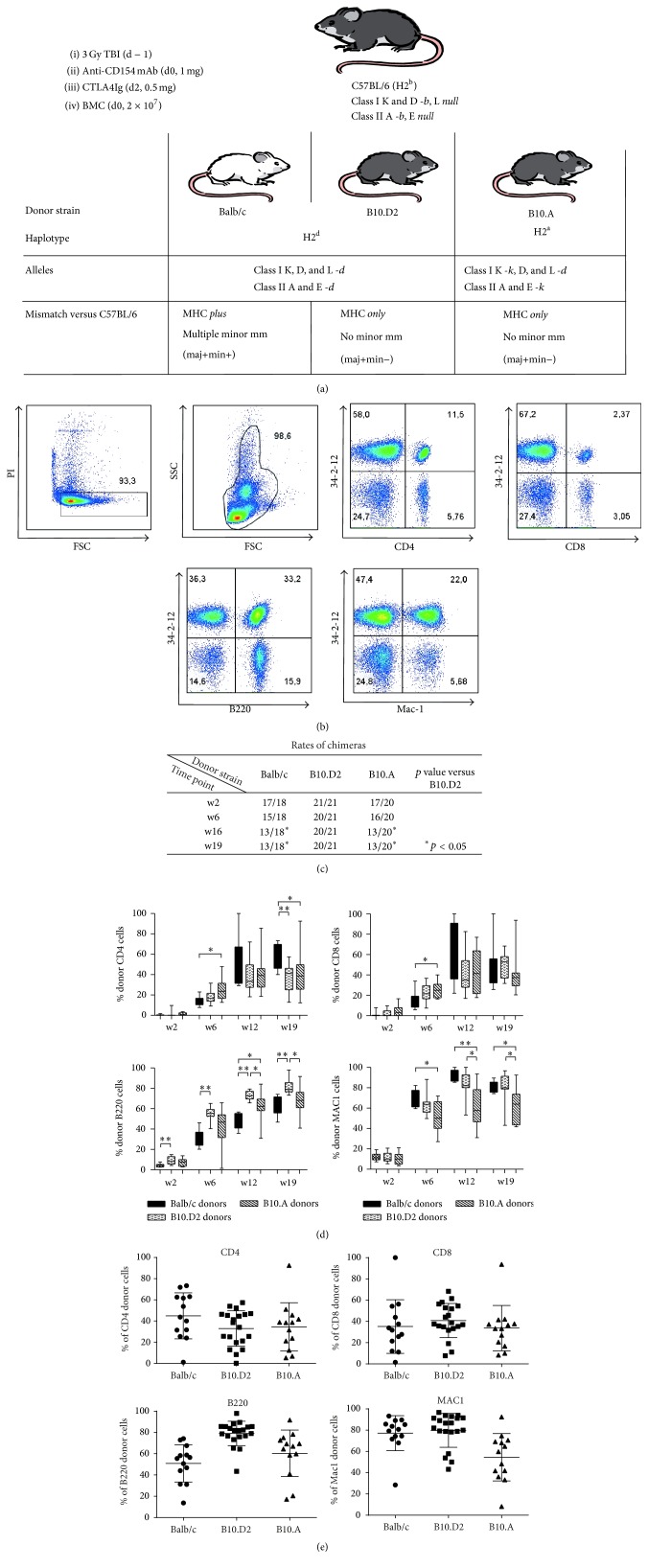
Mixed chimerism and tolerance can be successfully induced in rodents through allogeneic bone marrow transplantation (BMT) with costimulation blockade (CB), but varying success rates have been reported with distinct models and protocols. We therefore investigated the impact of minor antigen disparities on the induction of mixed chimerism and tolerance. C57BL/6 (H2b) mice received nonmyeloablative total body irradiation (3 Gy), costimulation blockade (anti-CD40L mAb and CTLA4Ig), and 2 × 10⁷ bone marrow cells (BMC) from either of three donor strains: Balb/c (H2d) (MHC plus multiple minor histocompatibility antigen (mHAg) mismatched), B10.D2 (H2d) or B10.A (H2a) (both MHC mismatched, but mHAg matched). Macrochimerism was followed over time by flow cytometry and tolerance was tested by skin grafting. 20 of 21 recipients of B10.D2 BMC but only 13 of 18 of Balb/c BMC and 13 of 20 of B10.A BMC developed stable long-term multilineage chimerism (p < 0.05 for each donor strain versus B10.D2). Significantly superior donor skin graft survival was observed in successfully established long-term chimeras after mHAg matched BMT compared to mHAg mismatched BMT (p < 0.05). Both minor and major antigen disparities pose a substantial barrier for the induction of chimerism while the maintenance of tolerance after nonmyeloablative BMT and costimulation blockade is negatively influenced by minor antigen disparities. .


Figures

Figure 1
Figure 1
mHAg disparities impede rates of chimerism achieved through BMT with CB. (a) depicts the BMT protocol used with distinct donor-recipient combinations. (b) FACS plots show total donor (34-2-12 mAb recognizes H-Dd) and recipient type cells in CD4, CD8, B cells, and myeloid cells of one representative recipient of Balb/c BMC at the end of observation period. Dead cells were excluded through propidium iodide (PI) staining. (c) Rates of chimeras were determined at different time points after BMT. Early multilineage chimerism was induced in almost all mice. Maintenance of chimerism was observed in all but one recipient mouse of MHC mismatched, but mHAg matched B10.D2 (H2d) bone marrow throughout the observation period. In contrast, chimerism rates dropped in mice, which were transplanted with BMC of MHC and mHAg mismatched Balb/c (H2d) mice over time similarly to mice, which received MHC mismatched but mHAg matched B10.A (H2a) BMC (p < 0.05 for both versus B10.D2 BMC recipients after week 16). (d) Chimerism levels among CD4 cells, CD8 cells, B cells, and myeloid cells were measured by FCM at different time points after BMT. All groups of recipients showed relatively similar chimerism levels among T cell lineages throughout the observation period. Significantly higher chimerism was observed in B cells among recipients of MHC mismatched, but mHAg matched B10.D2 (H2d) bone marrow compared to mice transplanted with MHC and mHAg mismatched Balb/c (H2d) BMC (∗∗ p < 0.01 at all measured time points). Myeloid cell chimerism was significantly lower in recipients of MHC mismatched, but mHAg matched B10.A (H2a) after week 6 after BMT compared to recipients of Balb/c (H2d) BMC ( p < 0.05 and ∗∗ p < 0.01 at indicated time points). Mean percent of chimerism, interquartile range (box), and SD (whiskers) of long-term chimeras are shown as box-and-whisker blots (representative data from 1 of 2 independent experiments). (e) Chimerism levels of long-term chimeras after transplantation of Balb/c BMC, B10.D2 BMC, or B10.A BMC, respectively, at the end of observation period. Individual percent, mean percent of chimerism, and SD (error bars) are shown as scatter plot (pooled data of two independent experiments).
Figure 2
Figure 2
mHAg are a barrier for induction of donor specific tolerance through BMT plus CB. (a) BL6 (H2b) recipients of bone marrow of fully MHC mismatched Balb/c (H2d, mHAg mismatched), B10.D2 (H2d), or B10.A (H2a, both mHAg matched) mice were tested for donor specific tolerance through transplantation of donor type and 3rd party skin 7 or 14 weeks after BMT. Treatment with 3 Gy TBI (d − 1): transplantation of 2 × 10⁷ BMC together with CB (1 mg anti-CD154mAb, d0; 0.5 mg CTLA4Ig, d2) leads to long lasting (>130 days) donor skin graft acceptance in long-term chimeras, which had received BMC from MHC and mHAg mismatched Balb/c (H2d) donors in 10 of 13 mice. Significantly better graft survival was observed in long-term chimeric recipients of MHC mismatched but mHAg identical bone marrow (28/29, B10.D2 and B10.A, p < 0.05; pooled data from 2 independent experiments). Skin graft survival was calculated according to the Kaplan-Meier product limit method and compared by using the log-rank test. p < 0.05 versus Balb/c donors. (b) Chimerism levels of long-term chimeras, which received Balb/c BMC were not significantly different in tolerant mice (circles) compared to mice, which rejected (squares ■) donor skin grafts. p = n.s. for all time points. (c) Macroscopical aspects of Balb/c (H2d, mHAg mismatched) donor skin grafts changed during the observation period with grafts showing shrinking, thickening, and loss of surface structure. In comparison, donor grafts of B10.D2 (H2d) and B10.A (H2a, both mHAg matched) mice stayed macroscopically unchanged from 7 days after skin grafting (when protecting bandage was removed) until the end of observation period. (d) Representative histology of donor skin grafts of Balb/c (left), B10.D2 (middle), and B10.A (right) 44 weeks after skin grafting. HE staining, magnification 160x, and Giemsa staining 160x (not shown) analyzed. (e) Classification of donor skin grafts 44 weeks after skin grafting according to The Banff 2007 Working Classification of Skin-Containing Composite Tissue Allograft Pathology [19] by a blinded expert pathologist. p < 0.05 versus Balb/c. Most of Balb/c donor skin grafts showed histologically moderate (grade 2, 2/3) signs of inflammation whereas all B10.D2 donor skin grafts were free of inflammatory infiltrates (grade 0, n = 4, p < 0.05). B10.A partially showed no (grade 0, 2/4) or mild signs of inflammation (grade 1, 2/4, p = n.s. versus Balb/c).
Figure 3
Figure 3
Deletion of donor-reactive T cells differs among donor strains. (a) Deletion of donor-reactive T cells was investigated through determination of the percentage of Vβ11+and Vβ8.1/2+CD4+PBL by 2-color flow cytometric analysis 4 weeks after BMT. Deletion of Vβ11+CD4+PBL in Balb/c (H2d, mHAg mismatched, n = 8) and B10.D2 (H2d, mHAg matched, n = 11) BMC recipients developed to a similar dimension irrespective of differing mHAg disparities of donor to recipient. In mice which were transplanted with BMC of B10.A (H2a, mHAg matched, n = 9) a significant increase of early deletion compared to BMT of Balb/c (H2d, mHAg mismatched) and B10.D2 (H2d, mHAg matched) donors was observed ( p < 0.05 versus Balb/c and B10.D2 BMC recipients). The percentage of Vβ8+CD4+ cells was not significantly reduced in any group compared to naïve BL6 mice indicating the specificity of the deletion for superantigens presented by the donor. (b) Ten weeks after BMT the degree of deletion was significantly enhanced in long time chimeras after Balb/c and B10.D2 BMT but still was significantly less pronounced than in recipients of B10.A BMC. Mean percentages of Vβ11+and Vβ8.1/2+CD4+PBL, interquartile range (box), and SD (whiskers) of long-term chimeras are shown as box-and-whisker blots. Representative data from one of two independent experiments. Statistical significance determined by log-rank test. p < 0.05 versus Balb/c donors; ∗∗ p < 0.01 versus week 4 after BMT (each group).

Similar articles

Cited by

References

    1. Pilat N., Wekerle T. Transplantation tolerance through mixed chimerism. Nature Reviews Nephrology. 2010;6(10):594–605. doi: 10.1038/nrneph.2010.110. - DOI - PubMed
    1. Wekerle T., Sayegh M. H., Hill J., et al. Extrathymic T cell deletion and allogeneic stem cell engraftment induced with costimulatory blockade is followed by central T cell tolerance. Journal of Experimental Medicine. 1998;187(12):2037–2044. doi: 10.1084/jem.187.12.2037. - DOI - PMC - PubMed
    1. Kurtz J., Raval F., Vallot C., Der J., Sykes M. CTLA-4 on alloreactive CD4 T cells interacts with recipient CD80/86 to promote tolerance. Blood. 2009;113(15):3475–3484. doi: 10.1182/blood-2008-01-133736. - DOI - PMC - PubMed
    1. Bigenzahn S., Blaha P., Koporc Z., et al. The role of non-deletional tolerance mechanisms in a murine model of mixed chimerism with costimulation blockade. American Journal of Transplantation. 2005;5(6):1237–1247. doi: 10.1111/j.1600-6143.2005.00862.x. - DOI - PubMed
    1. Blaha P., Bigenzahn S., Koporc Z., et al. The influence of immunosuppressive drugs on tolerance induction through bone marrow transplantation with costimulation blockade. Blood. 2003;101(7):2886–2893. doi: 10.1182/blood-2002-10-3014. - DOI - PubMed