Evaluation of solid breast lesions with power Doppler: value of penetrating vessels as a predictor of malignancy
- PMID: 27872938
- PMCID: PMC5331140
- DOI: 10.11622/smedj.2016001
Evaluation of solid breast lesions with power Doppler: value of penetrating vessels as a predictor of malignancy
Abstract
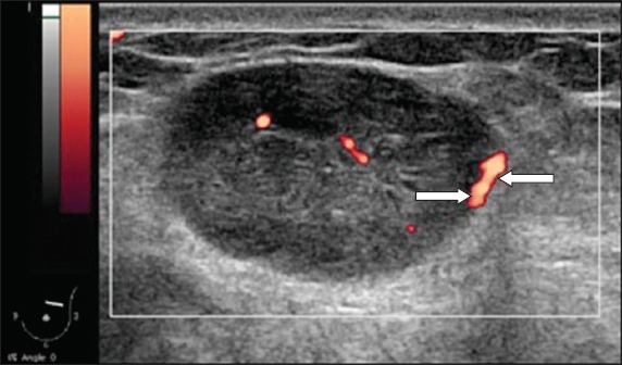
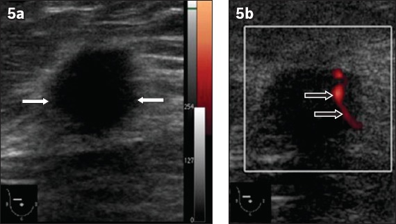
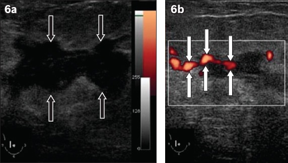
Introduction: This study aimed to evaluate the vascular pattern of solid breast lesions using power Doppler ultrasonography (PDUS) and assess whether the presence of intratumoural penetrating vessels can predict breast cancer malignancy.
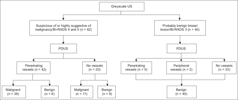
Methods: Greyscale ultrasonography (US) and PDUS were prospectively performed on 91 women in Malaysia with histopathologically proven breast lesions. The diagnostic accuracy of greyscale US, PDUS, and both greyscale US and PDUS was calculated and compared.
Results: The 91 women had 102 breast lesions (55 benign, 47 malignant). Of the 47 malignant lesions, 36 demonstrated intratumoural penetrating vessels. The sensitivity, specificity, positive predictive value (PPV) and negative predictive value (NPV) of greyscale US findings in diagnosing malignancy were 100.0%, 71.4%, 74.1% and 100.0%, respectively. The presence of calcification in the breast lesion and the margin, shape and posterior acoustic features of the lesion were significant parameters in predicting malignancy (p < 0.01). The sensitivity, specificity, PPV and NPV of the presence of intratumoural penetrating vessels in predicting malignancy were 76.5%, 80.0%, 76.5% and 80.0%, respectively. When both greyscale US and PDUS were used, there was a significant correlation in predicting malignancy (p < 0.05). The specificity and PPV values of the combined greyscale US and PDUS method (89.0% and 85.7%, respectively) were higher than those of greyscale US or PDUS alone.
Conclusion: Flow patterns revealed by PDUS can be useful for differentiating benign and malignant breast lesions. The visualisation of penetrating vessels in solid breast lesions can be used to complement greyscale US findings in predicting malignancy.
Keywords: breast cancer; penetrating vessel; power Doppler; ultrasonography.
Copyright: © Singapore Medical Association
Figures


Similar articles
-
Value of contrast-enhanced power Doppler sonography using a microbubble echo-enhancing agent in evaluation of small breast lesions.J Clin Ultrasound. 2003 Jun;31(5):227-38. doi: 10.1002/jcu.10172. J Clin Ultrasound. 2003. PMID: 12767017 Clinical Trial.
-
Power Doppler sonography: anything to add to BI-RADS US in solid breast masses?Eur J Radiol. 2009 Apr;70(1):77-85. doi: 10.1016/j.ejrad.2007.12.007. Epub 2008 Feb 19. Eur J Radiol. 2009. PMID: 18243623
-
Power Doppler sonography: evaluation of solid breast lesions and correlation with lymph node metastasis.Clin Imaging. 2008 May-Jun;32(3):167-71. doi: 10.1016/j.clinimag.2007.12.004. Clin Imaging. 2008. PMID: 18502342
-
Differentiating benign from malignant solid breast masses: value of shear wave elastography according to lesion stiffness combined with greyscale ultrasound according to BI-RADS classification.Br J Cancer. 2012 Jul 10;107(2):224-9. doi: 10.1038/bjc.2012.253. Epub 2012 Jun 12. Br J Cancer. 2012. PMID: 22691969 Free PMC article.
-
Solid breast lesions: evaluation with power Doppler US.Radiology. 1997 Apr;203(1):164-8. doi: 10.1148/radiology.203.1.9122386. Radiology. 1997. PMID: 9122386
Cited by
-
Ultrasound microflow patterns help in distinguishing malignant from benign thyroid nodules.Cancer Imaging. 2024 Jan 25;24(1):18. doi: 10.1186/s40644-024-00663-1. Cancer Imaging. 2024. PMID: 38268031 Free PMC article.
-
Radiopathological characteristics and outcomes of phyllodes tumor of the breast in Malaysian women.Medicine (Baltimore). 2018 Aug;97(31):e11412. doi: 10.1097/MD.0000000000011412. Medicine (Baltimore). 2018. PMID: 30075507 Free PMC article.
-
Diagnostic Accuracy of Spectral Doppler Indices and Sonoelastography in Predicting Malignancy in Breast Imaging Reporting and Database System 3 Breast Lesions With Histopathology as the Reference Standard.Cureus. 2024 Nov 11;16(11):e73481. doi: 10.7759/cureus.73481. eCollection 2024 Nov. Cureus. 2024. PMID: 39677136 Free PMC article.
-
Ultrasound-stimulated microbubbles enhanced vascular disruption in fractionated radiotherapy-treated tumours via ASMase activation.Dis Model Mech. 2023 Jun 1;16(6):dmm049531. doi: 10.1242/dmm.049531. Epub 2023 Jun 6. Dis Model Mech. 2023. PMID: 37278613 Free PMC article.
-
Evaluation of diagnostic value of Doppler ultrasound in the diagnosis of malignant breast masses.Eur J Transl Myol. 2024 Mar 26;34(2):12372. doi: 10.4081/ejtm.2024.12372. Eur J Transl Myol. 2024. PMID: 38536011 Free PMC article.
References
-
- Smith RA, Saslow D, Sawyer KA, et al. American Cancer Society High-Risk Work Group; American Cancer Society Screening Older Women Work Group; American Cancer Society Mammography Work Group; American Cancer Society Physical Examination Work Group; American Cancer Society New Technologies Work Group; American Cancer Society Breast Cancer Advisory Group. American Cancer Society guidelines for breast cancer screening: update 2003. CA Cancer J Clin. 2003;53:141–69. - PubMed
-
- Hisham AN, Yip CH. Spectrum of breast cancer in Malaysian women: overview. World J Surg. 2003;27:921–3. - PubMed
-
- Yip CH, Taib NA, Mohamed I. Epidemiology of breast cancer in Malaysia. Asian Pac J Cancer Prev. 2006;7:369–74. - PubMed
-
- Folkman J. Tumor angiogenesis: therapeutic implications. N Engl J Med. 1971;285:1182–6. - PubMed
-
- Milz P, Lienemann A, Kessler M, Reiser M. Evaluation of breast lesions by power Doppler sonography. Eur Radiol. 2001;11:547–54. - PubMed
MeSH terms
Substances
LinkOut - more resources
Full Text Sources
Other Literature Sources
Medical