

Can MRI predict meningioma consistency?: a correlation with tumor pathology and systematic review
- PMID: 27873040
- PMCID: PMC5438899
- DOI: 10.1007/s10143-016-0801-0
Can MRI predict meningioma consistency?: a correlation with tumor pathology and systematic review
Abstract
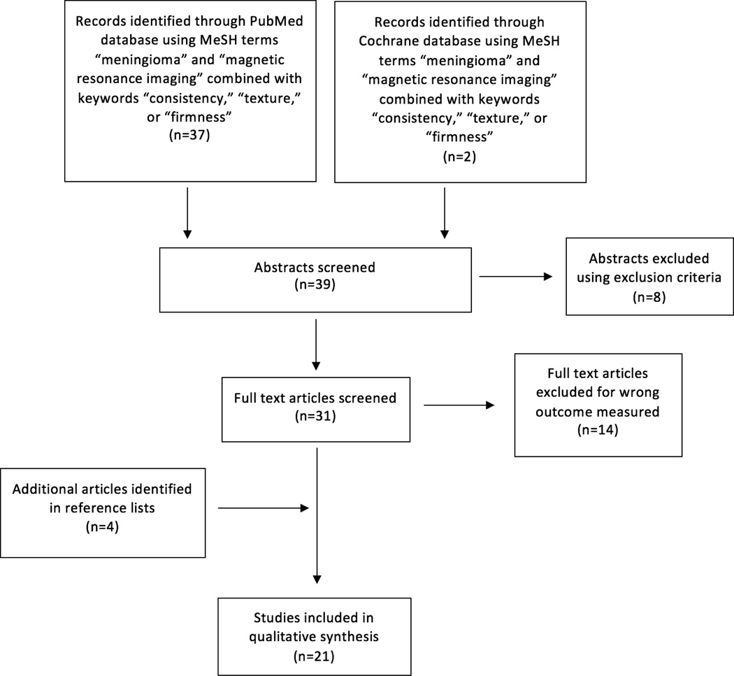
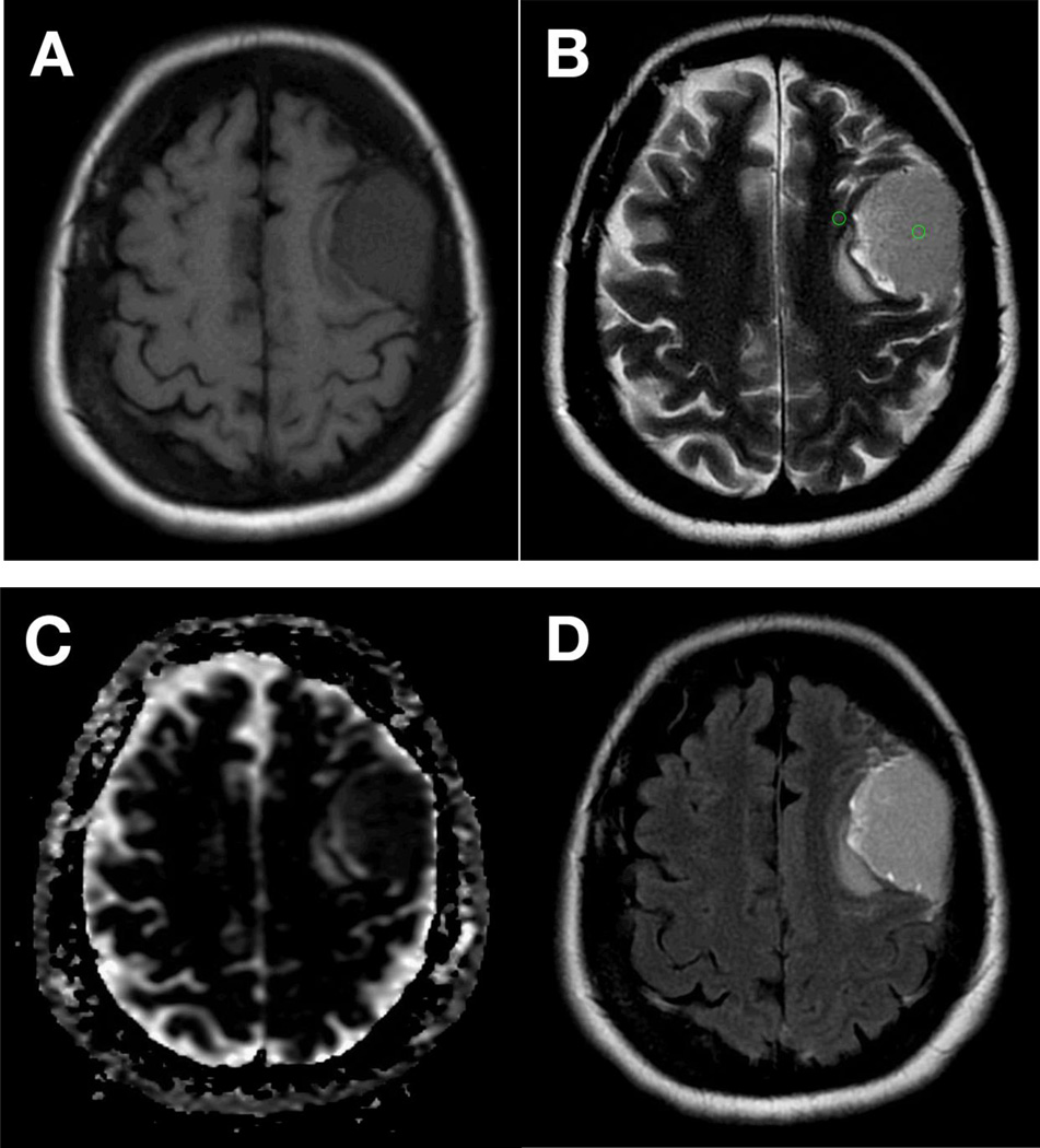
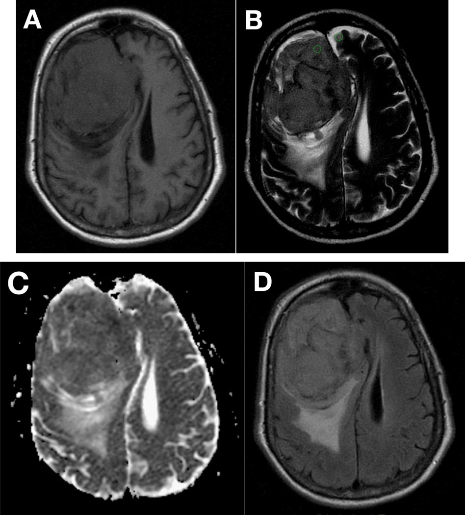
Tumor consistency is a critical factor that influences operative strategy and patient counseling. Magnetic resonance imaging (MRI) describes the concentration of water within living tissues and as such, is hypothesized to predict aspects of their biomechanical behavior. In meningiomas, MRI signal intensity has been used to predict the consistency of the tumor and its histopathological subtype, though its predictive capacity is debated in the literature. We performed a systematic review of the PubMed database since 1990 concerning MRI appearance and tumor consistency to assess whether or not MRI can be used reliably to predict tumor firmness. The inclusion criteria were case series and clinical studies that described attempts to correlate preoperative MRI findings with tumor consistency. The relationship between the pre-operative imaging characteristics, intraoperative findings, and World Health Organization (WHO) histopathological subtype is described. While T2 signal intensity and MR elastography provide a useful predictive measure of tumor consistency, other techniques have not been validated. T1-weighted imaging was not found to offer any diagnostic or predictive value. A quantitative assessment of T2 signal intensity more reliably predicts consistency than inherently variable qualitative analyses. Preoperative knowledge of tumor firmness affords the neurosurgeon substantial benefit when planning surgical techniques. Based upon our review of the literature, we currently recommend the use of T2-weighted MRI for predicting consistency, which has been shown to correlate well with analysis of tumor histological subtype. Development of standard measures of tumor consistency, standard MRI quantification metrics, and further exploration of MRI technique may improve the predictive ability of neuroimaging for meningiomas.
Keywords: Magnetic resonance imaging; Meningioma; Pathology; Tumor consistency.
Conflict of interest statement
The authors declare that they have no conflict of interest.
Figures
Similar articles
-
Signs and symptoms to determine if a patient presenting in primary care or hospital outpatient settings has COVID-19.Cochrane Database Syst Rev. 2022 May 20;5(5):CD013665. doi: 10.1002/14651858.CD013665.pub3. Cochrane Database Syst Rev. 2022. PMID: 35593186 Free PMC article.
-
Magnetic resonance perfusion for differentiating low-grade from high-grade gliomas at first presentation.Cochrane Database Syst Rev. 2018 Jan 22;1(1):CD011551. doi: 10.1002/14651858.CD011551.pub2. Cochrane Database Syst Rev. 2018. PMID: 29357120 Free PMC article.
-
Comparison of cellulose, modified cellulose and synthetic membranes in the haemodialysis of patients with end-stage renal disease.Cochrane Database Syst Rev. 2001;(3):CD003234. doi: 10.1002/14651858.CD003234. Cochrane Database Syst Rev. 2001. Update in: Cochrane Database Syst Rev. 2005 Jul 20;(3):CD003234. doi: 10.1002/14651858.CD003234.pub2. PMID: 11687058 Updated.
-
Cost-effectiveness of using prognostic information to select women with breast cancer for adjuvant systemic therapy.Health Technol Assess. 2006 Sep;10(34):iii-iv, ix-xi, 1-204. doi: 10.3310/hta10340. Health Technol Assess. 2006. PMID: 16959170
-
Comparison of the effectiveness of inhaler devices in asthma and chronic obstructive airways disease: a systematic review of the literature.Health Technol Assess. 2001;5(26):1-149. doi: 10.3310/hta5260. Health Technol Assess. 2001. PMID: 11701099
Cited by
-
Clinical Significance of Stiffness during Endoscopic Surgery for Intracerebral Hemorrhage: A Retrospective Study.Neurol Med Chir (Tokyo). 2023 Dec 15;63(12):563-570. doi: 10.2176/jns-nmc.2023-0043. Epub 2023 Nov 8. Neurol Med Chir (Tokyo). 2023. PMID: 37940569 Free PMC article.
-
Can an Imaging Marker of Consistency Predict Intraoperative Experience and Clinical Outcomes for Vestibular Schwannomas? A Retrospective Review.J Neurol Surg B Skull Base. 2021 Apr;82(2):251-257. doi: 10.1055/s-0039-1697026. Epub 2019 Sep 24. J Neurol Surg B Skull Base. 2021. PMID: 33777640 Free PMC article.
-
Consistency of pituitary adenomas: Amounts of collagen types I and III and the predictive value of T2WI MRI.Exp Ther Med. 2021 Nov;22(5):1255. doi: 10.3892/etm.2021.10690. Epub 2021 Sep 3. Exp Ther Med. 2021. PMID: 34603523 Free PMC article.
-
Fully automated detection and segmentation of meningiomas using deep learning on routine multiparametric MRI.Eur Radiol. 2019 Jan;29(1):124-132. doi: 10.1007/s00330-018-5595-8. Epub 2018 Jun 25. Eur Radiol. 2019. PMID: 29943184 Free PMC article.
-
Tumor to Cerebellar Peduncle T2-Weighted Imaging Intensity Ratio Fails to Predict Pituitary Adenoma Consistency.J Neurol Surg B Skull Base. 2019 Jun;80(3):252-257. doi: 10.1055/s-0038-1668516. Epub 2018 Aug 24. J Neurol Surg B Skull Base. 2019. PMID: 31143567 Free PMC article.
References
-
- Romani R, et al. Diffusion tensor magnetic resonance imaging for predicting the consistency of intracranial meningiomas. Acta Neurochir (Wien) 2014;156(10):1837–1845. - PubMed
-
- Kendall B, Pullicino P. Comparison of consistency of meningiomas and CT appearances. Neuroradiology. 1979;18(4):173–176. - PubMed
-
- Yamaguchi N, et al. Prediction of consistency of meningiomas with preoperative magnetic resonance imaging. Surg Neurol. 1997;48(6):579–583. - PubMed
-
- Sitthinamsuwan B, et al. Predictors of meningioma consistency: A study in 243 consecutive cases. Acta Neurochir (Wien) 2012;154(8):1383–1389. - PubMed
Publication types
MeSH terms
Grants and funding
LinkOut - more resources
Full Text Sources
Other Literature Sources
Medical
