Review
. 2008 Oct 23;8(10):6605-6641.
doi: 10.3390/s8106605.

Molecular Recognition and Specific Interactions for Biosensing Applications

Affiliations
Review

Molecular Recognition and Specific Interactions for Biosensing Applications

Dong Chung Kim et al. Sensors (Basel). .

Abstract

Molecular recognition and specific interactions are reliable and versatile routes for site-specific and well-oriented immobilization of functional biomolecules on surfaces. The control of surface properties via the molecular recognition and specific interactions at the nanoscale is a key element for the nanofabrication of biosensors with high sensitivity and specificity. This review intends to provide a comprehensive understanding of the molecular recognition- and specific interaction-mediated biosensor fabrication routes that leads to biosensors with well-ordered and controlled structures on both nanopatterned surfaces and nanomaterials. Herein self-assembly of the biomolecules via the molecular recognition and specific interactions on nanoscaled surfaces as well as nanofabrication techniques of the biomolecules for biosensor architecture are discussed. We also describe the detection of molecular recognition- and specific interaction-mediated molecular binding as well as advantages of nanoscale detection.

Keywords: Molecular recognition; biosensor; nanomaterial; nanopatterning; specific interaction.


Figures

Figure 1.
Figure 1.
(A) Chemical structures of MHDA, MHP, EG6OH and biotin disulfide. (B) AFM topographic images showing the growth of 3D microscale structures. (a, b) A gold surface patterned by μCP with 2 μm dot arrays of MHDA and back-filled with MHP was used as the template to grow micropillars through the LBL assembly of PLL and PSS after 7 (a) and 12 (b) cycles. Reproduced with permission of ACS [25]. (C) AFM topographic image of μCP-patterned SAMs with 2 μm biotin-thiol stripes separated by EG6OH stripes on gold surface, Reproduced with permission of RSC [3].
Figure 2.
Figure 2.
(A) Covalent modification of silicon. (B) The resulting monolayers of 100% EG3-ene, 50% Boc-N-ene + 50% EG3-ene, and 100% Boc-N-ene. Reproduced with permission of ACS [28].
Figure 3.
Figure 3.
Schematic of the LBL film deposition process. Gold electrodes were first functionalized with the negative charged thiol, 11-mercaptoundecanoic acid (MUA), and then alternatively incubated in solutions of positively charged redox polymer and negatively charged enzyme/SWNT. Reproduced with permission of ACS [38].
Figure 4.
Figure 4.
(A) LBL construction of GOx/PAA multilayer films via covalent bonding on the gold electrode surface. Reproduced with permission of Elsevier [61]. (B) Covalent attachment of FITC-labeled NH2-modified dsDNA to 10-undecenoic acid on NCD substrate using an EDC-mediated reaction. Reproduced with permission of Elsevier [62].
Figure 5.
Figure 5.
(A) Schematic of our approach to assemble 3D structures with active enzymes. (B) AFM topographic images showing the evolution of the avidin/biotin-HRP 3D structures. (a) A μCP-patterned surface; the EG6OH stripes are ∼2.4 nm taller than the biotin-thiol stripes. (b) After assembly of a bilayer of avidin/biotin-HRP, the protein stripes are ∼1.2 nm higher than the EG6OH stripes. (c) The same as b after a treatment with the surfactant; the protein stripes are ∼3.0 nm higher than the EG6OH stripes. (d) After assembly of 9 bilayers of avidin/biotin-HRP, the protein stripes are ∼12 nm higher than the EG6OH background [3].
Figure 6.
Figure 6.
Schematic of (A) DNA-directed immobilization of DNA-protein conjugates using complementary hybridization of DNA. Reproduced with permission of ACS [72], and (B) DNA-directed antibody immobilization by the protein G-DNA conjugate. Reproduced with permission of ACS [40].
Figure 7.
Figure 7.
(A) Schematic of fabrication of nanometer-sized patterns using EBL. (B) Fluorescence micrographs of Cy 5 labeled target hybridized to probe DNA immobilized within nanodot areas fabricated by EBL on ODS SAM: (a) complementary probe DNA (spot size: 500 nm); (b) complementary probe DNA (spot size: 250 nm); (c) non-complementary probe DNA. Reproduced with permission of RSC [83].
Figure 8.
Figure 8.
(A) Schematic of biomolecular nanopatterning using NIL in combination with molecular self-assembly. (B) AFM scans in tapping mode in air of 100 nm patterned stripes. (a) PLL-g-PEG/PEG-biotin stripes in an oxide background (after lift-off, stage VI in Figure 8A-b). (b) After PLL-g-PEG backfill the pattern is still visible due to the longer PEG chains supporting the biotin molecules (stage VII in Figure 8A-b). Reproduced with permission of ACS [104].
Figure 9.
Figure 9.
3D AFM topographic images showing the process of creating and growing the 3D protein nanostructures by electrostatic immobilization and biospecific interaction. (a) Two nanopatches (200 × 200 and 400 × 400 nm2) created by nanografting of MHP into the SAM of EG6OH had a depth of 2 nm. (b)After incubation with protein G, the patches were completely filled by protein G with an average height of ca. 1 nm. (c) After treatment with rabbit IgG, the height of the protein nanofeatures increased to 8-9 nm. (d) After subsequent treatment with goat anti-rabbit IgG, the height of the protein nanofeatures increased to 19-20 nm. Reproduced with permission of ACS [108].
Figure 10.
Figure 10.
(A) Schematic representation of the process used to obtain biologically active antibodies on the protein A/G, which is covalently bound to NHSC11SH nanoscale features patterned using DPN. (B) IgG was immobilized on protein A/G nanoarrays. (a) Topographical TMAFM image and its corresponding height profile of fluorescein isothiocyanate Alexa Fluor 594-labeled human IgG nanoarrays immobilized onto protein A/G templates. (b) Representative fluorescence microscopy image of Alexa Fluor 594-labeled antibody nanoarray patterns. Reproduced with permission of Wiley [112].
Figure 11.
Figure 11.
Real-time quartz crystal microbalance (QCM) and electronic sensing of specific biological recognition on nanotubes. (A) Scheme for streptavidin (SA) recognition with a nanotube coated with biotinylated Tween. (B) QCM signal; Biotin-nanotubes binds SA specifically but not other proteins. (C) Conductance; Specific binding of SA is detected electronically. (D) Scheme for IgG recognition with a nanotube coated with a SpA–Tween conjugate. (E) QCM signal; SpA-nanotubes binds IgG specifically but not unrelated proteins. Note that 10 nM IgG concentration approaches the lower detection limit of the instrument, whereas 100 nM approaches surface saturation of the sample; thus, the response does not show a full proportionality to the concentration. (F) Conductance; Specific binding of IgG is detected electronically. Reproduced with permission of APS [125].
Figure 12.
Figure 12.
(A) Schematic diagram of preparation of the dispersed film of gold nanorods and fabrication of bio-architecture with HRP activity on gold nanorod. (B) The catalytic activity of LBL-assembled HRP on gold nanorods compared to that on a flat gold surface. Closed square, on vertical array of gold nanorods; Closed circle, on dispersed film of gold nanorods; Open triangle, on flat gold surface. Reproduced with permission of KPS [126].
Figure 13.
Figure 13.
(A) Schematic illustration of the principle of the nanoparticle-based DNA biosensor for visual detection of GMO. The first red line is formed in the test zone (TZ), as the immobilized streptavidin captures the hybrids. The second red line is developed on the control zone (CZ), when the excess of conjugated gold nanoparticles hybridize with immobilized oligo(dA) probe. Au, conjugated gold nanoparticles; B, biotin; SA, streptavidin. (B) The hybridization assay of amplified lectin DNA by using the biosensor. PCR negative (N) and positive samples (S) are shown. TZ, test zone and CZ, control zone. Reproduced with permission of Elsevier [141].
Figure 14.
Figure 14.
(A) Assembly of the SWCNT electrically contacted GOx electrode. (B) Cyclic voltammograms corresponding to the electrocatalyzed oxidation of different concentrations of glucose by the GOx reconstituted on the 25 nm long FAD-functionalized CNTs assembly: a) 0, b) 20, c) 60, d) 160 mM glucose. (C) Calibration curve corresponding to the amperometric responses of the reconstituted GOx/CNTs electrode in the presence of different concentrations of glucose. Reproduced with permission of I. Willner [124].
Figure 15.
Figure 15.
(A) Binding of thrombin on a SWCNT-FET-based aptamer sensor (B) A real-time conductance measurement obtained from the thrombin aptamer immobilized SWCNT-FET. (C) The selectivity of thrombin aptamer immobilized SWCNT-FET. Arrow indicates the point of adding protein solution. Reproduced with permission of ACS [137].

Similar articles

Cited by

References

    1. McFadden P. Broadband biodetecion: holmes on a chip. Science. 2002;297:2075–2076. - PubMed
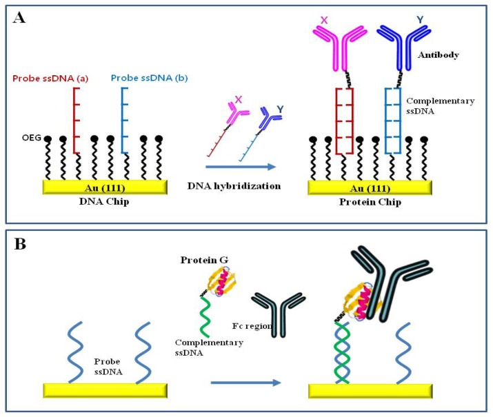
    1. Jung Y., Jeong J.Y., Chung B.H. Recent advances in immobilization methods of antibodies on solid supports. Analyst. 2008;133:697–701. - PubMed
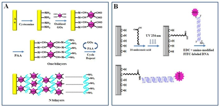
    1. Rauf S., Zhou D., Abell C., Klenerman D., Kang D.J. Building three-dimensional nanostructures with active enzymes by surface template layer-by-layer assembly. Chem. Commun. 2006:1721–1723. - PubMed
    1. Sun Y., Yan F., Yang W., Sun C. Multilayered construction of glucose oxidase and silica nanoparticles on Au electrodes based on layer-by-layer covalent attachment. Biomaterials. 2006;27:4042–4049. - PubMed
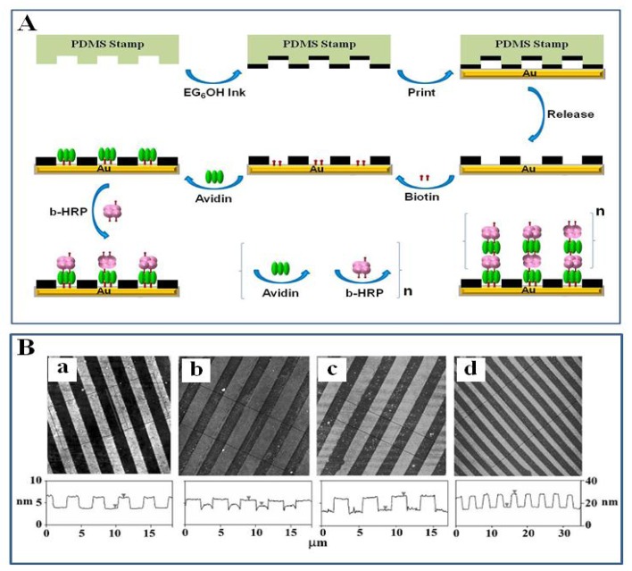
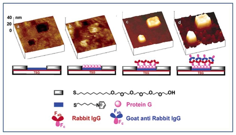
    1. Miyake T., Tanii T., Kato K., Zako T., Funatsu T., Ohdomari I. Selective improvement in protein nanopatterning with a hydroxyl-terminated self-assembled monolayer template. Nanotechnology. 2007;18:305304.

LinkOut - more resources