. 2016 Nov 22:7:13505.
doi: 10.1038/ncomms13505.

The Hippo signalling pathway coordinates organ growth and limits developmental variability by controlling dilp8 expression

Affiliations

The Hippo signalling pathway coordinates organ growth and limits developmental variability by controlling dilp8 expression

Emilie Boone et al. Nat Commun. .

Abstract

Coordination of organ growth during development is required to generate fit individuals with fixed proportions. We recently identified Drosophila Dilp8 as a key hormone in coupling organ growth with animal maturation. In addition, dilp8 mutant flies exhibit elevated fluctuating asymmetry (FA) demonstrating a function for Dilp8 in ensuring developmental stability. The signals regulating Dilp8 activity during normal development are not yet known. Here, we show that the transcriptional co-activators of the Hippo (Hpo) pathway, Yorkie (Yki, YAP/TAZ) and its DNA-binding partner Scalloped (Sd), directly regulate dilp8 expression through a Hpo-responsive element (HRE) in the dilp8 promoter. We further demonstrate that mutation of the HRE by genome-editing results in animals with increased FA, thereby mimicking full dilp8 loss of function. Therefore, our results indicate that growth coordination of organs is connected to their growth status through a feedback loop involving Hpo and Dilp8 signalling pathways.


Figures
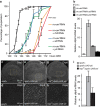
Figure 1
Figure 1. Yki regulates dilp8 expression independently of JNK signalling.
(a) Reducing Yki or Sd levels rescues the delay of rn>avl RNAi animals. The percentage of larvae that have pupariated at the indicated hours AED is shown (n>90, triplicate experiments, error bars represent s.e.m.). (b) Silencing of Yki or Sd efficiently suppresses the upregulation of dilp8 mRNA levels observed in rn>avl RNAi animals. Relative dilp8 mRNA levels were measured using qPCR on whole larvae of the indicated genotypes (triplicate experiments, error bars represent s.e.m.). (c,e,g) Yki induces dilp8 expression independent of JNK signalling. (d,f,h) represent higher magnification images of the area shown in the dashed lines. Larval wing discs of the indicated genotypes were dissected 116 h AED and stained for Dilp8. (i) qPCRs on the indicated genotypes showing that the Yki-dependent induction of dilp8 is still significant in a hep75 mutant background (triplicate experiments, error bars represent s.e.m.).
Figure 2
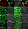
Figure 2. Yki regulates dilp8 transcription through Sd.
Wing imaginal disc carrying RFP-labelled yki-expressing clones (ad), GFP-labelled wt clones (e,f), GFP-labelled yki-expressing clones (g,h), GFP-labelled yki+sd RNAi clones (i,j), or GFP-negative clones mutant for ex (k,l), hpo (m,n) or wts (o,p), all dissected 116 h AED and stained for Dilp8 (c,f,h,j,l,n,p). In b, dilp8 expression is followed by GFP using the dilp8-GFP insertion allele (see the ‘Genotypes' section under the ‘Methods' section).
Figure 3
Figure 3. Yki directly regulates dilp8 expression through a HRE in the dilp8 promoter.
(a) Schematic of the dilp8 promoter region and the HRE harbouring the three putative Sd-binding sites (indicated as green squares). The dilp8 promoter fragments used to study Yki-dependent regulation of dilp8 expression in vivo are shown. In dilp8-PFΔ123, mutations of the three putative Sd-binding sites are indicated as red squares. (b) DNA pull-down experiments show that binding of Sd to the dilp8-PF is mediated by the three Sd-binding sites. The indicated DNA fragments were incubated with lysates from S2 cells transfected with Sd-Flag. Band intensities represents the average of three independent experiments: for diap-PF: 3.8±0.7 (positive control), for act: 1±0.2 (negative control), for dilp8-PF: 5.1±1.0, and for dilp8-PF Δ123: 1.9±0.4. (c) Luciferase assay showing that Yki/Sd activate gene expression through the HRE in the dilp8-PF. S2 cells were transfected with Yki and Sd. The ability of Yki/Sd to induce gene expression from the indicated promoter fragments was measured (triplicate samples, error bars represent s.e.m.). (ds) Yki induces dilp8 transcription through the HRE in vivo. Wing imaginal discs carrying GFP-labelled Yki-expressing clones were dissected from transgenic flies carrying the indicated dilp8 promoter fragments fused to the LacZ-encoding sequence. The full dilp8 promoter and dilp8-PF, but not dilp8-intron1 and dilp8-PFΔ123, induces lacZ expression as detected by β-gal staining (d,h,l,p in red) in the GFP-labelled yki-overexpressing clones (e,i,m,n in green). In each condition, yki overexpression leads to elevated levels of endogenous Dilp8 protein (f,j,n,r in white).
Figure 4
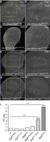
Figure 4. Yki-dependent regulation of Dilp8 adjusts organ growth and limits developmental instability.
(a,c,e,g) JNK-dependent regulation of dilp8 expression is not affected in dilp8-PFΔ123 mutants. (b,d,f,h) represent higher magnification images of the area shown in the dashed lines. Wing discs were dissected from the indicated genotypes and stained for Dilp8. (i) Mutation of the three Sd-binding sites in the dilp8 promoter by gene editing induces FA. Bar histograms shows FA indices (FAi) of left and right wing areas measured on individuals of the indicated genotypes. ***P<0.0001, F-test for unequal distributions.

References

    1. Twitty V. C. & Schwind J. L. The growth of eyes and limbs transplanted heteroplastically between two species of Amblystoma. J. Exp. Zool. 59, 61–86 (1931).
    1. Bryant P. J. & Simpson P. Intrinsic and extrinsic control of growth in developing organs. Q. Rev. Biol. 59, 387–415 (1984). - PubMed
    1. Halme A., Cheng M. & Hariharan I. K. Retinoids regulate a developmental checkpoint for tissue regeneration in Drosophila. Curr. Biol. 20, 458–463 (2010). - PMC - PubMed
    1. Parker N. F. & Shingleton A. W. The coordination of growth among Drosophila organs in response to localized growth-perturbation. Dev. Biol. 357, 318–325 (2011). - PubMed
    1. Jaszczak J. S., Wolpe J. B., Dao A. Q. & Halme A. Nitric oxide synthase regulates growth coordination during Drosophila melanogaster imaginal disc regeneration. Genetics 200, 1219–1228 (2015). - PMC - PubMed

Publication types

MeSH terms

Substances

LinkOut - more resources