. 2016 Nov 22:6:37391.
doi: 10.1038/srep37391.

Lymphocytic Microparticles Modulate Angiogenic Properties of Macrophages in Laser-induced Choroidal Neovascularization

Affiliations

Lymphocytic Microparticles Modulate Angiogenic Properties of Macrophages in Laser-induced Choroidal Neovascularization

Houda Tahiri et al. Sci Rep. .

Abstract

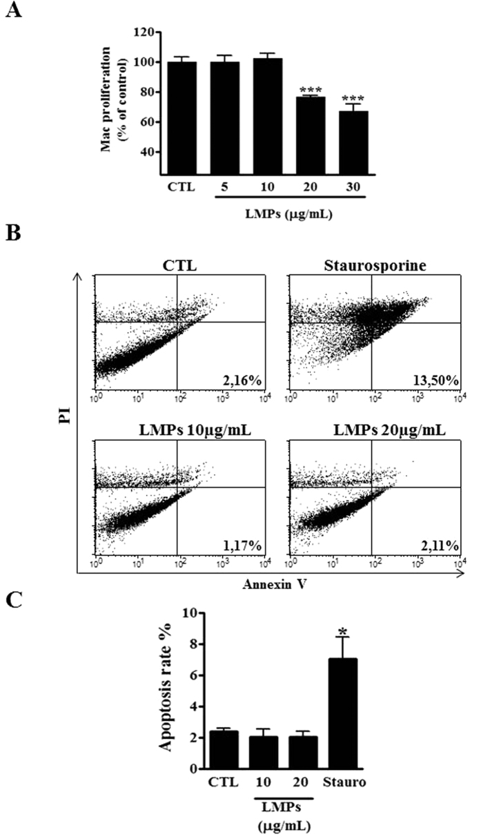
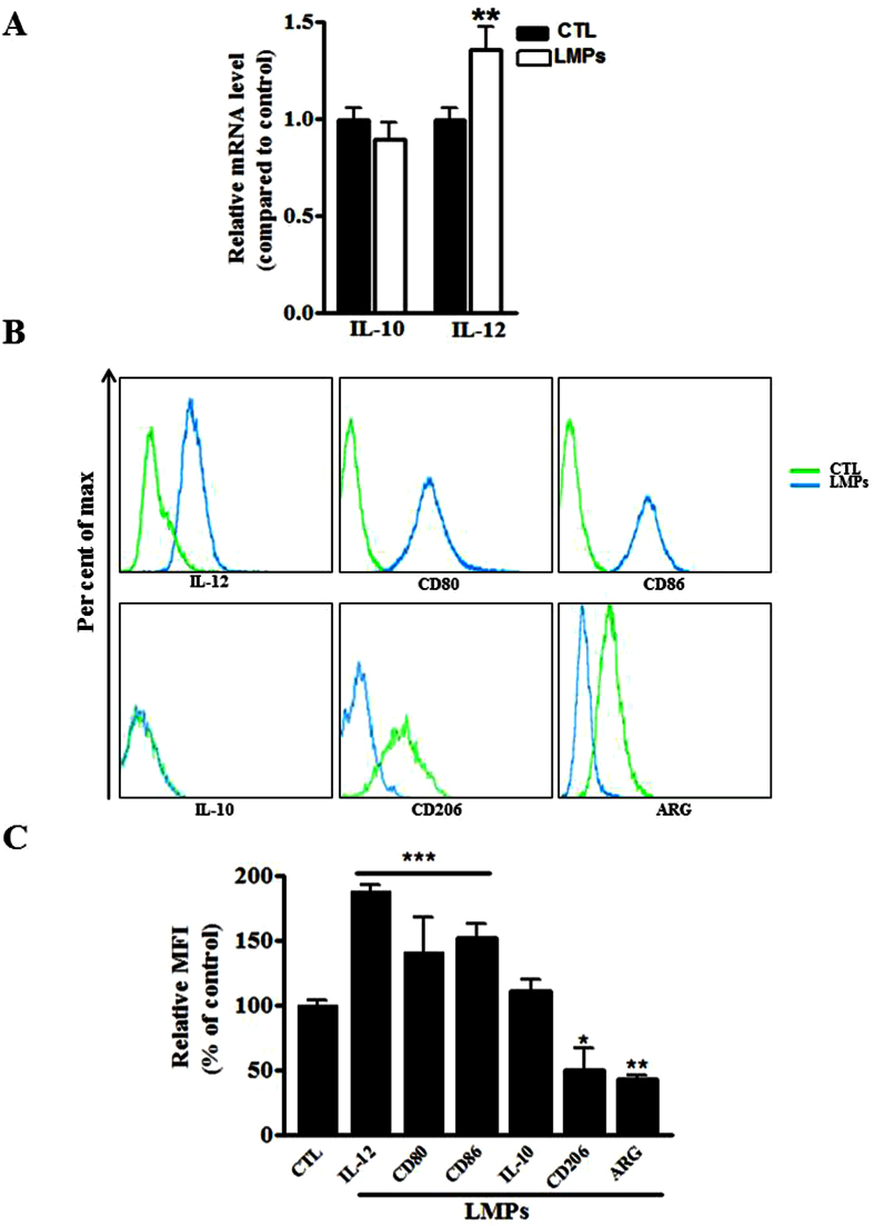
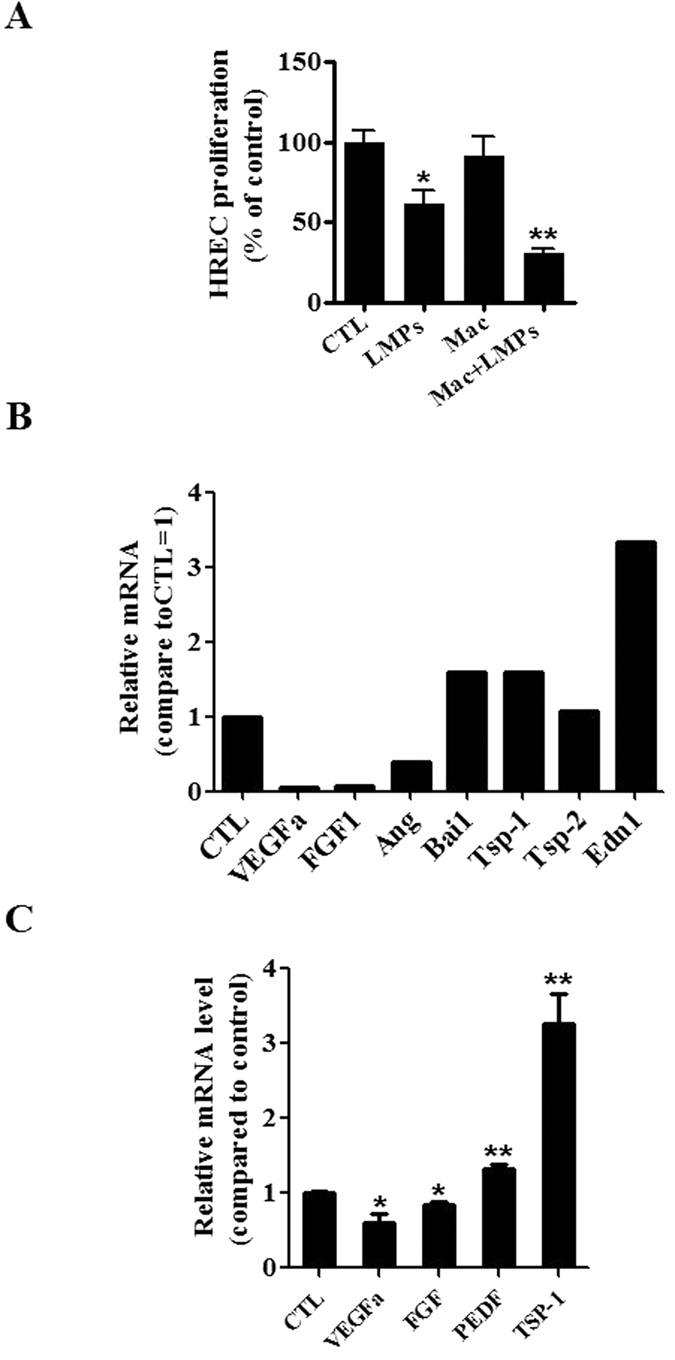
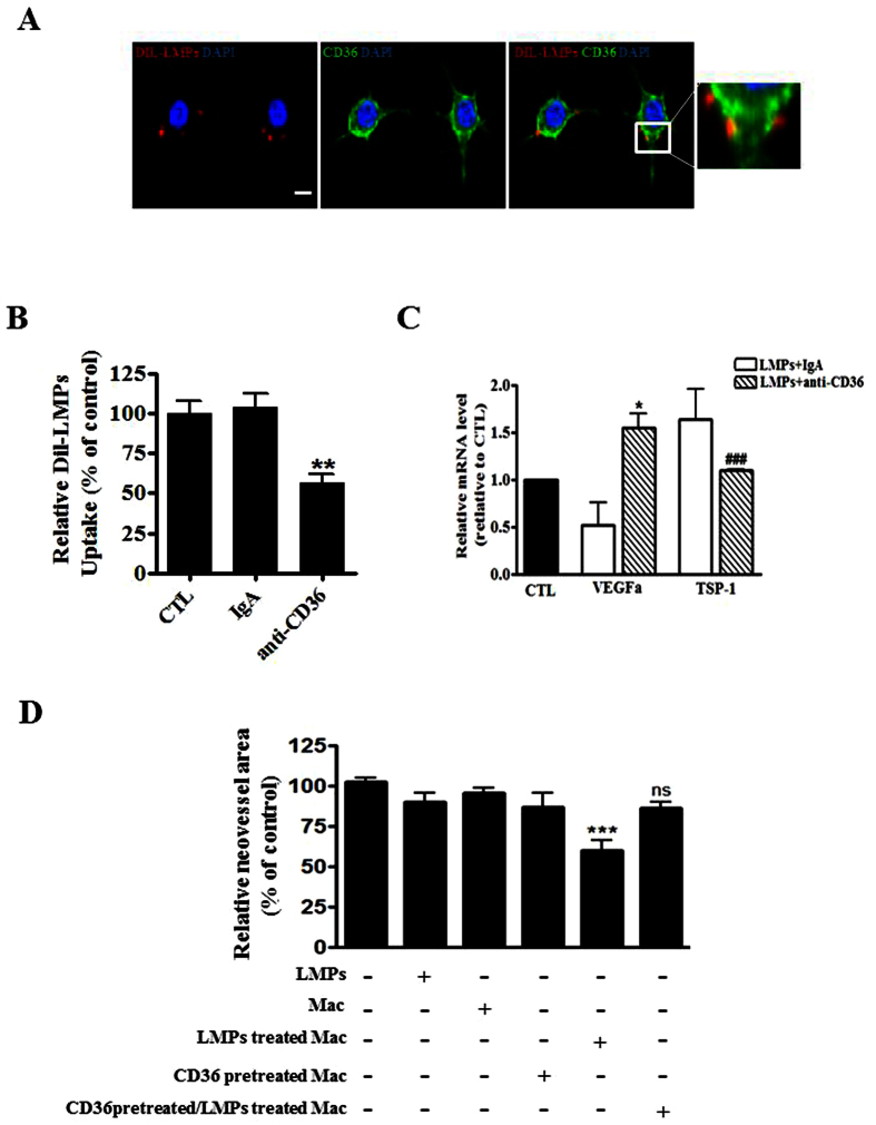
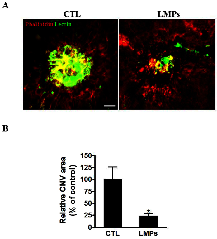
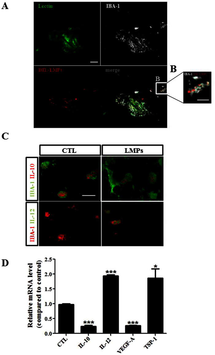
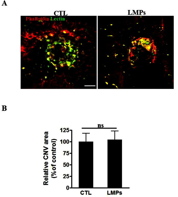
Pathological choroidal neovascularization (CNV) is the common cause of vision loss in patients with age-related macular degeneration (AMD). Macrophages possess potential angiogenic function in CNV. We have demonstrated that human T lymphocyte-derived microparticles (LMPs) exert a potent antiangiogenic effect in several pathological neovascularization models. In this study, we investigated the alteration of proangiogenic properties of macrophages by LMPs treatment in vitro and in vivo models. LMPs regulated the expression of several angiogenesis-related factors in macrophages and consequently stimulated their antiangiogenic effects evidenced by the suppression of the proliferation of human retinal endothelial cells in co-culture experiments. The involvement of CD36 receptor in LMPs uptake by macrophages was demonstrated by in vitro assays and by immunostaining of choroidal flat mounts. In addition, ex vivo experiments showed that CD36 mediates the antiangiogenic effect of LMPs in murine and human choroidal explants. Furthermore, intravitreal injection of LMPs in the mouse model of laser-induced CNV significantly suppressed CNV in CD36 dependent manner. The results of this study suggested an ability of LMPs to alter the gene expression pattern of angiogenesis-related factors in macrophages, which provide important information for a new therapeutic approach for efficiently interfering with both vascular and extravascular components of CNV.


Conflict of interest statement

The authors declare no competing financial interests.

Figures

Figure 1
Figure 1. LMPs dose-dependently inhibited macrophages proliferation.
(A) Indicated concentrations of LMPs were incubated with macrophages (RAW 264.7) for 24 hours. The proliferation of macrophages was determined using 3H-thymidine incorporation and values were presented as a percentage of control (CTL). ***P < 0.001 vs. CTL. (B) Representative results of flow cytometry analysis of macrophage cell apoptosis after 24-hours of treatment with indicated concentrations of LMPs, or staurosporine (positive control). FACS analysis was performed after macrophages were stained with Annexin-FITC and propidium iodide using Vybrant Apoptosis assay kit. Note that the staurosporin-treated cells undergo apoptosis and become PI+/Annexin V+ in the later stages of apoptosis. No difference is observed in LMPs-treated and control macrophages for Annexin V staining. (C) The apoptosis rates were presented as the percentage of apoptotic cells relative to the total number of cells. Values are means ± SEM. *P < 0.05 vs. CTL. Mac and Stauro indicate macrophages and staurosporin, respectively.
Figure 2
Figure 2. LMPs altered the expression of M1 and M2 markers of macrophages.
(A) The mRNA expression levels of IL-10 and IL-12 were quantified by quantitative RT-PCR after macrophages (RAW 264.7) were treated with 10 μg/mL of LMPs for 24 hours. The values were presented as fold changes relative to the control group (CTL) set as 1. **P < 0.01 vs. CTL (B) Representative FACS analysis of the expression of IL-10, IL-12, CD80, Cd86, ARG, and CD206 in macrophages after 24-hour treatment with 10 μg/mL of LMPs. (C) The numbers of cells expressing of IL-10, IL-12, CD80, CD86, ARG, or CD206 were calculated respectively and presented as a percentage of CTL (set as 100%). ***P < 0.001, **P < 0.01, *P < 0.05 vs. CTL.
Figure 3
Figure 3. LMPs modulated angiogenesis-related factor expression in macrophages.
(A) Cell proliferation of human retinal microvascular endothelial cells (HREC) was assessed after cells were co-cultured for 24 hours with LMPs, control medium (CTL), macrophages (RAW 264.7 pre-treated with the control medium, Mac), or LMPs pre-treated macrophages (Mac + LMPs). The relative proliferation rates of HREC were presented as a percentage of CTL. *P < 0.05, **P < 0.01 vs. CTL. (B) The mRNA levels of a panel of angiogenesis-related factors in macrophages (RAW 264.7) were evaluated by using a commercial PCR array after treatment with LMPs (10 μg/mL) for 24 hours. The relative mRNA level of each factor was calculated and expressed as fold change compared to control (CTL). (C) The gene expression of VEGFa, FGF, PEDF and TSP-1 in LMPs-treated macrophages was quantified by real-time PCR. The relative mRNA levels are presented as fold change relative to control macrophages (CTL). *P < 0.05, **P < 0.01 vs. CTL.
Figure 4
Figure 4. CD36 was involved in the uptake of LMPs by macrophages and mediated the effect of LMPs.
(A) Representative images of immunohistochemistry staining of CD36 (in green) and localization of LMPs in macrophages (RAW 264.7) after 4 hours of incubation with DiI-LMPs (in red). Bar: 25 μm. The merged images on the right indicate the co-localization of CD36 and LMPs. (B) After a 4 h incubation of DiI-LMPs (10 μg/ml) with macrophages (control), the isotype-matched control antibody pre-treated (IgA) macrophages, or anti-CD36 antibody pre-treated macrophages, the uptake of DiI-LMPs was measured and expressed as percentage of control (CTL, set as 100%). **P < 0.01 anti-CD36 vs. CTL. (C) RAW 264.7 were pre-treated with control antibody (IgA) or anti-CD36 before 24 hours LMPs treatment. The mRNA levels of VEGFa and TSP-1 in the macrophages were determined by quantitative PCR. *P < 0.05, ###P < 0.001 vs. LMPs + IgA. (D) The RPE-free choroidal explants were cultured for developing new vessels in the first 5 days, and then explants were incubated for another 48 hours with 50 μg/mL of LMPs, macrophages RAW 264.7(Mac), LMPs-treated Mac, anti-CD36 pre-treated Mac or anti-CD36 pre-treated + LMPs-treated Mac. The neovascularized areas in each condition were measured and presented as a percentage of control (explants without any treatment set as 100%). ***P < 0.001 vs. CTL.
Figure 5
Figure 5. LMPs suppressed laser-induced CNV in vivo.
(A) Representative images of laser-induced CNV on choroidal flat-mounts 7 days after the second intravitreal injection of 50 μg/mL of LMPs. The choroidal flat-mounts were stained with FITC-lectin (marker of endothelial cells, green) and Phalloidin (red). CTL represent the control mice that received intravitreal injections of the control medium. (B) CNV areas were quantified using computer-assisted semi-quantitative assay and normalized to control. Bar: 50 μm, *P < 0.05 vs. CTL. Values are means ± SEM from 10 eyes (each eye contains 3 impact laser-induced CNV) for each condition.
Figure 6
Figure 6. LMPs modulated gene expression in macrophages in vivo.
(A,B) Representative images of LMPs uptake by macrophages in vivo. Immunofluorescence staining of mouse choroidal flat-mounts 7 days after the second intravitreal injections of DiI-LMPs (red) in laser-induced CNV model. The vessels were stained with FITC-lectin (green) and macrophage with IBA-1 in grey. Bar: 50 μm in (A) and 25 μm in (B). (C) Representative images of expression of IL-12 (green) and IL-10 (red) in macrophages in CNV areas after LMPs treatments. Bar: 15 μm. CTL represent the control mice received intravitreal injections of control medium. (D) Relative mRNA expression level of IL-10, IL-12, VEGFa and TSP-1 in CNV areas. Tissues from the CNV areas were collected by laser-capture microdissection. The mRNA levels of the genes of interest were quantified by quantitative RT-PCR. The values were presented as mean fold changes relative to the control (CTL) values (set as 1). *P < 0.05, ***P < 0.001 vs. CTL.
Figure 7
Figure 7. The antiangiogenic effect of LMPs was attenuated in CD36 knockout mice.
(A) Representative images of choroidal flat-mounts of laser-induced CNV in CD36−/− mice. The images were taken 7 days after the second LMPs intravitreal injection. Choroidal flat-mounts were immunostained with FITC-lectin (green) and phalloidin (red). Bar: 50 μm. (B) The CNV areas were quantified with computer-assisted semi-quantitative assay. Values were presented as percentage of control (set as 100%). Values are means ± SEM of 6 eyes (each eye containing 3 impact laser-induced CNV) for each condition. P > 0.05 between the two groups.

Similar articles

Cited by

References

    1. Carpintero R. et al.. HDL interfere with the binding of T cell microparticles to human monocytes to inhibit pro-inflammatory cytokine production. PLoS One 5, e11869, doi: 10.1371/journal.pone.0011869 (2010). - DOI - PMC - PubMed
    1. Martin S. et al.. Shed membrane particles from T lymphocytes impair endothelial function and regulate endothelial protein expression. Circulation 109, 1653–1659, doi: 10.1161/01.CIR.0000124065.31211.6E (2004). - DOI - PubMed
    1. Yang C. et al.. Lymphocytic microparticles inhibit angiogenesis by stimulating oxidative stress and negatively regulating VEGF-induced pathways. Am J Physiol Regul Integr Comp Physiol 294, R467–R476, doi: 10.1152/ajpregu.00432.2007 (2008). - DOI - PubMed
    1. Yang C., Gagnon C., Hou X. & Hardy P. Low density lipoprotein receptor mediates anti-VEGF effect of lymphocyte T-derived microparticles in Lewis lung carcinoma cells. Cancer Biol Ther 10, 448–456 (2010). - PMC - PubMed
    1. Yang C. et al.. Role of receptor-mediated endocytosis in the antiangiogenic effects of human T lymphoblastic cell-derived microparticles. Am J Physiol Regul Integr Comp Physiol 302, R941–R949, doi: 10.1152/ajpregu.00527.2011 (2012). - DOI - PubMed

Publication types

Grants and funding