. 2016 Dec 23;291(52):26658-26669.
doi: 10.1074/jbc.M116.761643. Epub 2016 Nov 14.

Single Binding Mode Integration of Hemicellulose-degrading Enzymes via Adaptor Scaffoldins in Ruminococcus flavefaciens Cellulosome

Affiliations

Single Binding Mode Integration of Hemicellulose-degrading Enzymes via Adaptor Scaffoldins in Ruminococcus flavefaciens Cellulosome

Pedro Bule et al. J Biol Chem. .

Abstract

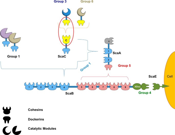
The assembly of one of Nature's most elaborate multienzyme complexes, the cellulosome, results from the binding of enzyme-borne dockerins to reiterated cohesin domains located in a non-catalytic primary scaffoldin. Generally, dockerins present two similar cohesin-binding interfaces that support a dual binding mode. The dynamic integration of enzymes in cellulosomes, afforded by the dual binding mode, is believed to incorporate additional flexibility in highly populated multienzyme complexes. Ruminococcus flavefaciens, the primary degrader of plant structural carbohydrates in the rumen of mammals, uses a portfolio of more than 220 different dockerins to assemble the most intricate cellulosome known to date. A sequence-based analysis organized R. flavefaciens dockerins into six groups. Strikingly, a subset of R. flavefaciens cellulosomal enzymes, comprising dockerins of groups 3 and 6, were shown to be indirectly incorporated into primary scaffoldins via an adaptor scaffoldin termed ScaC. Here, we report the crystal structure of a group 3 R. flavefaciens dockerin, Doc3, in complex with ScaC cohesin. Doc3 is unusual as it presents a large cohesin-interacting surface that lacks the structural symmetry required to support a dual binding mode. In addition, dockerins of groups 3 and 6, which bind exclusively to ScaC cohesin, display a conserved mechanism of protein recognition that is similar to Doc3. Groups 3 and 6 dockerins are predominantly appended to hemicellulose-degrading enzymes. Thus, single binding mode dockerins interacting with adaptor scaffoldins exemplify an evolutionary pathway developed by R. flavefaciens to recruit hemicellulases to the sophisticated cellulosomes acting in the gastrointestinal tract of mammals.

Keywords: cellulase; cellulose; cellulosome; cohesin; dockerin; protein structure; protein-protein interaction.


Figures

FIGURE 1.
FIGURE 1.
Group-specific interactions that contribute to cellulosome assembly in R. flavefaciens strain FD-1. The scheme is color-coded to highlight the four subgroups of cohesin-dockerin specificities as follows: dockerins and cognate cohesin counterparts of the different groups are shown in light blue (group 1 dockerins), yellow (groups 3 and 6), green (groups 2 and 4), and red (group 5), respectively. Group 2 dockerins are truncated derivatives of group 4 and are not represented in the figure for simplification. The red oval marks the complex of the group 3 interaction, whose structure is reported here.
FIGURE 2.
FIGURE 2.
Structure and cohesin-dockerin interface in the R. flavefaciens CohScaC-Doc3 complex. A, structure of CohScaC-Doc3 complex with the dockerin in green and the cohesin in blue. Phe-902, Arg-908, and His-943 that dominate cohesin recognition are labeled and shown as stick configuration. Ca2+ ions are depicted as purple spheres. B, polar interactions at the complex interface. C, hydrophobic interactions at the complex interface. The most important residues in both types of interaction are shown as sticks. The transparent gray disk in B and C marks the plane defined by the 8-3-6-5 β-sheet, where the β-strands form a distinctive dockerin interacting plateau.
FIGURE 3.
FIGURE 3.
Overlay of the R. flavefaciens CohScaC-Doc3 complex with the A. cellulolyticus type I cohesin-dockerin complex. A, overlay of CohScaC-Doc3 (depicted in blue) with the AcScaCCoh3-ScaBDoc type I complex from A. cellulolyticus (depicted in green, PDB code 4UYP), with the dockerins rotated 180° relative to each other, showing the high degree of overall similarity. B, overlay of both cohesins isolated from the complexes and rotated ∼90° down and right relatively to A, with the dockerin interacting plateau in the first plane. This view highlights the main differences between the two cohesins that consist of the large β-flap extension that interrupts β-strand 8 (dark blue box) and the well defined α-helix connecting β-strands 4 and 5 (red box). These two structural elements together with the loop formed by the distal part of β-strand 8 and the proximal section of β-strand 9 that is tilted toward the dockerin form a claw-like interaction interface.
FIGURE 4.
FIGURE 4.
Significant differences between the two cohesin-binding interfaces do not allow the dual binding mode of type I dockerin from R. flavefaciens. A, overlay of the two dockerin repeats observed in Doc3 showing that the structures are similar (r.m.s.d. of 0.82 Å) in the main chain atoms but have considerable differences in the side chains. B, two interacting helices of Doc3, helix 1 (bright green) and helix 3 (blue), with the most important cohesin recognition residues displayed as sticks. C, comparison of the two putative binding surfaces by overlaying Doc3 with a version of itself rotated by 180° (pink) shows a lack of conservation in the key contacting residues. Lack of internal symmetry in Doc3 and the involvement of the two helices in cohesin recognition suggest that Doc3 displays a single cohesin-binding platform.
FIGURE 5.
FIGURE 5.
Determination of Doc3 Phe-902, Arg-908, and His-943 importance for CohScaC recognition. A, representative binding isotherms of the interaction between CohScaC/Doc3, CohScaC/Doc3 R908A mutant, CohScaC/Doc3 R908A/H943A double mutant, and CohScaC/Doc3 F902A/R908A double mutant. The upper part of each panel shows the raw heats of binding, and the lower parts include the integrated heats after correction for heat of dilution. The curve represents the best fit to a single-site binding model. The corresponding thermodynamic parameters are shown in Table 3. B, non-denaturing gel electrophoretic analysis of CohScaC-Doc3 interaction. In the 1st lane, both gels were loaded with the cohesin (Coh). Adjacent lanes were loaded with the dockerin (D3) and with both the cohesin and dockerin modules together after a 60-min incubation at equimolar concentrations. The appearance of a band with a different migration pattern in lanes containing the complex represents a positive result (e.g. D3 WT), whereas a negative result (e.g. D3 FR) is given by the appearance of only the individual dockerin and cohesin bands. A faint cohesin band is seen even in the lanes where there is complex formation that results from excess cohesin probably due to not all the dockerin in solution being active.
FIGURE 6.
FIGURE 6.
Alignment of group 3 dockerins. All group 3 dockerins were aligned using Clustal Omega multiple sequence alignment software and are organized according to their affinity to CohScaC, from the highest Ka value to the lowest, as determined by ITC. Docs 381 and 1425 were aligned in opposite orientation relative to the other members by switching the N-terminal half with the C-terminal half of the sequence. This resulted in the N-terminal interface (blue residues) of Docs 381 and 1425 being perfectly aligned with the C-terminal interface (green residues) of the remaining group 3 members and vice versa, supporting the theory that they will bind the cohesin in an opposite orientation. The top line matches the protein secondary structure (red cylinders) to the primary structures, as observed in the Doc3 structure, and also points to the calcium coordinating residues (blue triangles). All residues involved in the Doc3 interaction with CohScaC are highlighted according to the color code displayed at the bottom. Conservation of key residues for cohesin recognition along the group is highlighted with black boxes. To some extent, this conservation pattern seems to correlate to the CohScaC affinity profile of the group.
FIGURE 7.
FIGURE 7.
Binding affinity of group 6 dockerins to CohScaC determined by ITC. Representative binding isotherms are displayed in A: CohScaC/Doc903, CohScaC/Doc1369, and CohScaC/Doc1965. The upper part of each panel shows the raw heats of binding, and the lower parts include the integrated heats after correction for heat of dilution. The curve represents the best fit to a single-site binding model. The corresponding thermodynamic parameters are shown in Table 4. B, alignment of tested group 6 dockerins with a version of Doc3 in which the C-terminal half was switched with the N-terminal half (Doc3_180), resulting in the N-terminal interface (blue residues) of Doc3 being perfectly aligned with the C-terminal interface (green residues) of the group 6 members and vice versa, supporting the theory that they will bind the cohesin in opposite orientation. Residues involved in Ca2+ binding are pointed out by the blue triangles at the top. All residues involved in the Doc3 interaction with CohScaC are highlighted according to the color code displayed at the bottom. Conservation of key residues for cohesin recognition is highlighted with black boxes.

References

    1. Burton R. A., and Fincher G. B. (2014) Evolution and development of cell walls in cereal grains. Front. Plant Sci. 5, 456. - PMC - PubMed
    1. Gilbert H. J. (2010) The biochemistry and structural biology of plant cell wall deconstruction. Plant Physiol. 153, 444–455 - PMC - PubMed
    1. Bayer E. A., Lamed R., and Himmel M. E. (2007) The potential of cellulases and cellulosomes for cellulosic waste management. Curr. Opin. Biotechnol. 18, 237–245 - PubMed
    1. Fontes C. M., and Gilbert H. J. (2010) Cellulosomes: highly efficient nanomachines designed to deconstruct plant cell wall complex carbohydrates. Annu. Rev. Biochem. 79, 655–681 - PubMed
    1. Bayer E. A., Belaich J.-P., Shoham Y., and Lamed R. (2004) The cellulosomes: multienzyme machines for degradation of plant cell wall polysaccharides. Annu. Rev. Microbiol. 58, 521–554 - PubMed

MeSH terms

LinkOut - more resources