. 2016 Nov 22;11(11):e0166400.
doi: 10.1371/journal.pone.0166400. eCollection 2016.

Palmitoylated APP Forms Dimers, Cleaved by BACE1

Affiliations

Palmitoylated APP Forms Dimers, Cleaved by BACE1

Raja Bhattacharyya et al. PLoS One. .

Erratum in

Abstract

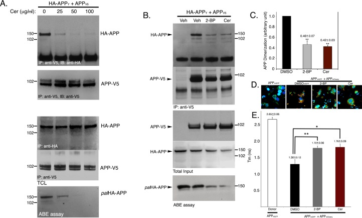
A major rate-limiting step for Aβ generation and deposition in Alzheimer's disease brains is BACE1-mediated cleavage (β-cleavage) of the amyloid precursor protein (APP). We previously reported that APP undergoes palmitoylation at two cysteine residues (Cys186 and Cys187) in the E1-ectodomain. 8-10% of total APP is palmitoylated in vitro and in vivo. Palmitoylated APP (palAPP) shows greater preference for β-cleavage than total APP in detergent resistant lipid rafts. Protein palmitoylation is known to promote protein dimerization. Since dimerization of APP at its E1-ectodomain results in elevated BACE1-mediated cleavage of APP, we have now investigated whether palmitoylation of APP affects its dimerization and whether this leads to elevated β-cleavage of the protein. Here we report that over 90% of palAPP is dimerized while only ~20% of total APP forms dimers. PalAPP-dimers are predominantly cis-oriented while total APP dimerizes in both cis- and trans-orientation. PalAPP forms dimers 4.5-times more efficiently than total APP. Overexpression of the palmitoylating enzymes DHHC7 and DHHC21 that increase palAPP levels and Aβ release, also increased APP dimerization in cells. Conversely, inhibition of APP palmitoylation by pharmacological inhibitors reduced APP-dimerization in coimmunoprecipitation and FLIM/FRET assays. Finally, in vitro BACE1-activity assays demonstrate that palmitoylation-dependent dimerization of APP promotes β-cleavage of APP in lipid-rich detergent resistant cell membranes (DRMs), when compared to total APP. Most importantly, generation of sAPPβ-sAPPβ dimers is dependent on APP-palmitoylation while total sAPPβ generation is not. Since BACE1 shows preference for palAPP dimers over total APP, palAPP dimers may serve as novel targets for effective β-cleavage inhibitors of APP as opposed to BACE1 inhibitors.


Conflict of interest statement

The authors have declared that no competing interests exist.

Figures

Fig 1
Fig 1. palAPP dimerizes ~4.5 times more efficiently compared totAPP and in cis-orientiation.
A. Cells expressing APPV5 or APPV5 plus HA-APPY were subjected to co-immunoprecipitation assays to detect APPV5/HA-APPY interaction or APP-dimerization. APPV5 was immunoprecipitated with an anti-V5 antibody. Immunoprecipitates were probed with an anti-HA antibody to detect pull-down of HA-APPY. Subsequently the immunoprecipitates were subjected to mABE assay to detect palAPPV5/HA-APPY interaction (or palAPP-dimerization). PalAPPV5 pulled down both palAPPV5 (Mwt ~102 kD) and palHA-APPY (Mwt ~150 kD) from cells expressing APPV5 plus HA-APPY but not from cells expressing only APPV5. B. TotAPP-dimers (APPV5/HA-APPY) only form in cells expressing both APPV5 and HA-APPY. C. Quantitation of palAPP-dimers (palAPPV5/palHA-APPY) versus totAPP-dimers (APPV5/HA-APPY). Error bars show the s.e.m. (**p<0.01). D. palAPP dimerizes is cis-orientiation. Cells expressing HA-APPY and cells expressing mycAPP were co-cultured in absence or presence of 1mM cell-impermeable cross-linker DTSSP. Cell extracts were subjected to a pull-down assay, using an anti-HA antibody to immunoprecipitate HA-APPY. To test for APP-dimerization, the precipitates were probed with an anti-myc antibody (panel b, co-culture). Cells co-expressing HA-APPY and mycAPP were also subjected to a co-IP assay using the anti-HA antibody to pull-down mycAPP with HA-APPY.(panel b, co-expression). To detect palAPP-dimerization, the immunoprecipitates were also subjected to mABE assay to detect co-IP of palHA-APPY with pal-mycAPP (panel a). The experiment is a representative of three independent experiments.
Fig 2
Fig 2. Palmitoylation-prone APP mutants exhibit increased APP dimerization compared to wtAPP.
A. Schematic representation of the Cys to Ser mutants of APP used for the following co-immunoprecipitation assays. B. Co-immunoprecipitation assay in cells co-expressing APPV5 and HA-APPY and its mutants containing indicated Cys to Ser substitution. HA-APPY pulls down APPV5, indicating APP-APP dimerization. APP(C133S) and APP(C158S) show 2 fold increase in dimerization, while APP(C186S) and APP(C187S) fail to dimerize. APP(C186S) and APP(C187S) generated trace amounts of palmitoylation-independent dimers (* and **). C. ABE assay of cells overexpressing indicated APP mutants show 2 fold increased palmitoylation of APP(C133S) and APP(C158S), where as APP(C186S) and APP(C187S) were defective in palmitoylation.
Fig 3
Fig 3. DHHC7 equally increases palmitoylation and dimerization of APP.
A. Co-IP assays to detect dimerization of APPV5 and HA-APPY in presence or absence of indicated DHHC proteins. APPV5 pulled-down HA-APPY, indicating APP dimerization. Overexpression of DHHC7 or DHHC21 increased co-IP of APPV5 and HA-APPY, suggesting increased dimerization of APP in presence of these two palmitoylating enzymes. No effect on APP dimerization was observed in presence of DHHC1. EV represents empty vector. B. ABE analysis detected increased level of in palAPP (both palHA-APPY and palAPPV5), as expected. C. Quantitation of dimerization assays (n = 3) detects 2.3 ± 0.17 fold increase of APP dimerization and ~2 fold (not shown) increase in APP palmitoylation in presence of DHHC7. Error bars show the s.e.m.
Fig 4
Fig 4. Palmitoylation inhibitors impair APP dimerization in CHO cells.
A. Co-immunoprecipitation assay of CHO cells expressing HA-APPY and APPV5 in presence of increasing amounts of cerulenin (Cer) (0–100 μg/ml), where 0 μg/ml represents DMSO-treatment. Cerulenin decreased HA-APPY/APPV5 interaction (APP-dimerization) in dose dependent manner similar to cerulenin’s effect on APP-palmitoylation. B. 25 μg/ml cerulenin and 50 μM 2-BP reduced both APP-palmitoylation and APP-dimerization in HA-APPY/APPV5-expressing cells compared to DMSO (Veh)-treated cells. C. Quantitation showed 54 and 58% decrease of APP-APP dimerization by cerulenin (25 μg/ml) and 2-BP (50 μM), respectively. D. Naïve CHO cells were either transiently transfected with an expression plasmid encoding APPmGFP or co-transfected with expression plasmids encoding APPmGFP and APPmCherry. After 24 h, cells were either treated with DMSO or with indicated palmitoylation inhibitors cerulenin or 2-BP for 6 h prior to formalin treatment. FRET/FLIM analysis was employed to measure decay time constant Tm of APPmEGFP in cells expressing APPmEGFP (n = 54), and APPmGFP and APPmCherry (n = 46). Tm of life-time decay of APPmEGFP in APPmEGFP/APPmCherry co-expressing cells was monitored in absence and in presence of palmitoylation inhibitors (25 μg/ml cerulenin or 50 μM 2-BP). Quantitation revealed that inhibitors increased Tm values by ~1.5 fold, indicating disruption of dimerization between APPmGFP and APPmCherry.
Fig 5
Fig 5. Bimolecular Fluorescence Complementation (BiFC) assay detects APP dimers.
A. Fluorescence microscopy of cells transiently expressing APP C-terminal Split GFP 1–10 (APP(1–10)) and APP C-terminal split GFP 11 (APP(11)), FACS sorted for equal intensity. Cells showing green fluorescence represents the APP dimers. These cells were sorted in a fluorescence activated cell sorter (FACS) to obtain equal intensity cells. Next the sorted cells were grown on coverslips for 18 h before treating with 0 (DMSO), 25, 50 and 100μg/ml cerulenin for 6 h showed little or no change in fluorescence intensities. B. Cells expressing APP(1–10) or APP(1–10)+APP(11) were subjected to ABE assay. Probing the samples with anti-GFP detected palmitoylaed APP(1–10) (palAPP(1–10)). Anti-Flotillin antibody detected palmitoylated flotillin (palFlotillin) in ABE assay. C. BiFC intensities of the cells were quantitated using ImageJ software. Intensities of more than 50 cells were measured for each treatment. Average intensities are plotted in percent (%) change in dimerization, using no treatment as 100% describing changes in APP-dimerization percent by increasing amount of cerulenin (solid line). The discontinuous line represents decrease in palAPP levels obtained from ABE analysis upon cerulenin treatment. Error bars show the s.e.m.
Fig 6
Fig 6. Palmitoylation inhibitors specifically impair ectodomain-dependent APP dimerization but not APP-CTF-dimerization.
A. Naïve CHO cells co-expressing APPV5 and APPHA were subjected to a co-IP assay in presence of DMSO (0 μg/ml) or increasing concentrations of cerulenin (25, 50 and 100 μg/ml). flAPPV5 (APPV5) pulled down fl- as well as the C-terminal fragments of APPHA (APPHA and CTFHA, respectively) in DMSO-treated (0 μg/ml cerulenin) cells. In presence of cerulenin, co-IP of flAPPV5 (APPV5) with flAPPHA (APPHA) decreased in a dose-dependent manner. Little or no co-IP of flAPP observed upon treatment with100 μg/ml cerulenin. In contrast, cerulenin had no effect on CTFHA pull-down even at the highest concentration (100 μg/ml). Cerulenin reduced palAPPHA levels in a dose-dependent manner (ABE assay) reaching complete inhibition at 100 μg/ml concentration. B. co-IP assay using an antibody specific for mGFP (anti-mGFP) to pull-down full-length (fl) APPmGFP with APPmCherry from differentiated neuronal cells (RenVM) co-expressing APPmGFP+APPmCherry. Anti-mGFP also pulled-down CTFmCherry with CTFmGFP. Cerulenin (25 μg/ml) treatment of the cells prior to co-IP assay dramatically decreased flAPPmGFP-flAPPmCherry interaction, but not that of CTFmGFP-CTFmCherry.
Fig 7
Fig 7. In vitro BACE-activity assay on palAPP-dimers in detergent resistant membranes (DRMs).
A. In vitro BACE-activity assay on DRMs isolated from CHO cells expressing HA-APPY and myc-APP, treated with increasing amounts of cerulenin (Cer) prior to membrane preparation. The in vitro BACE-activity assay was followed by co-IP analysis as described in Materials and Methods. Myc-sAPPβ pulled down HA-sAPPβ in absence of cerulenin. In presence of cerulenin, pull-down of HA-sAPPβ with myc-sAPPβ decreased in a dose dependent manner. 2-bromopalmitate (100μM) dramatically reduced co-IP of HA-sAPPβ and myc-sAPPβ. B. ABE assay of cells co-expressing HA-APPY and myc-APP. pal HA-APPY and palmyc-APP decreased upon cerulenin-treatment in a dose-dependent manner. C. Representation of dose-dependent decrease in HA-sAPPβ pull-down with myc-sAPPβ in presence of cerulenin.

References

    1. Walsh DM, Selkoe DJ. Deciphering the molecular basis of memory failure in Alzheimer's disease. Neuron. 2004;44(1):181–93. 10.1016/j.neuron.2004.09.010 - DOI - PubMed
    1. Haass C, Selkoe DJ. Soluble protein oligomers in neurodegeneration: lessons from the Alzheimer's amyloid beta-peptide. Nat Rev Mol Cell Biol. 2007;8(2):101–12. 10.1038/nrm2101 - DOI - PubMed
    1. Tanzi RE, Moir RD, Wagner SL. Clearance of Alzheimer's Abeta peptide: the many roads to perdition. Neuron. 2004;43(5):605–8. 10.1016/j.neuron.2004.08.024 - DOI - PubMed
    1. Bhattacharyya R, Barren C, Kovacs DM. Palmitoylation of amyloid precursor protein regulates amyloidogenic processing in lipid rafts. J Neurosci. 2013;33(27):11169–83. 10.1523/JNEUROSCI.4704-12.2013 - DOI - PMC - PubMed
    1. Bettayeb K, Chang JC, Luo W, Aryal S, Varotsis D, Randolph L, et al. delta-COP modulates Abeta peptide formation via retrograde trafficking of APP. Proc Natl Acad Sci U S A. 2016;113(19):5412–7. 10.1073/pnas.1604156113 . - DOI - PMC - PubMed

Substances

LinkOut - more resources