. 2016 Nov 22;16(1):83.
doi: 10.1186/s12896-016-0314-5.

Generation of monoclonal antibodies against native viral proteins using antigen-expressing mammalian cells for mouse immunization

Affiliations

Generation of monoclonal antibodies against native viral proteins using antigen-expressing mammalian cells for mouse immunization

Natalie Rose et al. BMC Biotechnol. .

Abstract

Background: Due to their rising incidence and progressive geographical spread, infections with mosquito-borne viruses, such as dengue (DENV), chikungunya and zika virus, have developed into major public health challenges. Since all of these viruses may cause similar symptoms and can occur in concurrent epidemics, tools for their differential diagnosis and epidemiological monitoring are of urgent need.

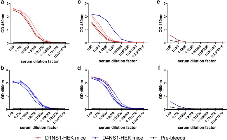
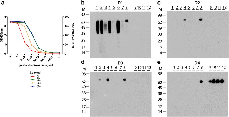
Results: Here we report the application of a novel strategy to rapidly generate monoclonal antibodies (mAbs) against native viral antigens, exemplified for the DENV nonstructural glycoprotein 1 (NS1). The described system is based on the immunization of mice with transfected mammalian cells expressing the target antigens in multiple displays on their cell surface and thereby presenting them efficiently to the host immune system in their native conformation. By applying this cell-based approach to the DENV NS1 protein of serotypes 1 (D1NS1) and 4 (D4NS1), we were able to rapidly generate panels of DENV NS1 serotype cross-reactive, as well as D1NS1- and D4NS1 serotype-specific mAbs. Our data show that the generated mAbs were capable of recognizing the endogenous NS1 protein in DENV-containing biological samples.

Conclusion: The use of this novel immunization strategy, allows for a fast and efficient generation of hybridoma cell lines, producing mAbs against native viral antigens. Envisaged applications of the mAbs include the development of test platforms enabling a differentiation of the DENV serotypes and high resolution immunotyping for epidemiological studies.

Keywords: Dengue virus; HEK cells; Hybridoma technology; Monoclonal antibodies; Mouse immunization; NS1 protein; Transfection.


Figures

Fig. 1
Fig. 1
Expression of D1NS1 and D4NS1 by transfected HEK cells. HEK cell lysates were analyzed by aqua-staining and Western blot analysis after separation on NuPAGE Novex 4–12 % Bis-Tris Gels under reducing conditions (addition of reducing agent and heating of the samples). While aqua-staining of lysates prepared from HEK-derived cell lines expressing D1NS1 (D1) and D4NS1 (D4) showed no additional band of the predicted size of the NS1 proteins, as compared to the lysates of untransfected HEK cells (−) (a), Western blot analysis using anti-hexa-His tag antibodies confirmed the expression of D1NS1 and D4NS1 by the HEK cells (b). Western blotting using anti-tubulin antibodies was performed as a control for the amount of cellular proteins of the untransfected and transfected HEK cell lysates loaded on the gels (c). M = molecular weight marker in kDa
Fig. 2
Fig. 2
Localization of D1NS1 and D4NS1 on the surface of transfected HEK cells. IFA of methanol fixed D1NS1-HEK, D4NS1-HEK and untransfected HEK cells using mouse anti-hexa-His tag mAbs and Alexa568-labelled anti-mouse IgG antibodies. Nuclei were stained with DAPI
Fig. 3
Fig. 3
ELISAs showing humoral immune responses of mice immunized with D1NS1-HEK and D4NS1-HEK cells. a and b: Mouse antisera of D1NS1-HEK (red, panel a) and D4NS1-HEK (blue, panel b) immunized mouse groups (4 mice each) were analyzed on recombinant D1NS1 and recombinant D4NS1, respectively. c and d: Serotype cross-reactive humoral immune responses of D1NS1-HEK (red, panel c) and D4NS1-HEK (blue, panel d) mouse groups were analyzed on recombinant D4NS1 and recombinant D1NS1, respectively. Blue line (panel c) and red line (panel d) = pool of D4NS1-HEK and D1NS1-HEK mouse antisera, respectively, to enable direct comparison with the level of serotype-specific immune responses. e and f: Analysis of pooled mouse sera (red = D1NS1-HEK (e), blue = D4NS1-HEK (f)) on an unrelated hexa-His tagged protein (MUL_3720) as a control for humoral responses against the hexa-His tag. Black line in all panels = pool of mouse sera before immunization (pre-bleed)
Fig. 4
Fig. 4
IFA of selected generated anti-NS1 mAbs on HEK cells. IFA of methanol fixed D1NS1-HEK, D4NS1-HEK and untransfected HEK cells after staining with selected generated anti-NS1 mAbs and Alexa568-labelled anti-mouse IgG antibodies. a: D1NS1 and D4NS1 serotype cross-reactive mAb NR1.8. b: D1NS1 serotype-specific mAb NR1.1. c: D4NS1 serotype-specific mAb NR4.4. Nuclei were stained with DAPI
Fig. 5
Fig. 5
ELISAs of D1NS1-HEK - D4NS1-HEK cell whole protein lysates with the generated mAbs. DENV NS1 serotype-specificity of the 12 generated mAbs was assessed by capturing the NS1 protein in D1NS1 - D4NS1 HEK cell lysates using anti-hexa-His tag mAbs and detecting the protein with the anti-NS1 mAbs NR1.1 - NR1.8 and NR4.1 - NR4.4. a: While analyses with mAbs NR1.1 - NR1.3, NR1.6 and NR1.7 exhibited a high degree of D1NS1-specificity in the ELISA, mAbs NR1.4, NR1.5 and NR1.8 showed a certain degree of NS1 cross-reactivity among different DENV serotypes. b: MAbs NR4.1 - NR4.4 were highly specific for D4NS1, since they did not recognize D1NS1 - D3NS1 in the HEK cell lysates. M = molecular weight marker in kDa
Fig. 6
Fig. 6
ELISA and Western blot analysis of lysates derived from DENV-infected VeroE6 cell lines. a: NS1 relative units were determined in lysates of VeroE6 cells infected with DENV serotypes 1–4 by NS1 capture ELISA (EUROIMMUN, Luzern, Switzerland). NS1 relative units were calculated in accordance with the manufacturer’s instructions and approximate values are shown. Gels for Western blot analysis were loaded with DENV serotype 1–4 infected VeroE6 cell lysates containing 40.000 relative units of NS1, each under non-reducing conditions. The 12 generated mAbs were tested on serotype 1 (b), serotype 2 (c), serotype 3 (d) and serotype 4 (e) virus-infected cell lysates

References

    1. Cao-Lormeau VM, Musso D. Emerging arboviruses in the pacific. Lancet. 2014;384(9954):1571–1572. doi: 10.1016/S0140-6736(14)61977-2. - DOI - PubMed
    1. Guzman A, Isturiz RE. Update on the global spread of dengue. Int J Antimicrob Agents. 2010;36(Suppl 1):S40–42. doi: 10.1016/j.ijantimicag.2010.06.018. - DOI - PubMed
    1. Murray NE, Quam MB, Wilder-Smith A. Epidemiology of dengue: past, present and future prospects. Clin Epidemiol. 2013;5:299–309. - PMC - PubMed
    1. Weaver SC, Lecuit M. Chikungunya virus and the global spread of a mosquito-borne disease. N Engl J Med. 2015;372(13):1231–1239. doi: 10.1056/NEJMra1406035. - DOI - PubMed
    1. Musso D, Cao-Lormeau VM, Gubler DJ. Zika virus: following the path of dengue and chikungunya? Lancet. 2015;386(9990):243–244. doi: 10.1016/S0140-6736(15)61273-9. - DOI - PubMed

Publication types