. 2016 Nov 23:6:37267.
doi: 10.1038/srep37267.

The N-terminal loop of IRAK-4 death domain regulates ordered assembly of the Myddosome signalling scaffold

Affiliations

The N-terminal loop of IRAK-4 death domain regulates ordered assembly of the Myddosome signalling scaffold

Anthony C G Dossang et al. Sci Rep. .

Abstract

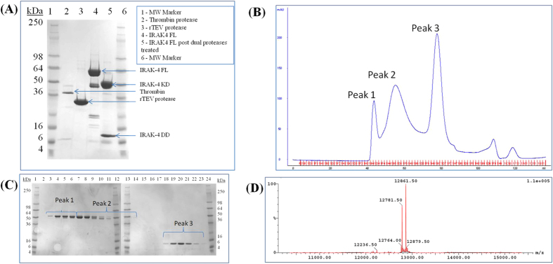
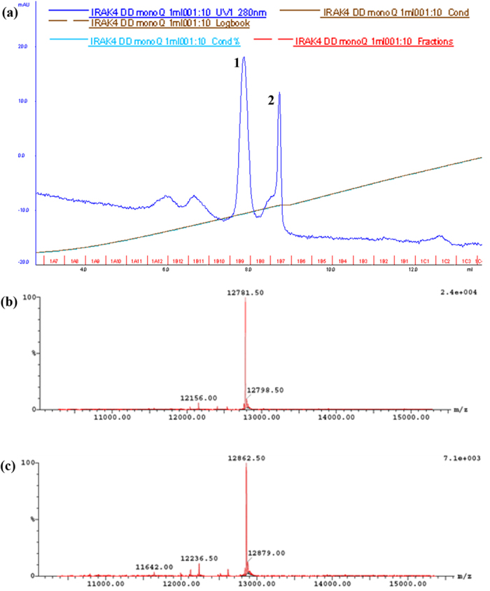
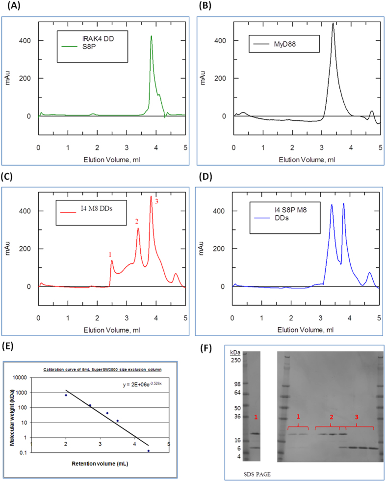
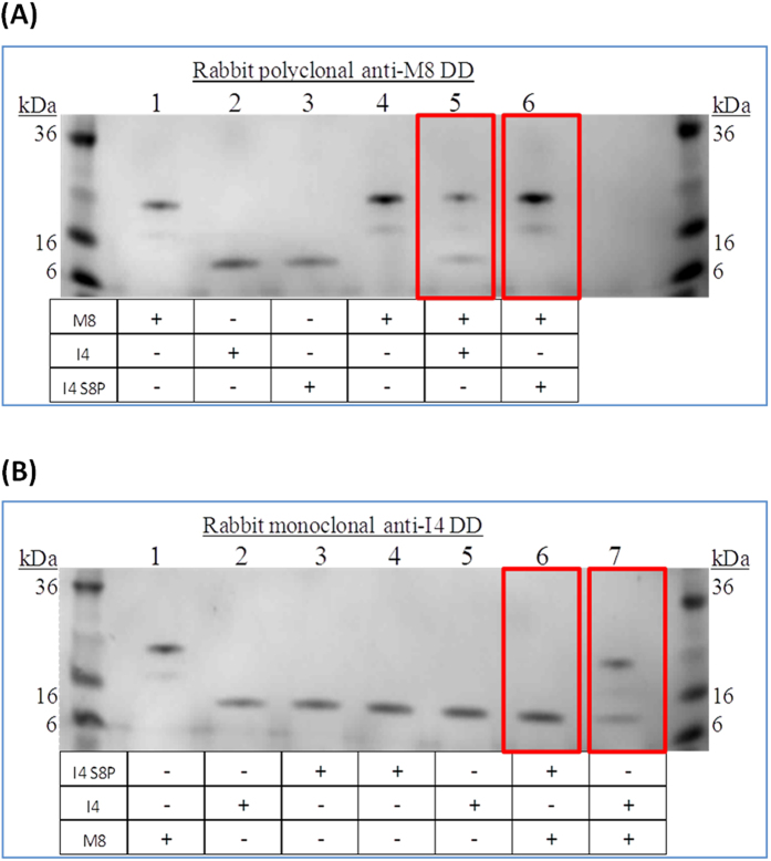
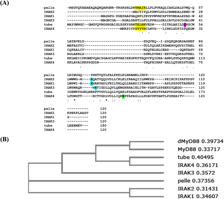
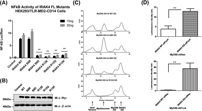
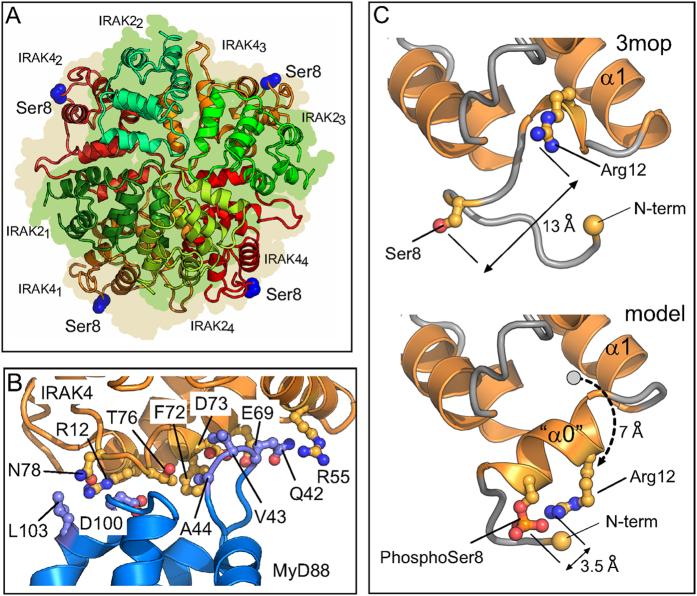
Activation of Toll-like receptors induces dimerization and the recruitment of the death domain (DD) adaptor protein MyD88 into an oligomeric post receptor complex termed the Myddosome. The Myddosome is a hub for inflammatory and oncogenic signaling and has a hierarchical arrangement with 6-8 MyD88 molecules assembling with exactly 4 of IRAK-4 and 4 of IRAK-2. Here we show that a conserved motif in IRAK-4 (Ser8-X-X-X-Arg12) is autophosphorylated and that the phosphorylated DD is unable to form Myddosomes. Furthermore a mutant DD with the phospho-mimetic residue Asp at this position is impaired in both signalling and Myddosome assembly. IRAK-4 Arg12 is also essential for Myddosome assembly and signalling and we propose that phosphorylated Ser8 induces the N-terminal loop to fold into an α-helix. This conformer is stabilised by an electrostatic interaction between phospho-Ser8 and Arg12 and would destabilise a critical interface between IRAK-4 and MyD88. Interestingly IRAK-2 does not conserve this motif and has an alternative interface in the Myddosome that requires Arg67, a residue conserved in paralogues, IRAK-1 and 3(M).


Figures

Figure 1
Figure 1. Purification of IRAK-4 death domain.
(A) IRAK-4 full length was cleaved with thrombin and TEV proteases and analysed by XX% SDS-PAGE (B) Cleaved IRAK-4 was fractionated by gel filtration on 16/60 Superdex™ 75 pg (C) SDS-PAGE analysis of gel filtration peaks 1–3. (D) LC/MS spectrum showing deconvoluted masses of IRAK-4 DD purified in peak 3. The mass of 12782 Da corresponded to IRAK-4 death domain residues (−7) to 107 (see Methods). The mass of 12862 Da represents IRAK-4 death domain with a phosphorylated residue (+80 Da) (C).
Figure 2
Figure 2. Separation of singly phosphorylated Ser8 IRAK-4 death domain.
(a) Absorbance elution spectrum at 280 nm of IRAK-4 DD using a monoQ anion exchange column. Peaks 1 and 2 eluted at approximately 70 mM and 120 mM NaCl respectively. (b) LC/MS spectrum showing deconvoluted masses of IRAK-4 death domain corresponding to peak 1 fraction. The mass of 12782 Da is non-phosphorylated IRAK-4 death domain residues (−7) to 107. (c) LC/MS spectrum showing deconvoluted masses of IRAK-4 death domain corresponding to peak 2. The mass of 12863 Da corresponded to singly phosphorylated IRAK-4 death domain residues (−7) to 107.
Figure 3
Figure 3. Phosphorylated Ser8 IRAK-4 death domain interferes with the Myddosome formation.
(A–D) Absorbance Elution spectrum of a TSKgel SuperSW3000 analytical size exclusion column at 280 nm. (A) 0.5 mg.ml−1 of IRAK-4 death domain only. (B) 0.5 mg.ml−1 of MyD88 DD only. (C) 1:1 mix of MyD88 and non-phosphorylated IRAK-4 death domains concentrated to 0.5 mg.ml−1 prior to loading. (D) 1:1 mix of MyD88 and Ser8 phosphorylated IRAK-4 death domains concentrated to 0.5 mg.ml−1 prior loading. (E) Calibration of gel filtration using protein standard markers (F) 4–20% reducing Tris-Glycine SDS PAGE of fractions corresponding to peak 1, 2 and 3 of figure 15a. Fractions corresponding to peak 1 were concentrated using a 3 kDa MW cut-off Amicon Spin Filters for better visualisation of samples. One of three repeats is shown.
Figure 4
Figure 4. Reciprocal pull down assays confirm inability of Ser8-P death domain to interact with MyD88.
(A) 4–20% reducing Tris-Glycine SDS PAGE of samples precipitated using a rabbit polyclonal anti-human MyD88 death domain antibody. Lane 1 to 3 are control lanes each loaded with 3 μg of the protein samples: lane 1-MyD88 DD, lane 2-non-phosphorylated IRAK-4 death domain, lane 3-Ser8 phosphorylated IRAK-4 death domain. Lanes 4 to 6 were loaded with the pull down experiment samples as indicated. (B) 4–20% reducing Tris-Glycine SDS PAGE of samples precipitated using a rabbit monoclonal anti-human IRAK-4 death domain antibody. Lane 1 to 3 were control lanes each loaded with 3 μg of the protein samples: lane 1 corresponded to MyD88 DD, lane 2-non-phosphorylated IRAK-4 death domain, lane 3-Ser8 phosphorylated IRAK-4 death domain. Lanes 4 to 7 were loaded with the pull down experiment samples as indicated. One repeat of three biological replicates is shown.
Figure 5
Figure 5. Specificity and regulation of Myddosome assembly mediated by Type II interfaces.
(A) Sequence alignment of human MyD88 and IRAKs and Drosophila homologues dMyD88, tube and pelle. The conserved N-terminal loop motif is highlighted in yellow, tube Glu50 in purple, Arg67 of IRAK-2 in cyan and IRAK-4 Glu92 in green. (B) Phylogenetic relationships of DDs from Drosophila and human IRAK kinases and MyD88 adaptors.
Figure 6
Figure 6. Regulatory function of the IRAK-4 N-terminal loop.
(A) HEK293/TLR4-MD2-CD14 cells were transiently transfected with 10 ng or 20 ng of Myc-tagged IRAK4 constructs. Cells were harvested 24 hours post-transfection. NF-κB activation was quantified by dual luciferase assay. One representative of three independent experiments is shown. Statistical Analysis—p values were determined using multiple t test and designated with p < 0.01 (**) and p < 0.05 (*) One experiment of three biological replicates is shown. (B) Expression of all IRAK4 mutants is comparable with wild type IRAK4. HEK293/TLR4-MD2-CD14 cells were transfected with Myc-tagged IRAK4 constructs and analyzed by immunoblot (IB). One experiment of three biological replicates is shown. (C) Lumier assays: MyD88 and IRAK were tagged with renilla (Ren) or protein A (ProA) at the N- or C-terminal, as indicated. KD = kinase dead. One representative out of three identical experiments shown. **p < 0.01 measured by Student’s t-test.
Figure 7
Figure 7. Specificity and regulation of Myddosome assembly mediated by Type II interfaces.
(A) Cross section of the Myddosome structure (pdb:3mop) showing the four IRAK4 subunits (orange/red) and four IRAK 2 subunits (green/bluegreen). The peripheral location of IRAK4 Ser8 (blue atoms) is indicated. (B) Arg 12 forms part of a Type IIa DD-DD interface. The interaction between IRAK4 (g chain, pdb 3mop, orange ribbons and sidechains) and MyD88 (d chain, pdb 3mop, blue ribbons and sidechains). IRAK4 Arg12 and MyD88 Asp100 interact. (C) Modelled helix at N-terminus of IRAK4 stabilized by electrostatic interaction between phospho-serine 8 and arginine 12. Upper view shows N-terminus of pdb 3mop with 13 Å units between Arg12 and Ser8. Lower view shows modelled helix at N-terminus. Arg12 Calpha is moved 7 Å units from its position in pdb 3mop (dotted arrow). Arg12 sidechain can approach within 3.5 Å units of the modelled Phospho-Ser8 and is displaced from its interaction with MyD88 Asp100.

References

    1. Gay N. J., Symmons M. F., Gangloff M. & Bryant C. E. Assembly and localization of Toll-like receptor signalling complexes. Nature Rev. Immunol. 14, 546–558 (2014). - PubMed
    1. Kang J. Y. & Lee J. O. Structural biology of the Toll-like receptor family. Annu. Rev. Biochem. 80, 917–941 (2011). - PubMed
    1. Gay N. J., Gangloff M. & Weber A. N. Toll-like receptors as molecular switches. Nature Rev. Immunol. 6, 693–698 (2006). - PubMed
    1. Motshwene P. G. et al.. An oligomeric signaling platform formed by the Toll-like receptor signal transducers MyD88 and IRAK-4. J. Biol. Chem. 284, 25404–25411 (2009). - PMC - PubMed
    1. Lin S. C., Lo Y. C. & Wu H. Helical assembly in the MyD88-IRAK4-IRAK2 complex in TLR/IL-1R signalling. Nature 465, 885–890 (2010). - PMC - PubMed

Publication types

MeSH terms

Substances