. 2016 Nov 23:6:37635.
doi: 10.1038/srep37635.

Overexpression of SerpinE2/protease nexin-1 Contribute to Pathological Cardiac Fibrosis via increasing Collagen Deposition

Affiliations

Overexpression of SerpinE2/protease nexin-1 Contribute to Pathological Cardiac Fibrosis via increasing Collagen Deposition

Xuelian Li et al. Sci Rep. .

Abstract

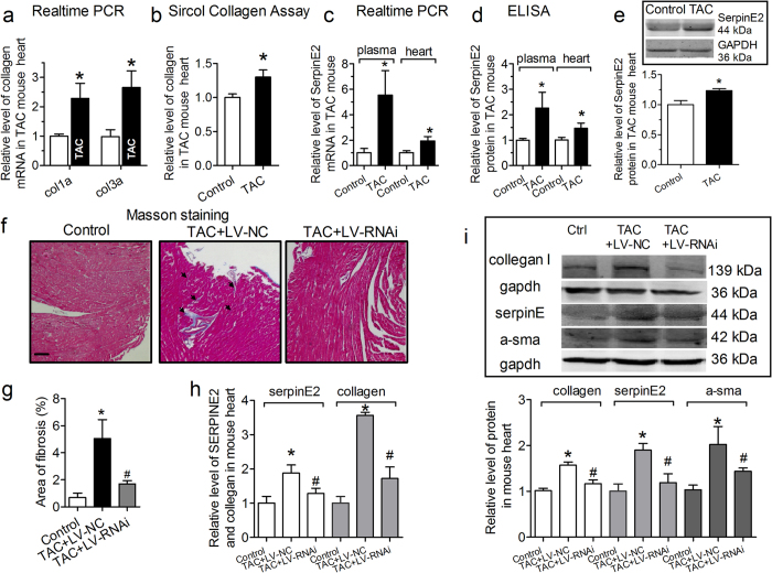
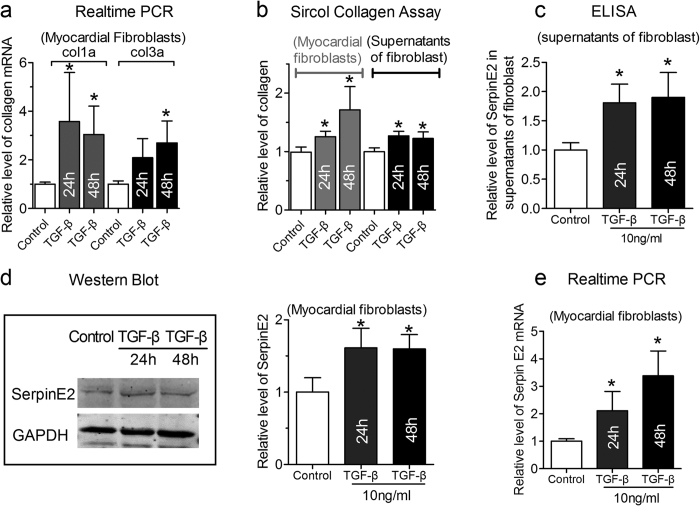
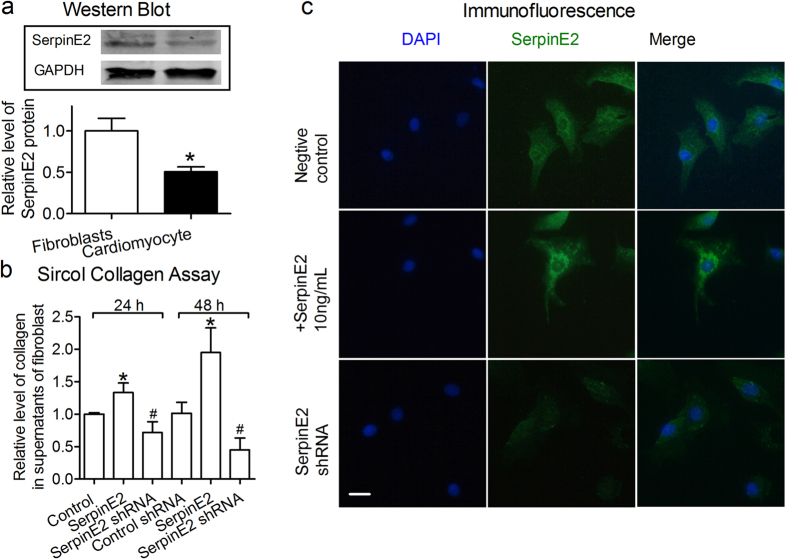
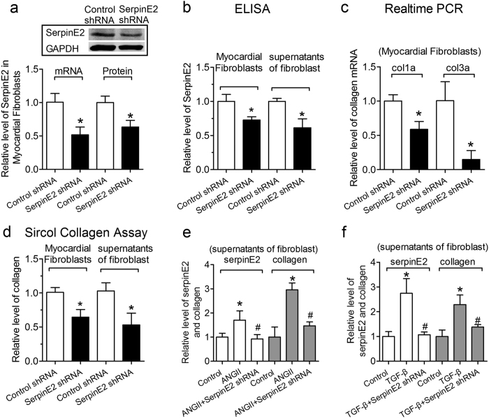
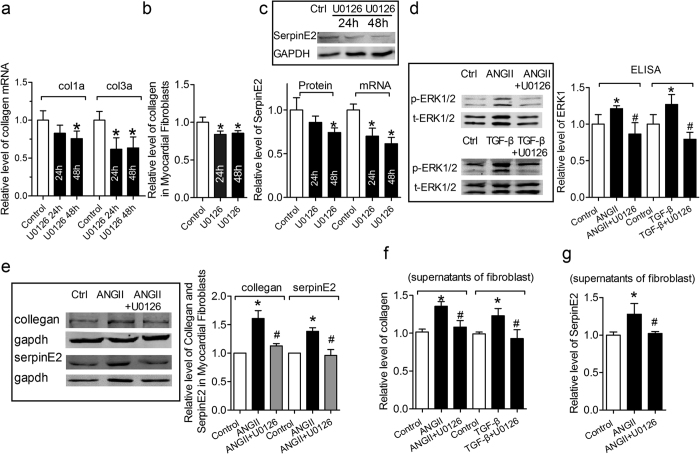
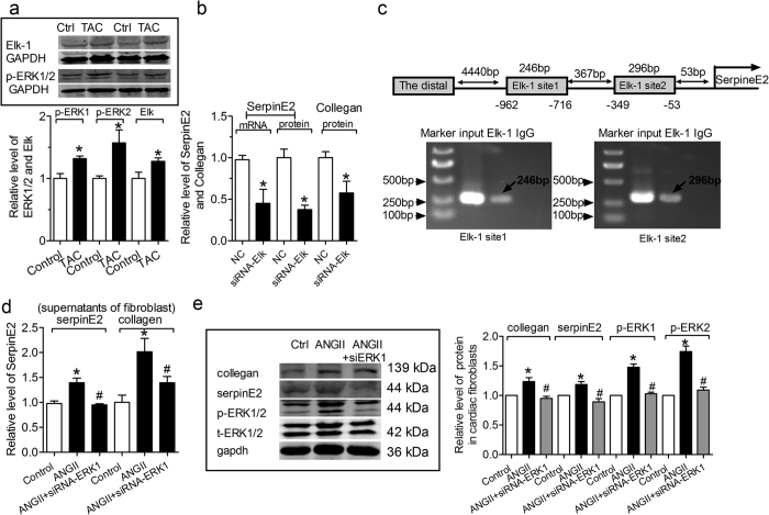
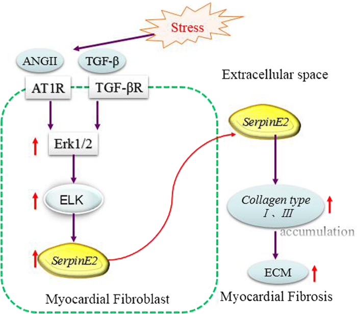
Although increases in cardiovascular load (pressure overload) are known to elicit ventricular remodeling including cardiomyocyte hypertrophy and interstitial fibrosis, the molecular mechanisms of pressure overload or AngII -induced cardiac interstitial fibrosis remain elusive. In this study, serpinE2/protease nexin-1 was over-expressed in a cardiac fibrosis model induced by pressure-overloaded via transverse aortic constriction (TAC) in mouse. Knockdown of serpinE2 attenuates cardiac fibrosis in a mouse model of TAC. At meantime, the results showed that serpinE2 significantly were increased with collagen accumulations induced by AngII or TGF-β stimulation in vitro. Intriguingly, extracellular collagen in myocardial fibroblast was reduced by knockdown of serpinE2 compared with the control in vitro. In stark contrast, the addition of exogenous PN-1 up-regulated the content of collagen in myocardial fibroblast. The MEK1/2- ERK1/2 signaling probably promoted the expression of serpinE2 via transcription factors Elk1 in myocardial fibroblast. In conclusion, stress-induced the ERK1/2 signaling pathway activation up-regulated serpinE2 expression, consequently led accumulation of collagen protein, and contributed to cardiac fibrosis.


Figures

Figure 1
Figure 1. The expression of SerpinE2 increases in transverse aortic constriction (TAC)-induced mouse cardiac myocardial fibrosis model.
(a) Collagen I/III (col1a/col3a) mRNA levels was detected by qRT-PCR. n = 8. *P < 0.05 vs. control. (b) Sircol™ Collagen Assay was used to quantify the total collagen contents in myocardial tissue from TAC models. n = 10. *P < 0.05 vs. control. (c) The expression of serpinE2 mRNA in plasma (n = 5, *P < 0.05 vs. control) and myocardium (n = 8, *P < 0.05 vs. control) of TAC mice using qRT-PCR. (d) The level of serpinE2 protein in plasma (n = 7, *P < 0.05 vs. control) and myocardium (n = 10, *P < 0.01 vs. control) of TAC mice using ELISA. (e) Western blot analysis of serpinE2 protein and statistical analysis (n = 6, *P < 0.01 vs. control). Full-length blots/gels are presented in Supplementary Figure 1. (f) Representative sections of heart with Masson staining. The fibrotic tissues are stained as blue, as indicated by the arrows. Scale bars: 200 μm. (g) Collagen deposition was quantified by automated image analysis and expressed as percentage of tissue area. n = 6. (h) The relative protein level of serpinE2 and collagen was detected by ELISA and Sircol™ Collagen Assay in Knockdown -serpinE2 mice. n = 6. (i) The protein of collagen, serpinE2 and α-sma were decreased in Knockdown-serpinE2 mice. Full-length blots/gels are presented in Supplementary Figure 2 and Supplementary Figure 3 (n = 6, #P < 0.05 vs. TAC + LV-NC).
Figure 2
Figure 2. ANG-II-mediated increase in cardiac fibrosis and the serpinE2 expression.
(a) qRT-PCR assay was applied for the detection of collagen I and collagen III mRNA expression in myocardial fibroblasts (n = 6, *P < 0.05 vs. control) in 50 nM AngII induced collagen deposition; (b) Sircol™ Collagen Assay was used to quantify the total collagen concentration both in fibroblasts and in the supernatants of fibroblast induced by AngII (n = 6, *P < 0.05 vs. control); (c) The expression of serpinE2 was detected by ELISA in the supernatants of fibroblasts (n = 6, *P < 0.05 vs. control); (d) Western blot analysis of serpinE2 protein (left panel) and statistical analysis (right panel) (n = 6, *P < 0.01 vs. control). Full-length blots/gels are presented in Supplementary Figure 4; (e) qRT-PCR was also used to detect expression of serpinE2 mRNA in myocardial fibroblasts stimulated by AngII (n = 8, *P < 0.01 vs. control).
Figure 3
Figure 3. TGF-β-induced fibrosis and mediated increase in serpinE2 expression.
(a) qRT-PCR assay was applied for detection of collagen I and collagen III mRNA expression in myocardial fibroblast (n = 6, *P < 0.05 vs. control) in TGF-β-induced fibrosis; (b) Sircol™ Collagen Assay was used to quantify the total collagen concentration both in fibroblasts and in the supernatants of fibroblasts (n = 6, *P < 0.05 vs. control); (c) The expression of serpinE2 was detected by ELISA in the supernatants of fibroblast (n = 6, *P < 0.05 vs. control); (d). Western blot analysis of serpinE2 protein (left panel) and statistical analysis (right panel) (n = 6, *P < 0.01 vs. control). Full-length blots/gels are presented in Supplementary Figure 5; (e). qRT-PCR was also used to detect expression of serpinE2 mRNA in myocardial fibroblasts stimulated by TGF-β (n = 8, *P < 0.01 vs. control).
Figure 4
Figure 4. The changes of collagen content induced by exogenous serpinE2.
(a) Differential expression of serpinE2 in myocardial fibroblast and myocardial cell. n = 3. *P < 0.05 vs. control. Full-length blots/gels are presented in Supplementary Figure 6. (b) Relative level of collagen in supernatants of fibroblast by over-expression of serpinE2 or knock-down of serpinE2. n = 6. *P < 0.05 vs. control, #P < 0.05 vs. serpinE2 group. (c) The Immunofluorescence for serpinE2 in myocardial fibroblast. Scale bar:10 μm.
Figure 5
Figure 5. Knock-down of serpinE2 reduces the collagen deposition in supernatant of fibroblasts.
(a) After 24 h transfection with serpinE2 shRNA, the mRNA level of serpinE2 was detected by qRT-PCR in cardiac fibroblasts. n = 10, *P < 0.05 vs. control; the protein level of serpinE2 was detected by western blot in preparation. n = 6, *P < 0.05 vs. control. Full-length blots/gels are presented in Supplementary Figure 7. (b) After transfection with serpinE2 shRNA, the protein level of serpinE2 was also detected by ELISA assay both in the fibroblast and the supernatants. n = 6, *P < 0.05 vs. control. (c) The mRNA expression for collagen was measured by qRT-PCR after transfection with serpinE2 shRNA. n = 10, *P < 0.05 vs. control. (d) Collagen expression were also measured by Sircol™ Collagen Assay both in the fibroblasts and the supernatants. n = 7, *P < 0.05 vs. control. (e) Knock-down of serpinE2 can inhibit the collagen deposition induced by ANG-II. n = 6. *P < 0.05 vs. control, #P < 0.05 vs. AngII group. (f) Knock-down of serpinE2 suppressed the collagen deposition induced by TGF-β. n = 6. *P < 0.05 vs. control, #P < 0.05 vs. TGF-β group.
Figure 6
Figure 6. Potential involvement of ERK 1/2 signal pathway in regulating serpinE2 and collagen deposition.
(a) Collagen mRNA levels measured by qRT-PCR after 24 and 48 h treatment with U0126 (The inhibitor of ERK 1/2 signal pathway). n = 10, *P < 0.05 vs. control. (b) Collagen expression levels detected by Sircol™ Collagen Assay after 24 and 48 h treatment with U0126. n = 6, *P < 0.05 vs. control. (c) After treatment with U0126, the protein and mRNA level of serpinE2 was detected in cardiac fibroblasts. n = 6, *P < 0.05 vs. control. Full-length blots/gels are presented in Supplementary Figure 8. (d) Effect of U0126 on expression of phosphor-ERK1/2 (p-ERK1/2) and total ERK1/2 (t-ERK1/2) in myocardial fibroblasts inducd by AngII or TGF-β. n = 8, *P < 0.05 vs. control, #P < 0.05 vs. AngII or TGF-β. Left, western blot. Right, Elisa assay. Full-length blots/gels are presented in Supplementary Figure 9 and 10. (e) Effect of U0126 on expression of serpinE2 and collagen in myocardial fibroblasts inducd by ANG-II. n = 4, *P < 0.05 vs. control, #P < 0.05 vs. ANG-II. Full-length blots/gels are presented in Supplementary Figure 11 and 12. (f) Effect of U0126 on the collagen content inducd by AngII or TGF-β in supernatants of fibroblasts. n = 6, *P < 0.05 vs. control, #P < 0.05 vs. AngII or TGF-β. (g) Effect of U0126 on expression of serpinE2 inducd by AngII in supernatants of fibroblasts. n = 6, *P < 0.05 vs. control, #P < 0.05 vs. ANG-II.
Figure 7
Figure 7. ERK1/2 signal pathway via Elk1 in regulating serpinE2.
(a) The change of p-ERK1/2 and Elk1 in TAC mouse heart. n = 6, *P < 0.05 vs. control. Full-length blots/gels are presented in Supplementary Figure 13 and 14. (b) The mRNA level of serpinE2 was decreased by transfection with siRNA Elk in CFs; The level of serpinE2 was decreased by transfection with siRNA Elk in the supernatants of fibroblast; The level of collagen was decreased by transfection with siRNA Elk in the supernatants of fibroblast. n = 6, *P < 0.05 vs. control. (c) ChIP testing in vivo binding of Elk1 to promoter of Rattus norvegicus genes of serpin family E member 2 (SerpinE2). Top: Schematic representation of the two upstream region of the serpinE2. Bottom: PCR products of Elk1-binding sites following immunoprecipitation with anti- Elk1 antibody. The anti-IgG antibody and H2O treatment were used as negative control. The anti- Elk1 antibody was used to target specific immunoprecipitation. ChIP analysis of Elk1 binding to the promoter between-962 and-716 (PCR products 246 bp). Elk1 binding to target Elk1 site 1 activates serpinE2 promoter activity. ChIP analysis of in vivo Elk1 site2 binding to the promoter between -349 and -53 bp (PCR products 296 bp). ChIP assay showed that Elk1 binding to this target Elk1 site 2 also did activate serpinE2 promoter activity. (d) Effect of knockdown of ERK1 on serpinE2 and the collagen content in supernatants of fibroblasts inducd by ANG-II. n = 6, *P < 0.05 vs. control, #P < 0.05 vs. ANG-II. (e) Effect of knockdown of ERK1 on serpinE2, collagen and p-ERK1/2 in cardiac fibroblasts inducd by ANG-II. n = 6, *P < 0.05 vs. control, #P < 0.05 vs. ANG-II. Full-length blots/gels are presented in Supplementary Figure 15.
Figure 8
Figure 8. Model demonstrating possible molecular basis of SerpinE2 induced collagen deposition in myocardial fibrosis.
Pressure overload can trigger some response: activation of renin-angiotensin-aldosterone system, in particular, systemic and local production of Ang II. Elevated AngII and TGF-β promote fibrotic responses of the heart, and then activates MAPK-ERK1/2 signaling. Phospho-ERK1/2 can further activate ERK-dependent transcription activator Elk1 in myocardial fibroblasts which in turn activates SerpinE2.

Similar articles

Cited by

References

    1. Bishop J. E. & Lindahl G. Regulation of cardiovascular collagen synthesis by mechanical load. Cardiovasc Res 42, 27–44 (1999). - PubMed
    1. Spinale F. G. Matrix metalloproteinases: regulation and dysregulation in the failing heart. Circ Res 90, 520–530 (2002). - PubMed
    1. Tang M. et al.. Differential regulation of collagen types I and III expression in cardiac fibroblasts by AGEs through TRB3/MAPK signaling pathway. Cell Mol Life Sci 65, 2924–2932, doi: 10.1007/s00018-008-8255-3 (2008). - DOI - PMC - PubMed
    1. Sommer J. et al.. cDNA sequence coding for a rat glia-derived nexin and its homology to members of the serpin superfamily. Biochemistry 26, 6407–6410 (1987). - PubMed
    1. Farrell D. H., Wagner S. L., Yuan R. H. & Cunningham D. D. Localization of protease nexin-1 on the fibroblast extracellular matrix. J Cell Physiol 134, 179–188, doi: 10.1002/jcp.1041340203 (1988). - DOI - PubMed

Publication types

MeSH terms

LinkOut - more resources