

Why one-size-fits-all vaso-modulatory interventions fail to control glioma invasion: in silico insights
- PMID: 27876890
- PMCID: PMC5120360
- DOI: 10.1038/srep37283
Why one-size-fits-all vaso-modulatory interventions fail to control glioma invasion: in silico insights
Abstract
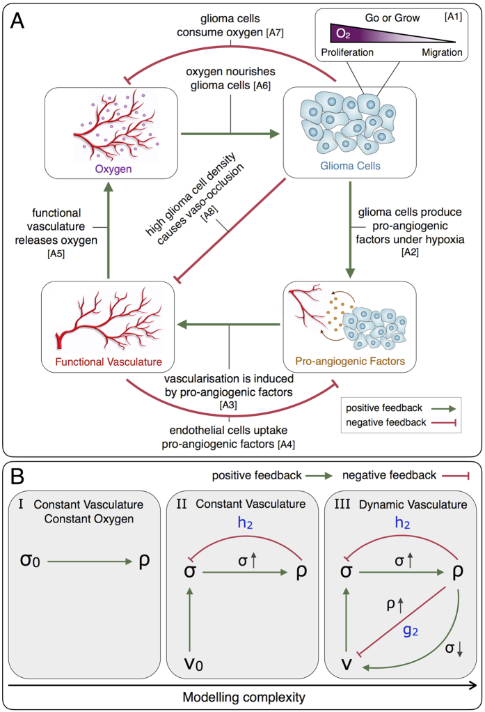
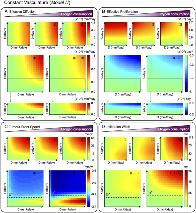
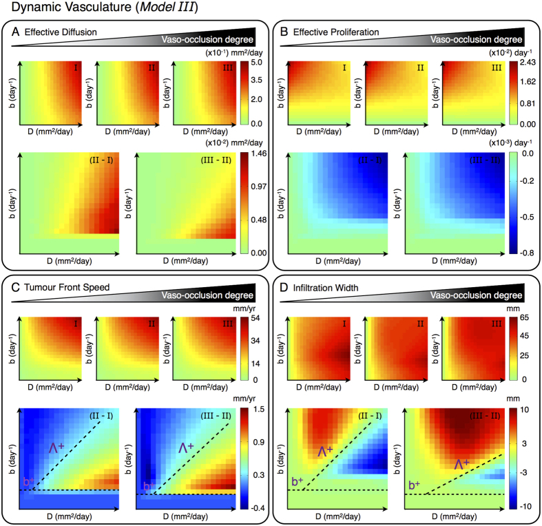
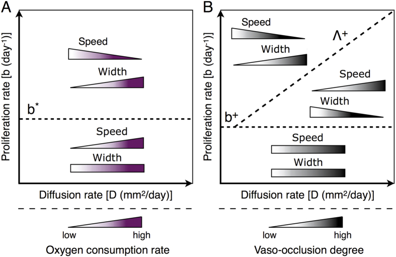
Gliomas are highly invasive brain tumours characterised by poor prognosis and limited response to therapy. There is an ongoing debate on the therapeutic potential of vaso-modulatory interventions against glioma invasion. Prominent vasculature-targeting therapies involve tumour blood vessel deterioration and normalisation. The former aims at tumour infarction and nutrient deprivation induced by blood vessel occlusion/collapse. In contrast, the therapeutic intention of normalising the abnormal tumour vasculature is to improve the efficacy of conventional treatment modalities. Although these strategies have shown therapeutic potential, it remains unclear why they both often fail to control glioma growth. To shed some light on this issue, we propose a mathematical model based on the migration/proliferation dichotomy of glioma cells in order to investigate why vaso-modulatory interventions have shown limited success in terms of tumour clearance. We found the existence of a critical cell proliferation/diffusion ratio that separates glioma responses to vaso-modulatory interventions into two distinct regimes. While for tumours, belonging to one regime, vascular modulations reduce the front speed and increase the infiltration width, for those in the other regime, the invasion speed increases and infiltration width decreases. We discuss how these in silico findings can be used to guide individualised vaso-modulatory approaches to improve treatment success rates.
Figures
Similar articles
-
The biology and mathematical modelling of glioma invasion: a review.J R Soc Interface. 2017 Nov;14(136):20170490. doi: 10.1098/rsif.2017.0490. J R Soc Interface. 2017. PMID: 29118112 Free PMC article. Review.
-
Novel anti-angiogenic therapies for malignant gliomas.Lancet Neurol. 2008 Dec;7(12):1152-60. doi: 10.1016/S1474-4422(08)70260-6. Lancet Neurol. 2008. PMID: 19007739 Review.
-
LIM domain only 2 induces glioma invasion via cytosolic p27(KIP1).Tumour Biol. 2016 Feb;37(2):2473-80. doi: 10.1007/s13277-015-4072-0. Epub 2015 Sep 18. Tumour Biol. 2016. PMID: 26383528
-
Human and rat glioma growth, invasion, and vascularization in a novel chick embryo brain tumor model.Clin Exp Metastasis. 2005;22(3):225-36. doi: 10.1007/s10585-005-7889-x. Clin Exp Metastasis. 2005. PMID: 16158250
-
Gab2 expression in glioma and its implications for tumor invasion.Acta Oncol. 2013 Nov;52(8):1739-50. doi: 10.3109/0284186X.2012.750032. Epub 2012 Dec 11. Acta Oncol. 2013. PMID: 23231021
Cited by
-
Tumor Volume Dynamics as an Early Biomarker for Patient-Specific Evolution of Resistance and Progression in Recurrent High-Grade Glioma.J Clin Med. 2020 Jun 27;9(7):2019. doi: 10.3390/jcm9072019. J Clin Med. 2020. PMID: 32605050 Free PMC article.
-
New insights into targeted therapy of glioblastoma using smart nanoparticles.Cancer Cell Int. 2024 May 7;24(1):160. doi: 10.1186/s12935-024-03331-3. Cancer Cell Int. 2024. PMID: 38715021 Free PMC article. Review.
-
Multiscale modeling of glioma pseudopalisades: contributions from the tumor microenvironment.J Math Biol. 2021 Apr 12;82(6):49. doi: 10.1007/s00285-021-01599-x. J Math Biol. 2021. PMID: 33846838 Free PMC article.
-
On the Impact of Chemo-Mechanically Induced Phenotypic Transitions in Gliomas.Cancers (Basel). 2019 May 24;11(5):716. doi: 10.3390/cancers11050716. Cancers (Basel). 2019. PMID: 31137643 Free PMC article.
-
A combined experimental-computational approach uncovers a role for the Golgi matrix protein Giantin in breast cancer progression.PLoS Comput Biol. 2023 Apr 17;19(4):e1010995. doi: 10.1371/journal.pcbi.1010995. eCollection 2023 Apr. PLoS Comput Biol. 2023. PMID: 37068117 Free PMC article.
References
-
- Stupp R. et al.. Radiotherapy plus concomitant and adjuvant temozolomide for glioblastoma. New England Journal of Medicine 352, 987–996 (2005). - PubMed
-
- Weller M. et al.. Mgmt promoter methylation in malignant gliomas: ready for personalized medicine? Nature Reviews Neurology 6, 39–51 (2010). - PubMed
-
- Giese A., Bjerkvig R., Berens M. & Westphal M. Cost of migration: invasion of malignant gliomas and implications for treatment. Journal of Clinical Oncology 21, 1624–1636 (2003). - PubMed
-
- Westphal M. & Lamszus K. The neurobiology of gliomas: from cell biology to the development of therapeutic approaches. Nature Reviews Neuroscience 12, 495–508 (2011). - PubMed
Publication types
MeSH terms
LinkOut - more resources
Full Text Sources
Other Literature Sources
