

Rainbow glare after laser-assisted in situ keratomileusis: a review of literature
- PMID: 27877015
- PMCID: PMC5108617
- DOI: 10.2147/OPTH.S117971
Rainbow glare after laser-assisted in situ keratomileusis: a review of literature
Abstract
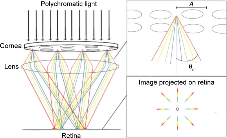
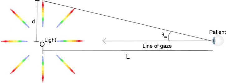
This article reviews the current literature pertaining to rainbow glare (RG), including incidence rate, clinical presentation, etiology, prognosis, and management. RG is a rare optical complication of femtosecond laser-assisted in situ keratomileusis that results in patients seeing an array of spectral bands surrounding point sources of light under mesopic and scotopic conditions. The mechanism is thought to be a consequence of the formation of a transmissive diffraction grating on the posterior surface of the corneal flap created by the FS laser. RG has a good prognosis and is usually self-limiting. Persistent RG with concomitant residual refractive error may warrant lifting the flap and photoablating the posterior surface of the flap. Patients with persistent RG and no residual refractive error should be considered candidates for phototherapeutic keratectomy on the posterior flap surface.
Keywords: LASIK; femtosecond; keratomileusis; phototherapeutic keratectomy; rainbow glare.
Conflict of interest statement
The authors report no conflicts of interest in this work.
Figures
References
-
- Stonecipher KG, Ignacio TS, Stonecipher KG, Thompson V. Management of Complications in Refractive Surgery: Femtosecond Laser. Berlin Heidelberg: Springer; 2008.
-
- Krueger RR, Thornton IL, Xu M, Bor Z, van den Berg TJ. Rainbow glare as an optical side effect of IntraLASIK. Ophthalmology. 2008;115(7):1187–1195. - PubMed
-
- Bamba S, Rocha KM, Ramos-Esteban JC, Krueger RR. Incidence of rainbow glare after laser in situ keratomileusis flap creation with a 60 kHz femtosecond laser. J Cataract Refract Surg. 2009;35(6):1082–1086. - PubMed
-
- Krueger RR, Rocha KM, Dupps WJ., Jr . Natural History and Incidence of Rainbow Glare and Light Scattering in Femtosecond LASIK. San Diego, CA: American Society of Cataract and Refractive Surgery; 2015. Paper 16815.
-
- Salz JJ. Difficult and Complicated Cases in Refractive Surgery: Suction Loss After Complete Raster Pattern and No Side Cut, No Flap Lift, and Rainbow Glare. Berlin Heidelberg: Springer; 2014.
Publication types
LinkOut - more resources
Full Text Sources
Other Literature Sources
