Review
. 2016 Nov 8:10:566.
doi: 10.3389/fnhum.2016.00566. eCollection 2016.

The Indispensable Roles of Microglia and Astrocytes during Brain Development

Affiliations
Review

The Indispensable Roles of Microglia and Astrocytes during Brain Development

Kitty Reemst et al. Front Hum Neurosci. .

Abstract

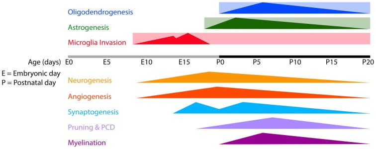
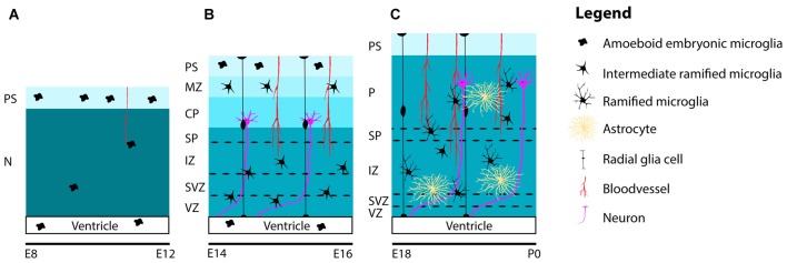
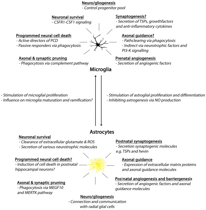
Glia are essential for brain functioning during development and in the adult brain. Here, we discuss the various roles of both microglia and astrocytes, and their interactions during brain development. Although both cells are fundamentally different in origin and function, they often affect the same developmental processes such as neuro-/gliogenesis, angiogenesis, axonal outgrowth, synaptogenesis and synaptic pruning. Due to their important instructive roles in these processes, dysfunction of microglia or astrocytes during brain development could contribute to neurodevelopmental disorders and potentially even late-onset neuropathology. A better understanding of the origin, differentiation process and developmental functions of microglia and astrocytes will help to fully appreciate their role both in the developing as well as in the adult brain, in health and disease.

Keywords: astrocytes; brain development; glial cells; microglia; neurodevelopmental disorders.


Figures

Figure 1
Figure 1
Timeline of microglia invasion, gliogenesis and several developmental processes in the developing mouse brain. Rectangles indicate the estimated periods during/from which microglia, astrocytes and oligodendrocytes remain present in the brain. Triangles indicate the onset and peaks of the indicated developmental processes. Abbreviations: E, embryonic; P, postnatal; PCD, programmed cell death.
Figure 2
Figure 2
Schematic representation of the distribution and maturation of microglia and the beginning of astrogenesis in the mouse cerebral cortex. (A) At early embryonic stages between E8 and E12, microglia are located at the PS in the meninges and in the lateral ventricles. Only a few highly, motile and proliferative cells can be found in the neuroepithelium. (B) Between E14 and E16, microglia ramify further to form intermediate ramified microglia that can be found in the VZ, SVZ, IZ, SP and MZ. At the PS and in the lateral ventricle, one can still detect amoeboid microglia. Remarkably, until E16, microglia are absent from the cortical plate. (C) From E18 and onwards, microglia can be found in the cortical layers, after migrating from the deeper to the more superficial layers. During this period, microglia are found in close association with radial glia, blood vessels and developing axons. Around E18 astrogenesis starts. Similar to microglia, astrocytes are also found near radial glia, blood vessels and axons. Along the course of embryonic brain development, many microglia ramify further and transform from intermediate ramified microglia into mature ramified microglia with long processes. Abbreviations: PS, pial surface; N, neuroepithelium; MZ, marginal zone; CP, cortical plate; SP, subplate; IZ, intermediate zone; SVZ, subventricular zone; VZ, ventricular zone; P, parenchyma.
Figure 3
Figure 3
Summary of developmental roles of microglia and astrocytes. Abbreviations: CSFR1, colony-stimulating factor 1 receptor; CSF1, colony-stimulating factor 1; PCD, programmed cell death; TSPs, thrombospondins; PI3-K, phosphoinositide-3 kinase; ROS, reactive oxygen species; MEGF10, Multiple EGF-like-domains 10; MERTK, MER Tyrosine Kinase.

References

    1. Aarum J., Sandberg K., Haeberlein S. L. B., Persson M. A. A. (2003). Migration and differentiation of neural precursor cells can be directed by microglia. Proc. Natl. Acad. Sci. U S A 100, 15983–15988. 10.1073/pnas.2237050100 - DOI - PMC - PubMed
    1. Abbott N. J. (2002). Astrocyte-endothelial interactions and blood-brain barrier permeability. J. Anat. 200, 629–638. 10.1046/j.1469-7580.2002.00064.x - DOI - PMC - PubMed
    1. Abbott N. J., Romero I. A. (1996). Transporting therapeutics across the blood-brain barrier. Mol. Med. Today 2, 106–113. 10.1016/1357-4310(96)88720-x - DOI - PubMed
    1. Abbott N. J., Rönnbäck L., Hansson E. (2006). Astrocyte-endothelial interactions at the blood-brain barrier. Nat. Rev. Neurosci. 7, 41–53. 10.1038/nrn1824 - DOI - PubMed
    1. Abbott N. J., Patabendige A. A. K., Dolman D. E. M., Yusof S. R., Begley D. J. (2010). Structure and function of the blood-brain barrier. Neurobiol. Dis. 37, 13–25. 10.1016/j.nbd.2009.07.030 - DOI - PubMed