Review
. 2016 Oct 8;6(13):2439-2457.
doi: 10.7150/thno.16088. eCollection 2016.

Recent Advances of Light-Mediated Theranostics

Affiliations
Review

Recent Advances of Light-Mediated Theranostics

Xiangzhao Ai et al. Theranostics. .

Abstract

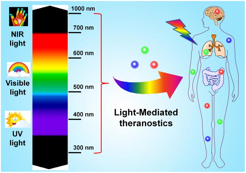
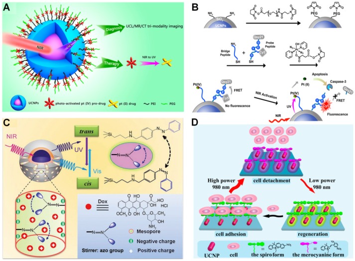
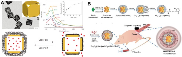
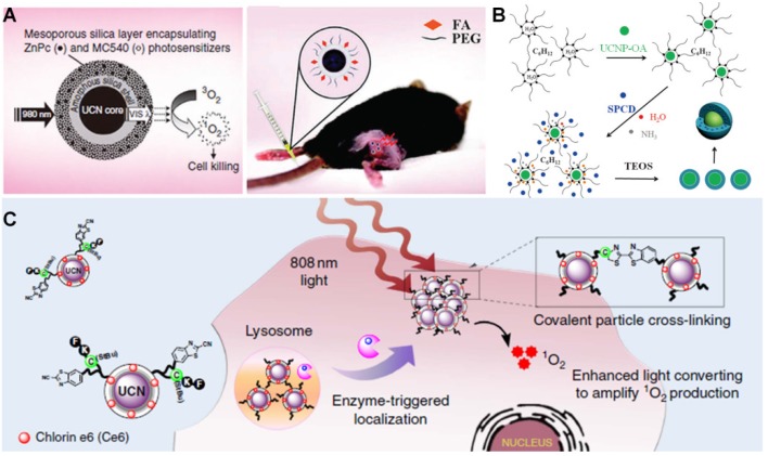
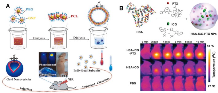
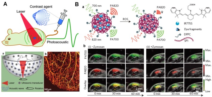
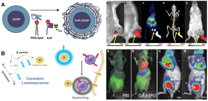
Currently, precision theranostics have been extensively demanded for the effective treatment of various human diseases. Currently, efficient therapy at the targeted disease areas still remains challenging since most available drug molecules lack of selectivity to the pathological sites. Among different approaches, light-mediated therapeutic strategy has recently emerged as a promising and powerful tool to precisely control the activation of therapeutic reagents and imaging probes in vitro and in vivo, mostly attributed to its unique properties including minimally invasive capability and highly spatiotemporal resolution. Although it has achieved initial success, the conventional strategies for light-mediated theranostics are mostly based on the light with short wavelength (e.g., UV or visible light), which may usually suffer from several undesired drawbacks, such as limited tissue penetration depth, unavoidable light absorption/scattering and potential phototoxicity to healthy tissues, etc. Therefore, a near-infrared (NIR) light-mediated approach on the basis of long-wavelength light (700-1000 nm) irradiation, which displays deep-tissue penetration, minimized photo-damage and low autofluoresence in living systems, has been proposed as an inspiring alternative for precisely phototherapeutic applications in the last decades. Despite numerous NIR light-responsive molecules have been currently proposed for clinical applications, several inherent drawbacks, such as troublesome synthetic procedures, low water solubility and limited accumulation abilities in targeted areas, heavily restrict their applications in deep-tissue therapeutic and imaging studies. Thanks to the amazing properties of several nanomaterials with large extinction coefficient in the NIR region, the construction of NIR light responsive nanoplatforms with multifunctions have become promising approaches for deep-seated diseases diagnosis and therapy. In this review, we summarized various light-triggered theranostic strategies and introduced their great advances in biomedical applications in recent years. Moreover, some other promising light-assisted techniques, such as photoacoustic and Cerenkov radiation, were also systemically discussed. Finally, the potential challenges and future perspectives for light-mediated deep-tissue diagnosis and therapeutics were proposed.

Keywords: Cerenkov radiation.; multifunctional nanomaterials; near-infrared light; photoacoustic; precision theranostics.


Conflict of interest statement

The authors have declared that no competing interest exists.

Figures

Figure 1
Figure 1
Illustration of light-mediated theranostics under UV, visible and NIR light irradiation.
Figure 2
Figure 2
UV light-mediated photocaged strategies. (A) Brief scheme of UV light-induced photocaged compounds: (a) o-nitrobenzyl, (b) pyrenylmethyl ester, (c) coumarinyl ester. (B) Schematic illustration of UV light-induced release of D-luciferin from the ONB photocaged D-luciferin derivatives structures for the detection of fLuc. Bioluminescence imaging was performed in mice bearing C6-fLuc tumors after injection with: (a) compound Lu-NPE without UV excitation; (b) compound Lu-NPE with UV excitation; (c) D-luciferin only, respectively. Reproduced with permission from reference . (C) Scheme of photo-activated drug release from assembled photosensitive DNA-drug nanoconjugates. Reproduced with permission from reference .
Figure 3
Figure 3
UV light-mediated photoisomerization strategies. (A) Commonly used molecular structures with UV light-triggered reversible conformational exchanges. (a) azobenzene, (b) spiropyran, (c) diarylethene. (B) Scheme of UV light-induced drug release from the self-assembled vesicles. TEM showed the reversible morphology change of vesicles in the presence of UV/visible light irradiation. Reproduced with permission from reference . (C) Scheme of azobezene-modified DNA-controlled reversible drug photorelease system. Reproduced with permission from reference .
Figure 4
Figure 4
Visible light-mediated drug delivery strategies. (A) Structures of vitamin B12 derivatives for tunable wavelength light-controlled molecules release. Reproduced with permission from reference . (B) Structures of photoactive [Ru(tpy)(5CNU)3]2+, tpy and 5CNU. Confocal images presented 5CNU ligands (green) could be released upon light irradiation in HeLa cells. Reproduced with permission from reference . (C) Scheme of visible light-controlled drug (PTX) delivery of Ru(II)-dppz complexes modified MSNs. Reproduced with permission from reference . (C) Scheme of light-induced Dox release from DNA-AuNPs nanocomplexes for targeted cancer therapy. Reproduced with permission from reference .
Figure 5
Figure 5
NIR light-triggered drug delivery strategies. (A) Scheme of NIR light-mediated UCNPs nanoplatform for Pt(IV) prodrug-based therapy and multimodal imaging. Reproduced with permission from reference . (B) Schematic illustration of 980 nm light-induced Pt(IV) prodrug photoactivation and intracellular apoptosis monitoring. Reproduced with permission from reference . (C) NIR light-triggered drug release from the mesopores of UCNPs@mSiO2 due to trans-cis azobenzene photoisomerization. Reproduced with permission from reference . (D) Scheme of power density controlled photoswitching for non-invasive cell adhesion/detachment. Reproduced with permission from reference .
Figure 6
Figure 6
NIR light-triggered heat-induced drug delivery approaches. (A) Schematic illustration of the smart thermal responsive system. TEM images showed the morphology of Au nanocages. The increased absorption spectrum presented the controlled dye release from the Au nanocages upon a pulsed NIR laser (10 mW/cm2) irradiation for 1, 2, 4, 8 and 16 min, respectively. Reproduced with permission from reference . (B) Scheme of the 808 nm light-mediated nanocomposites synthesis for therapy in vivo. Reproduced with permission from reference .
Figure 7
Figure 7
NIR light-mediated PDT strategies. (A) Scheme of FA/PEG modified UCNPs@mSO2 loaded with ZnPc and MC540 for PDT. Reproduced with permission from reference . (B) Schematic illustration of UCNPs coated with PS-doped silica shell for PDT. Reproduced with permission from reference . (C) Scheme of tumor microenvironment sensitive approach for covalent cross-linking of peptide-modified UCNs in cancer treatment. Reproduced with permission from reference .
Figure 8
Figure 8
NIR light-mediated PTT strategies. (A) Scheme of the formation of biodegradable gold vesicles for PTT therapy and PA imaging. Reproduced with permission from reference . (B) Scheme of self-assembled HAS-ICG-PTX nanocomposite and thermal images of tumor-bearing mice injected with HAS-ICG-PTX, HAS-ICG or PBS under the 808 nm laser irradiation. Reproduced with permission from reference .
Figure 9
Figure 9
PA imaging based diagnosis in vivo. (A) Brief scheme of the mechanism of PA signal production. (a) Structure of 128-element hemisphere array-based PA tomography. (b) PA imaging of blood vessels in a living mouse ear. Reproduced with permission from reference . (B) Design of photo-activated probe (a) for ROS monitoring with PA imaging in mice (b). Reproduced with permission from reference .
Figure 10
Figure 10
(A) Schematic design of the PoP-UCNP structure. The animal imaging shows the mice injected with PoP-UCNPs and imaged in six modalities after 1 h injection, including (a) FL, (b) UC imaging. (c) PET, (d) PET/CT and (e) CL imaging, respectively. Reproduced with permission from reference . (B) Scheme of CL generation and nanoparticle induced quenching of CL. The animal imaging presents fluorescent imaging (a, b) and CR imaging (b, d) of mice injected with PBS and Cy5.5 labeled probe. B; bladder. Reproduced with permission from reference .

References

    1. Reichard CA, Stephenson AJ, Klein EA. Applying precision medicine to the active surveillance of prostate cancer. Cancer. 2015;121:3403–11. - PMC - PubMed
    1. Friedman AA, Letai A, Fisher DE, Flaherty KT. Precision medicine for cancer with next-generation functional diagnostics. Nat Rev Cancer. 2015;15:747–56. - PMC - PubMed
    1. Arnedos M, Vicier C, Loi S, Lefebvre C, Michiels S, Bonnefoi H. et al. Precision medicine for metastatic breast cancer-limitations and solutions. Nat Rev Clin Oncol. 2015;12:693–704. - PubMed
    1. Lloyd KC, Meehan T, Beaudet A, Murray S, Svenson K, McKerlie C. et al. Precision medicine: Look to the mice. Science. 2015;349:390. - PMC - PubMed
    1. Mahoney KM, Rennert PD, Freeman GJ. Combination cancer immunotherapy and new immunomodulatory targets. Nat Rev Drug Discov. 2015;14:561–84. - PubMed

Publication types

MeSH terms