. 2013 May 15;14(3):035002.
doi: 10.1088/1468-6996/14/3/035002. eCollection 2013 Jun.

Tissue-engineered endothelial cell layers on surface-modified Ti for inhibiting in vitro platelet adhesion

Affiliations

Tissue-engineered endothelial cell layers on surface-modified Ti for inhibiting in vitro platelet adhesion

Xiupeng Wang et al. Sci Technol Adv Mater. .

Abstract

A tissue-engineered endothelial layer was prepared by culturing endothelial cells on a fibroblast growth factor-2 (FGF-2)-l-ascorbic acid phosphate magnesium salt n-hydrate (AsMg)-apatite (Ap) coated titanium plate. The FGF-2-AsMg-Ap coated Ti plate was prepared by immersing a Ti plate in supersaturated calcium phosphate solutions supplemented with FGF-2 and AsMg. The FGF-2-AsMg-Ap layer on the Ti plate accelerated proliferation of human umbilical vein endothelial cells (HUVECs), and showed slightly higher, but not statistically significant, nitric oxide release from HUVECs than on as-prepared Ti. The endothelial layer maintained proper function of the endothelial cells and markedly inhibited in vitro platelet adhesion. The tissue-engineered endothelial layer formed on the FGF-2-AsMg-Ap layer is promising for ameliorating platelet activation and thrombus formation on cardiovascular implants.

Keywords: 10.01; 10.03; apatite; ascorbate; endothelial cell; fibroblast growth factor-2; platelet adhesion.


Figures
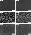
Figure 1.
Figure 1.
FE-SEM images of the bare Ti plate and Ap, FAp, AsAp and FAsAp coatings on Ti plates.
Figure 2.
Figure 2.
TEM images of Ap, FAp, AsAp and FAsAp coatings on Ti plates.
Figure 3.
Figure 3.
XRD patterns of Ti, Ap, FAp, AsAp and FAsAp coatings on Ti plates.
Figure 4.
Figure 4.
FTIR spectra of Ap, FAp, AsAp and FAsAp coatings on Ti plates. The spectra are offset vertically for clarity.
Figure 5.
Figure 5.
Amount of Ca and P immobilized on Ti plates.
Figure 6.
Figure 6.
Amount of FGF-2 and AsMg immobilized on Ti plates.
Figure 7.
Figure 7.
HUVEC proliferation on Ap, FAp, AsAp and FAsAp coated Ti plates after 3 days of culture.
Figure 8.
Figure 8.
Nitric oxide (NO) release from HUVECs on as-prepared Ti, Ap and FAsAp coated Ti plates after 3 days of culture.
Figure 9.
Figure 9.
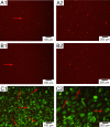
Fluorescence microscopy images of platelets adhered to Ti (A1, A2), Ap (B1, B2) and FAsAp + HUVEC coated Ti (C1, C2). HUVECs were stained for green fluorescence. Platelets were stained for red fluorescence (red arrows).
Figure 10.
Figure 10.
Number of platelets adhered to Ti, Ap and FAsAp + HUVEC coated Ti.

Similar articles

Cited by

References

    1. Lietz K. and Miller L W. Semin. Thorac. Cardiovasc. Surg. 2008;20:225. doi: 10.1053/j.semtcvs.2008.08.004. - DOI - PubMed
    1. Schillinger M, Sabeti S, Dick P, Amighi J, Mlekusch W, Schlager O, Loewe C, Cejna M, Lammer J. and Minar E. Circulation. 2007;115:2745. doi: 10.1161/CIRCULATIONAHA.107.688341. - DOI - PubMed
    1. Pagani F D.et al J. Am. Coll. Cardiol. 2009;54:312. doi: 10.1016/j.jacc.2009.03.055. - DOI - PubMed
    1. Toyoda K. Drugs. 2009;69:633. doi: 10.2165/00003495-200969060-00001. - DOI - PubMed
    1. Baigent C, Sudlow C, Collins R, Peto R. and Collaborat A T. Br. Med. J. 2002;324:71. doi: 10.1136/bmj.324.7329.71. - DOI - PMC - PubMed

LinkOut - more resources