

Accelerated colorimetric immunosensing using surface-modified porous monoliths and gold nanoparticles
- PMID: 27877588
- PMCID: PMC5090314
- DOI: 10.1088/1468-6996/14/4/044403
Accelerated colorimetric immunosensing using surface-modified porous monoliths and gold nanoparticles
Abstract
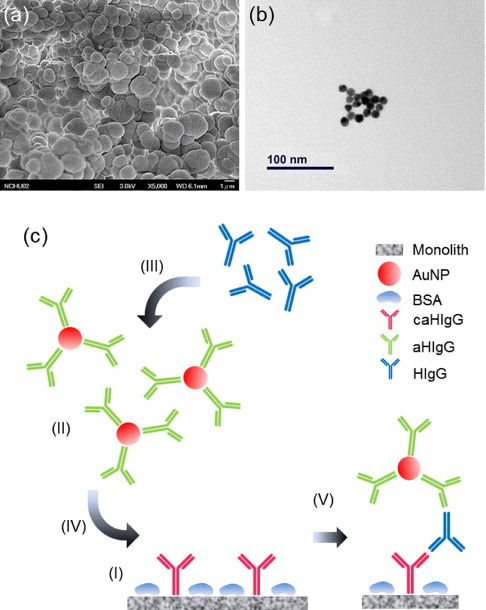
A rapid and sensitive immunoassay platform integrating polymerized monoliths and gold nanoparticles (AuNPs) has been developed. The porous monoliths are photopolymerized in situ within a silica capillary and serve as solid support for high-mass transport and high-density capture antibody immobilization to create a shorter diffusion length for antibody-antigen interactions, resulting in a rapid assay and low reagent consumption. AuNPs are modified with detection antibodies and are utilized as signals for colorimetric immunoassays without the need for enzyme, substrate and sophisticated equipment for quantitative measurements. This platform has been verified by performing a human IgG sandwich immunoassay with a detection limit of 0.1 ng ml-1. In addition, a single assay can be completed in 1 h, which is more efficient than traditional immunoassays that require several hours to complete.
Keywords: colorimetric assay; gold nanoparticles; immunoassay; porous monoliths.
Figures


Similar articles
-
Flow-through immunosensors using antibody-immobilized polymer monoliths.Biosens Bioelectron. 2010 Sep 15;26(1):182-8. doi: 10.1016/j.bios.2010.06.007. Epub 2010 Jun 11. Biosens Bioelectron. 2010. PMID: 20598520 Free PMC article.
-
Flow-through microfluidic immunosensors with refractive index-matched silica monoliths as volumetric optical detection elements.Sens Actuators B Chem. 2018 Jan;254:878-886. doi: 10.1016/j.snb.2017.07.137. Epub 2017 Jul 21. Sens Actuators B Chem. 2018. PMID: 29225421 Free PMC article.
-
Naked-eye sensitive ELISA-like assay based on gold-enhanced peroxidase-like immunogold activity.Anal Bioanal Chem. 2016 Feb;408(4):1015-22. doi: 10.1007/s00216-015-9219-8. Epub 2015 Dec 16. Anal Bioanal Chem. 2016. PMID: 26677026
-
Ultrasensitive and accelerated detection of ciguatoxin by capillary electrophoresis via on-line sandwich immunoassay with rotating magnetic field and nanoparticles signal enhancement.Anal Chim Acta. 2015 Aug 12;888:27-35. doi: 10.1016/j.aca.2015.06.018. Epub 2015 Jul 8. Anal Chim Acta. 2015. PMID: 26320955
-
Heteroassembled gold nanoparticles with sandwich-immunoassay LSPR chip format for rapid and sensitive detection of hepatitis B virus surface antigen (HBsAg).Biosens Bioelectron. 2018 Jun 1;107:118-122. doi: 10.1016/j.bios.2018.02.019. Epub 2018 Feb 7. Biosens Bioelectron. 2018. PMID: 29454301
References
-
- Edwards R. Immunodiagnostics: A Practical Approach. New York: Oxford University Press; 1999.
LinkOut - more resources
Full Text Sources
Other Literature Sources