Review
. 2015 Apr 28;16(2):023501.
doi: 10.1088/1468-6996/16/2/023501. eCollection 2015 Apr.

Recent progress on magnetic iron oxide nanoparticles: synthesis, surface functional strategies and biomedical applications

Affiliations
Review

Recent progress on magnetic iron oxide nanoparticles: synthesis, surface functional strategies and biomedical applications

Wei Wu et al. Sci Technol Adv Mater. .

Abstract

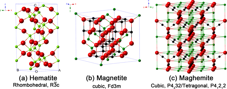
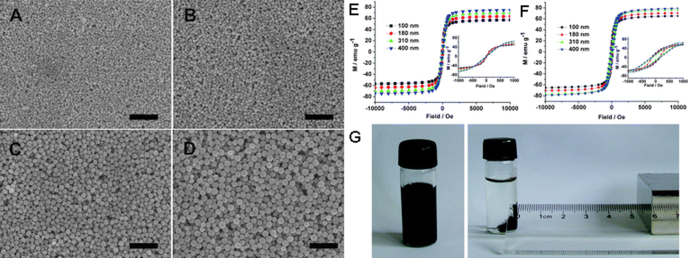
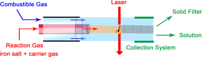
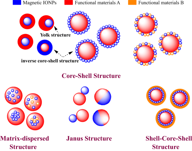
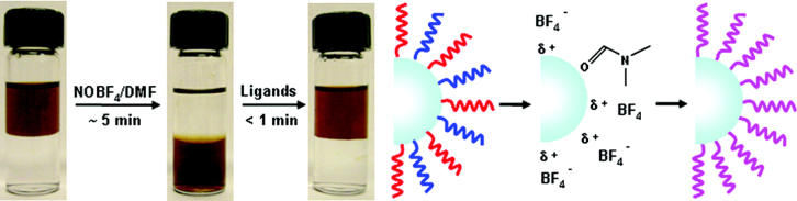
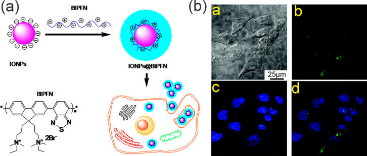
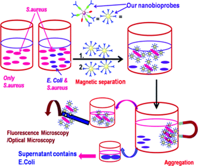
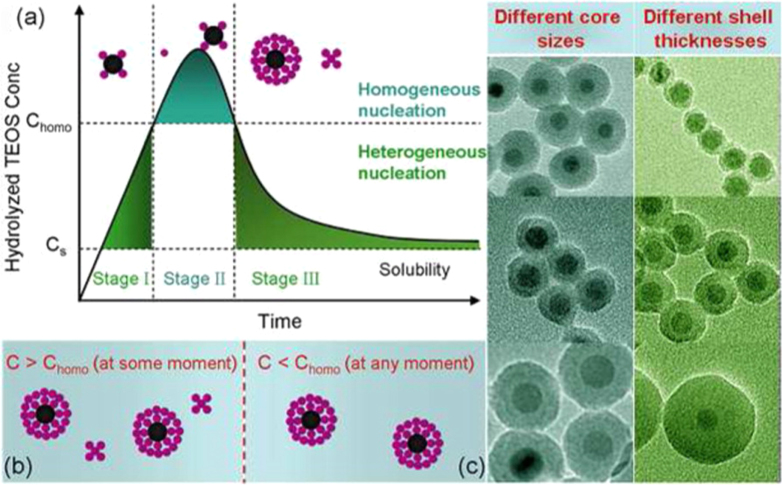
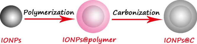
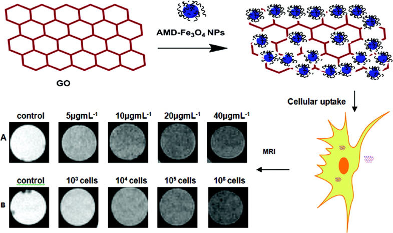
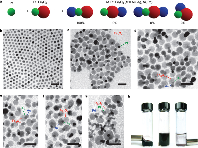
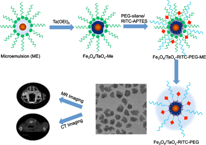
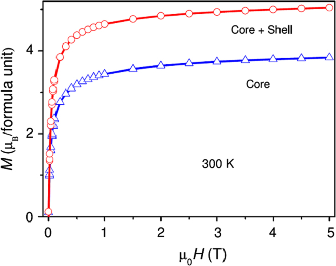
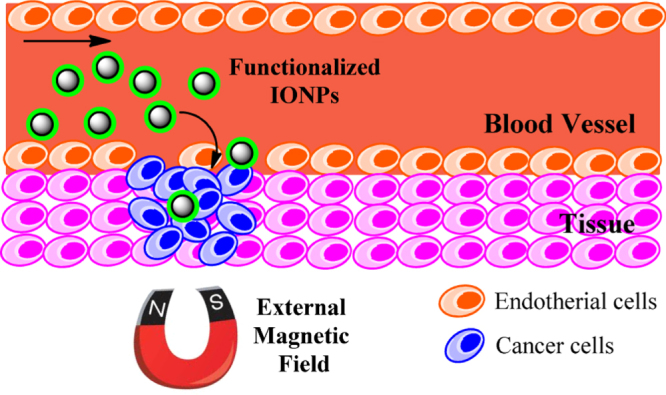
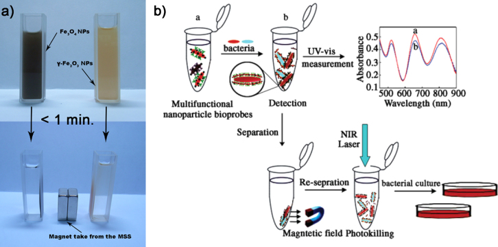
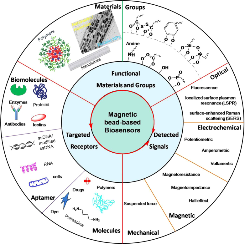
This review focuses on the recent development and various strategies in the preparation, microstructure, and magnetic properties of bare and surface functionalized iron oxide nanoparticles (IONPs); their corresponding biological application was also discussed. In order to implement the practical in vivo or in vitro applications, the IONPs must have combined properties of high magnetic saturation, stability, biocompatibility, and interactive functions at the surface. Moreover, the surface of IONPs could be modified by organic materials or inorganic materials, such as polymers, biomolecules, silica, metals, etc. The new functionalized strategies, problems and major challenges, along with the current directions for the synthesis, surface functionalization and bioapplication of IONPs, are considered. Finally, some future trends and the prospects in these research areas are also discussed.

Keywords: biomedical application; magnetic iron oxide nanoparticles; surface functional strategy.


Figures

Figure 1.
Figure 1.
Crystal structure and crystallographic data of the hematite, magnetite and maghemite (the black ball is Fe2+, the green ball is Fe3+ and the red ball is O2−).
Figure 2.
Figure 2.
The XRD peak lines from standard powder diffraction files of α-Fe2O3 (33–0664), Fe3O4 (19–0629) and γ-Fe2O3 (39–1346).
Figure 3.
Figure 3.
Schematic presentation of the typical hysteresis loops of IONPs (a); the ZFC/FC curves of γ-Fe2O3 at the different applied field (b).
Figure 4.
Figure 4.
Schematic showing the in situ co-precipitation synthesis process of IONPs in polymer. (Reprinted with permission from S K Suh et al 2012 J. Am. Chem. Soc. 134 7337. Copyright 2012 American Chemical Society.)
Figure 5.
Figure 5.
Metal–oleate precursors were prepared from the reaction of metal chlorides and sodium oleate. The thermal decomposition of the metal–oleate precursors in the high boiling solvent produced monodisperse nanocrystals (a). (Reprinted with permission from J Park et al 2004 Nat. Mater. 3 891. Copyright 2004 Nature Publishing Group.) Transmission electron microscopy (TEM) images of 6, 7, 8, 9, 10, 11, 12, and 13 nm-sized air-oxidized IONPs showing the one nanometer level increments in diameter (b). (Reprinted with permission from J Park et al 2005 Angew. Chem. Int. Edn 44 2872. Copyright 2005 John Wiley and Sons.)
Figure 6.
Figure 6.
Monodisperse IONPs with spherical and cubic morphologies are prepared by the thermal decomposition of FeOOH, and exhibit very different blocking temperatures. (Reprinted with permission from R Chalasani and S Vasudevan 2011 J. Phys. Chem. C 115 18088. Copyright 2010 American Chemical Society.)
Figure 7.
Figure 7.
Schematic illustration of the shape evolution for hematite nanostructures at different reaction times and different ferric concentrations. (Reprinted with permission from W Wu et al 2010 J. Phys. Chem. C 114 16092. Copyright 2010 American Chemical Society.)
Figure 8.
Figure 8.
Schematic diagram of the procedure for the encapsulation of Fe3O4 NPs and monomer droplet to latex particle conversion by the sonochemically driven miniemulsion polymerization pathway. (Reprinted with permission from B M Teo et al 2009 Langmuir 25 2593. Copyright 2009 American Chemical Society.)
Figure 9.
Figure 9.
Typical SEM images of the polyacrylic acid-Fe3O4 hybrid nanostructure synthesized using different initial iron amounts of 0.7 mmol (A), 1.5 mmol (B), 3.0 mmol (C), and 5.0 mmol (D). All of the scale bars are 2 μm. Magnetization curves of the hybrid nanostructure with different sizes at a temperature of 300 K and 1.8 K. Insets show the data around zero field with an expanded scale ranging from −1000 to 1000 Oe (E), (F). Photographs of a solution of the hybrid nanostructure with the diameter of 400 nm in the absence and presence of a magnet (G). (Reprinted with permission from S Liu et al 2011 CrystEngComm 13 2425. Copyright 2011 Royal Society of Chemistry.)
Figure 10.
Figure 10.
Detail of the reaction area where the laser interacts with the gas reactants and the influence of the collection system to obtain larger aggregates (solid filter) or well-dispersed ultrasmall IONPs (solution, the size is below 5 nm) under similar experimental conditions.
Figure 11.
Figure 11.
Typical morphologies of magnetic composite nanomaterials. Blue spheres represent magnetic IONPs, and the non-magnetic entities and matrix materials are displayed in other colors. The nonmagnetic entity may provide the composite material with further functionalities and properties, providing multifunctional hybrid systems.
Figure 12.
Figure 12.
A facile ligand-exchange approach, which enables sequential surface functionalization and phase transfer of colloidal NCs while preserving the NC size and shape. (Reprinted with permission from A Dong et al 2010 J. Am. Chem. Soc. 133 998. Copyright 2010 American Chemical Society.)
Figure 13.
Figure 13.
Schematic of the preparation of IONP@conjugated polymer (BtPFN) and internalization by cancer cells; confocal laser scanning microscopy (CLSM) images of Bel-7402 cells incubated with MP/BtPFN (green color) for 4 h at 37 °C, whereas cell nuclei are stained by Hoechst 33342 dye (blue color). (a) Bright-field image. (b)–(d) Fluorescence images of the green (b) and blue (c) channels, and a merged image (d). (Reprinted with permission from B Sun et al 2010 Macromolecules 43 10348. Copyright 2010 American Chemical Society.)
Figure 14.
Figure 14.
Schematic demonstration of pathogen detection by antibody-modified-fluorescent-MPA–Au–Fe3O4 nanocomposites. (Reprinted with permission from D Bhattacharya et al 2011 J. Mater. Chem. 21 17273. Copyright 2011 Royal Society of Chemistry.)
Figure 15.
Figure 15.
(a) La Mer-like diagram: hydrolyzed TEOS (monomers) concentration against time on homogeneous nucleation and heterogeneous nucleation, (b) the existence of Fe3O4@SiO2 core/shell NPs and SiO2 NPs in the reaction production when C > Chomo at some moment, (c) only the existence of Fe3O4@SiO2 core/shell NPs in the reaction production when C < Chomo at any moment. (Reprinted with permission from H L Ding et al 2012 Chem. Mater. 24 4572. Copyright 2012 American Chemical Society.)
Figure 16.
Figure 16.
Schematic illustration of the fabrication of IONP@C composites.
Figure 17.
Figure 17.
Schematic diagram showing preparation of Fe3O4–GO composites and using for cellular MRI. (Reprinted with permission from W H Chen et al 2011 ACS Appl. Mater. Interfaces 3 4085. Copyright 2011 American Chemical Society.)
Figure 18.
Figure 18.
Stepwise construction of M–Pt–Fe3O4 heterotrimers (M = Ag, Au, Ni, Pd). a, Schematic showing the multistep synthesis of M–Pt–Fe3O4 heterotrimers, along with the most significant possible products and their observed frequencies (expressed as the percentage of observed heterotrimers, not total yield). Representative TEM images show Pt nanoparticle seeds (b), Pt–Fe3O4 heterodimers (c) and Au–Pt–Fe3O4 (d), Ag–Pt–Fe3O4 (e), Ni–Pt–Fe3O4 (f) and Pd–Pt–Fe3O4 (g) heterotrimers. All scale bars are 25 nm. (h) Photographs of a vial that contains Au–Pt–Fe3O4 heterotrimers in hexane (left), which responds to an external Nd–Fe–B magnet, the same vial with Au–Pt–Fe3O4 heterotrimers in a larger volume of hexanes (middle) and the same vial after precipitation of the heterotrimers with ethanol (right). The precipitated heterotrimers collect next to the external magnet. (Reprinted with permission from M R Buck et al 2011 Nat. Chem. 4 37. Copyright 2011 Nature Publishing Group.)
Figure 19.
Figure 19.
Schematic illustration of synthesis and modification of Fe3O4–TaOx core–shell NPs, and application for simultaneous MRI and CT. (Reprinted with permission from N Lee et al 2012 J. Am. Chem. Soc. 134 10309. Copyright 2012 American Chemical Society.)
Figure 20.
Figure 20.
Magnetization versus field plots of bare core Fe3O4 and core–shell Fe3O4γ-Mn2O3 NPs at 300 K. (Reprinted with permission from P Manna et al 2011 J. Phys. Condens. Matter 23 506004. Copyright 2011 Institute of Physics.)
Figure 21.
Figure 21.
Schematic representation of magnetic nanoparticle-based drug delivery system: these magnetic carriers concentrate at the targeted site using an external high-gradient magnetic field. After accumulation of the magnetic carrier at the target tumor site in vivo, drugs are released from the magnetic carrier and effectively taken up by the tumor cells.
Figure 22.
Figure 22.
Detection of pulmonary metastases in a breast adenocarcinoma mouse model. (a) HP images (TE = 4 ms) from a control mouse, showing normal ventilation patterns. (b) Images from a human breast adenocarcinoma mouse model (TE = 4 ms) after injection of LHRH-SPIONs. A clear signal defect can be seen in the right lobe (yellow circles). All of the HP 3He lung MR images are formatted with 1 mm slice thickness. (Reprinted with permission from R T Branca et al 2010 Proc. Natl Acad. Sci. 107 3693.)
Figure 23.
Figure 23.
Ferrofluids containing IONPs are synthesized and characterized as possible agents for medical treatment and diagnosis. Specifically, novel iron-oxide-based NPs are investigated (i) as contrast agents for MRI, and (ii) for tumor treatment using the technique of magnetic hyperthermia where magnetic NPs are injected in the tumor and heated by applying a strong ac magnetic field. In the left picture the temperature increase of an extracranial tumor, after injecting a small quantity of ferrofluid and irradiating with low frequency radiofrequency waves (150 kHz).
Figure 24.
Figure 24.
(a) The SAR values of the different sized Fe3O4 NPs for different mPEG: 9 nm (orange), 19 nm (yellow), 31 nm (blue). (b) A schematic diagram of nanoparticle based hyperthermia agents with iron oxide core and varied mPEG coating. (Reprinted with permission from X L Liu et al 2012 J. Mater. Chem. 22 8235. Copyright 2012 Royal Society of Chemistry.)
Figure 25.
Figure 25.
(a) The Fe3O4 and γ-Fe2O3 hollow NPs (dispersed in ethanol solution) before and after magnetic separation by an external magnet. (b) The detection, separation, and thermal ablation of multiple bacterial targets. (Reprinted with permission from C G Wang and J Irudayar 2010 Small 6 283. Copyright 2010 John Wiley and Sons.)
Figure 26.
Figure 26.
The components for magnetic biosensor and detail information.

References

    1. Wu W, He Q G, Jiang C Z. Magnetic iron oxide nanoparticles: synthesis and surface functionalization strategies. Nanoscale Res. Lett. 2008;3:397. doi: 10.1007/s11671-008-9174-9. - DOI - PMC - PubMed
    1. Gupta A K, Gupta M. Synthesis and surface engineering of iron oxide nanoparticles for biomedical applications. Biomater. 2005;26:3995. doi: 10.1016/j.biomaterials.2004.10.012. - DOI - PubMed
    1. Gilchrist R, Medal R, Shorey W D, Hanselman R C, Parrott J C, Taylor C B. Selective inductive heating of lymph nodes. Ann. Surg. 1957;146:596. doi: 10.1097/00000658-195710000-00007. - DOI - PMC - PubMed
    1. Yang L. Development of receptor targeted magnetic iron oxide nanoparticles for efficient drug delivery and tumor imaging. J. Biomed. Nanotechnol. 2008;4:439. doi: 10.1166/jbn.2008.007. - DOI - PMC - PubMed
    1. Lee N, Hyeon T. Designed synthesis of uniformly sized iron oxide nanoparticles for efficient magnetic resonance imaging contrast agents. Chem. Soc. Rev. 2012;41:2575. doi: 10.1039/C1CS15248C. - DOI - PubMed