. 2016 Apr 8;17(1):136-148.
doi: 10.1080/14686996.2016.1145532. eCollection 2016.

Three dimensional printed macroporous polylactic acid/hydroxyapatite composite scaffolds for promoting bone formation in a critical-size rat calvarial defect model

Affiliations

Three dimensional printed macroporous polylactic acid/hydroxyapatite composite scaffolds for promoting bone formation in a critical-size rat calvarial defect model

Haifeng Zhang et al. Sci Technol Adv Mater. .

Abstract

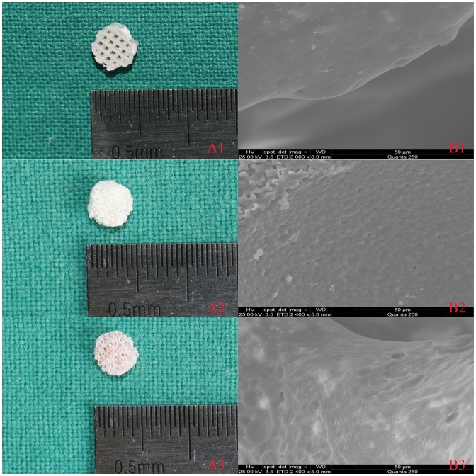
We have explored the applicability of printed scaffold by comparing osteogenic ability and biodegradation property of three resorbable biomaterials. A polylactic acid/hydroxyapatite (PLA/HA) composite with a pore size of 500 μm and 60% porosity was fabricated by three-dimensional printing. Three-dimensional printed PLA/HA, β-tricalcium phosphate (β-TCP) and partially demineralized bone matrix (DBM) seeded with bone marrow stromal cells (BMSCs) were evaluated by cell adhesion, proliferation, alkaline phosphatase activity and osteogenic gene expression of osteopontin (OPN) and collagen type I (COL-1). Moreover, the biocompatibility, bone repairing capacity and degradation in three different bone substitute materials were estimated using a critical-size rat calvarial defect model in vivo. The defects were evaluated by micro-computed tomography and histological analysis at four and eight weeks after surgery, respectively. The results showed that each of the studied scaffolds had its own specific merits and drawbacks. Three-dimensional printed PLA/HA scaffolds possessed good biocompatibility and stimulated BMSC cell proliferation and differentiation to osteogenic cells. The outcomes in vivo revealed that 3D printed PLA/HA scaffolds had good osteogenic capability and biodegradation activity with no difference in inflammation reaction. Therefore, 3D printed PLA/HA scaffolds have potential applications in bone tissue engineering and may be used as graft substitutes in reconstructive surgery.

Keywords: 102 Porous/Nanoporous/Nanostructured materials; 103 Composites; 211 Scaffold/Tissue engineering/Drug delivery; 30 Bio-inspired and biomedical materials; DBM; PLA/HA; Three-dimensional printing; biocompatibility; biomaterials; β-TCP.


Figures

None
Graphical abstract
Figure 1.
Figure 1.
3D printing machine using the new mini-deposition system.
Figure 2.
Figure 2.
(A) Optical and (B) SEM images for three scaffolds: 3D printed PLA/HA (1), β-TCP (2) and DBM (3).
Figure 3.
Figure 3.
BMSC responses to three scaffolds. (A) SEM images of the attachment of BMSCs on (1) PLA/HA, (2) β-TCP and (3) DBM scaffolds after culturing for seven days. (B, C) The fluorescence images of proliferation of BMSCs on (1) PLA/HA, (2) β-TCP and (3) DBM scaffolds after culturing for seven days. (← The state of proliferation of BMSCs; the selected areas in B corresponding to C. Length scales: (A) 50 μm, (B) 10 μm and (C) 8 μm).
Figure 4.
Figure 4.
Quantitative analysis of three scaffolds: (A) cell adhesion rate, (B) cell proliferation, (C) ALP activity, (D) osteogenic gene expression and (E) COL-1 of BMSCs cultured on different scaffolds (*p<0.05).
Figure 5.
Figure 5.
Micro CT images for three scaffolds. (A) Plane and profile images of (1) PLA/HA, (2) β-TCP and (3) DBM scaffolds before implanting. (B) Typical micro-CT images of (1) PLA/HA, (2) β-TCP and (3) DBM scaffolds as well as (4) bone defect without scaffold at four weeks in vivo. (C) Micro-CT images of (1) PLA/HA, (2) β-TCP and (3) DBM scaffolds as well as (4) bone defect without scaffold at eight weeks in vivo.
Figure 6.
Figure 6.
BV/TV and degradation rate in every group (A) Percentage of new bone formation at four and eight weeks. (B) Percentage of degradation of three scaffolds at four and eight weeks. (*p<0.05)
Figure 7.
Figure 7.
HE images of implanted and control group after four and eight weeks. (A) Histological images of implanted (1) PLA/HA, (2) β-TCP and (3) DBM scaffolds as well as (4) control group four weeks after implantation. (B) Histological images of (1) implanted PLA/HA, (2) β-TCP and (3) DBM scaffolds as well as (4) control group eight weeks after implantation. Scale bars 10 μm.
Figure 8.
Figure 8.
Immunohistochemical examination of osteocalcin and type I collagen were carried out in the margin of bone defect in different time (magnification ×100). (A) Osteocalcin expression images of implanted (1) PLA/HA, (2) β-TCP and (3) DBM scaffolds as well as (4) control group four weeks after implanting. (B) Osteocalcin expression images of implanted (1) PLA/HA, (2) β-TCP and (3) DBM scaffolds as well as (4) control group eight weeks after implanting. (C) Semi-quantitative scatter plot of osteocalcin expression. (D) Type I collagen expression images of implanted (1) PLA/HA, (2) β-TCP and (3) DBM scaffolds as well as (4) control group four weeks after implanting. (E) Type I collagen expression images of implanted (1) PLA/HA, (2) β-TCP and (3) DBM scaffolds as well as (4) control group eight weeks after implanting. (F) Semi-quantitative scatter plot of type I collagen expression (*p<0.05).

References

    1. John HD, Wenz B. Histomorphometric analysis of natural bone mineral for maxillary sinus augmentation. Int J Oral Maxillofac Implants. 2004;19:199–207. - PubMed
    1. Shalash MA, Rahman HA, Azim AA, et al. Evaluation of horizontal ridge augmentation using beta tricalcium phosphate and demineralized bone matrix: a comparative study. J Clin Exp Dent. 2013;5:253–9. - PMC - PubMed
    1. Dimitriou R, Jones E, McGonagle D, et al. Bone regeneration: current concepts and future directions. BMC Med. 2011;9:66. - PMC - PubMed
    1. McAllister BS, Haghighat K. Bone augmentation techniques. J Periodontol. 2007;78:377–96. - PubMed
    1. Browaeys H, Bouvry P, De Bruyn H. A literature review on biomaterials in sinus augmentation procedures. Clin Implant Dent Relat Res. 2007;9:166–77. - PubMed