. 2016 Jul 26;17(1):361-374.
doi: 10.1080/14686996.2016.1200948. eCollection 2016.

Lysosomal pH-inducible supramolecular dissociation of polyrotaxanes possessing acid-labile N-triphenylmethyl end groups and their therapeutic potential for Niemann-Pick type C disease

Affiliations

Lysosomal pH-inducible supramolecular dissociation of polyrotaxanes possessing acid-labile N-triphenylmethyl end groups and their therapeutic potential for Niemann-Pick type C disease

Atsushi Tamura et al. Sci Technol Adv Mater. .

Abstract

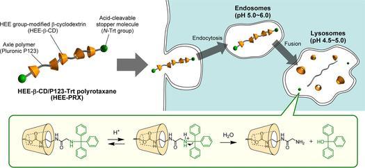
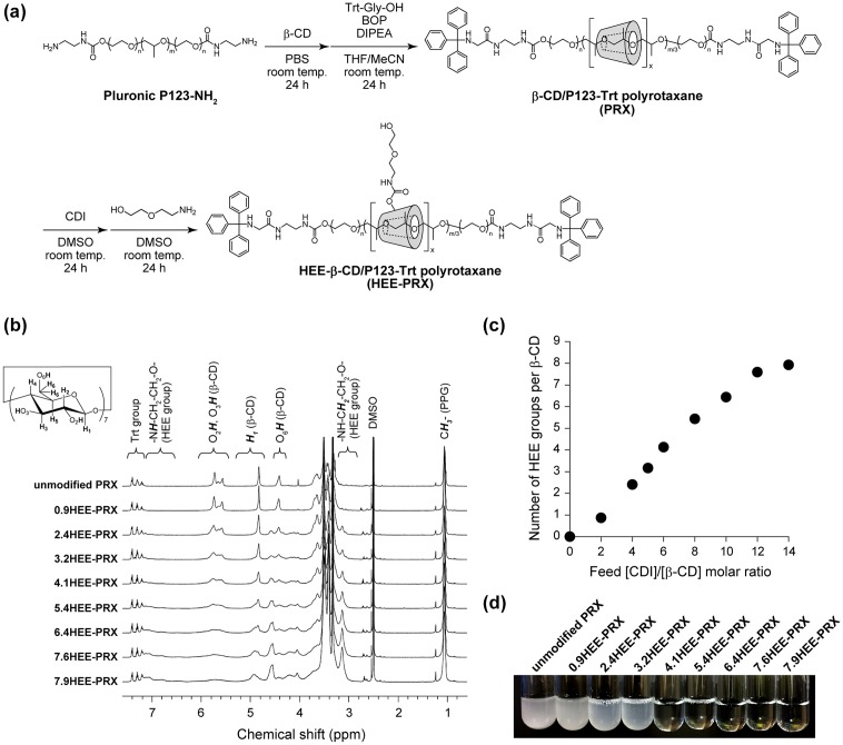
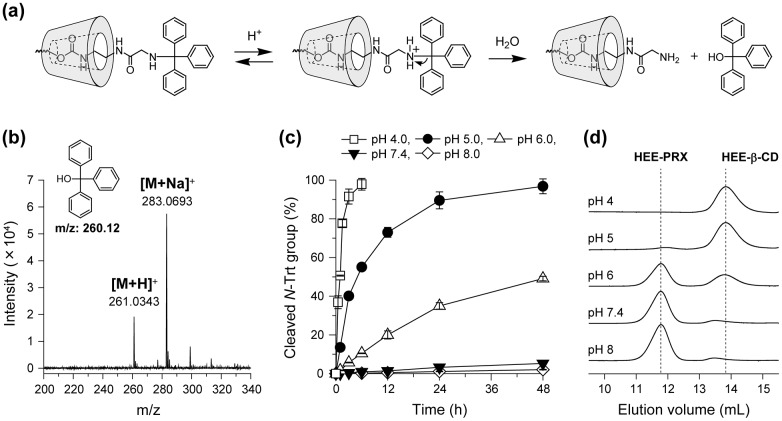
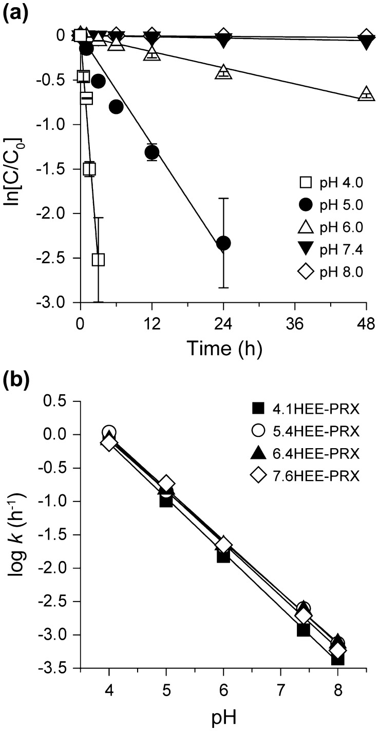
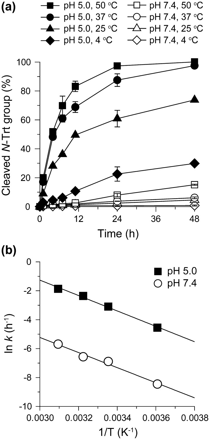
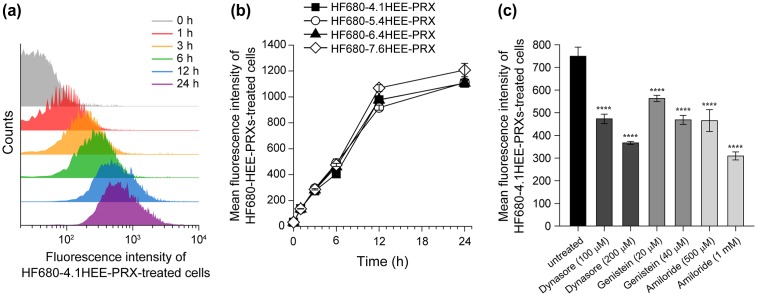
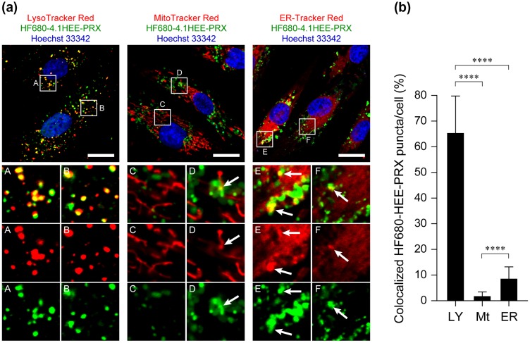
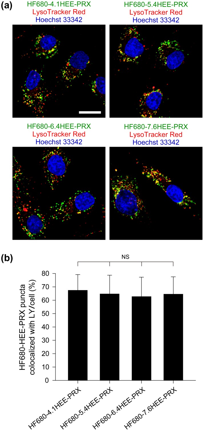
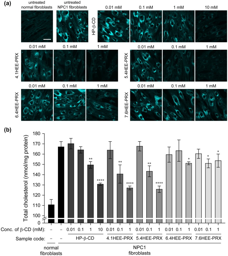
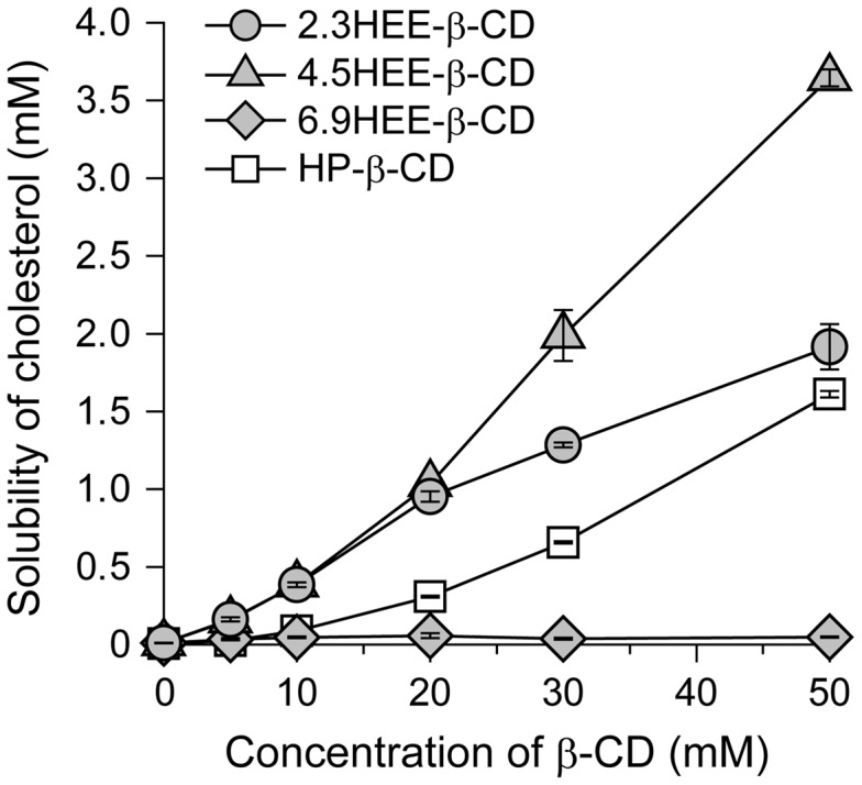
Niemann-Pick type C (NPC) disease is characterized by the accumulation of cholesterol in lysosomes. We have previously reported that biocleavable polyrotaxanes (PRXs) composed of β-cyclodextrins (β-CDs) threaded onto a linear polymer capped with bulky stopper molecules via intracellularly cleavable linkers show remarkable cholesterol reducing effects in NPC disease patient-derived fibroblasts owing to the stimuli-responsive intracellular dissociation of PRXs and subsequent β-CD release from the PRXs. Herein, we describe a series of novel acid-labile 2-(2-hydroxyethoxy)ethyl group-modified PRXs (HEE-PRXs) bearing terminal N-triphenylmethyl (N-Trt) groups as a cleavable component for the treatment of NPC disease. The N-Trt end groups of the HEE-PRXs underwent acidic pH-induced cleavage and led to the dissociation of their supramolecular structure. A kinetic study revealed that the number of HEE groups on the PRX did not affect the cleavage kinetics of the N-Trt end groups of the HEE-PRXs. The effect of the number of HEE groups of the HEE-PRXs, which was modified to impart water solubility to the PRXs, on cellular internalization efficiency, lysosomal localization efficiency, and cholesterol reduction ability in NPC disease-derived fibroblasts (NPC1 fibroblasts) was also investigated. The cellular uptake and lysosomal localization efficiency were almost equivalent for HEE-PRXs with different numbers of HEE groups. However, the cholesterol reducing ability of the HEE-PRXs in NPC1 fibroblasts was affected by the number of HEE groups, and HEE-PRXs with a high number of HEE groups were unable to reduce lysosomal cholesterol accumulation. This deficiency is most likely due to the cholesterol-solubilizing ability of HEE-modified β-CDs released from the HEE-PRXs. We conclude that the N-Trt group acts as a cleavable component to induce the lysosomal dissociation of HEE-PRXs, and acid-labile HEE-PRXs with an optimal number of HEE groups (4.1 to 5.4 HEE groups per single β-CD threaded onto the PRX) have great therapeutic potential for treating NPC disease.

Keywords: 101 Self-assembly/Self-organized materials; 211 Scaffold/Tissue engineering/Drug delivery; 30 Bio-inspired and biomedical materials; Niemann-Pick type C disease; Polyrotaxane; cholesterol; cyclodextrin; triphenylmethyl group.


Figures

None
Graphical abstract
Figure 1.
Figure 1.
Schematic illustration of HEE group-modified acid-labile polyrotaxanes (HEE-PRXs) and their intracellular dissociation in response to endosomal/lysosomal pH reduction.
Figure 2.
Figure 2.
(a) Synthetic scheme of the 2-(2-hydroxyethoxy)ethyl (HEE) group-modified PRXs (HEE-PRXs). (b) 1H NMR spectra of the unmodified PRX and the HEE-PRXs (0.9HEE-PRX, 2.4HEE-PRX, 3.2HEE-PRX, 4.1HEE-PRX, 5.4HEE-PRX, 6.4HEE-PRX, 7.6HEE-PRX, and 7.9HEE-PRX) in DMSO-d 6. (c) Relationship between the feed [CDI]/[β-CD] molar ratio and the average number of HEE groups per single β-CD threaded on the HEE-PRXs. (d) Photograph of the unmodified PRX and the HEE-PRXs in water. Concentration of the unmodified PRX and the HEE-PRXs adjusted to 10 mg ml–1.
Figure 3.
Figure 3.
(a) Reaction mechanism for the cleavage of N-Trt end groups in HEE-PRX under acidic pH conditions. (b) ESI-MS char of degraded products of HEE-PRX (4.1HEE-PRX) after a 24-h incubation at pH 4.0. (c) Representative time course of the cleavage of N-Trt end groups in the HEE-PRX (4.1HEE-PRX) under various pH conditions at 37 °C (open squares: pH 4.0, closed circles: pH 5.0, open triangles: pH 6.0, closed triangles: pH 7.4, and open diamonds: pH 8.0). The data are expressed as the mean ± standard deviation (n = 3). (d) Representative SEC charts of HEE-PRX (4.1HEE-PRX) after a 24-h incubation under various pH conditions at 37 °C.
Figure 4.
Figure 4.
(a) Representative first-order kinetic plots for the cleavage of N-Trt end groups in HEE-PRX (4.1HEE-PRX) under various pH conditions at 37 °C (open squares: pH 4.0, closed circles: pH 5.0, open triangles: pH 6.0, closed triangles: pH 7.4, open diamonds: pH 8.0). The data are expressed as the mean ± standard deviation (n = 3). (b) Logk-pH profiles for the cleavage of N-Trt end groups in HEE-PRXs at 37 °C (closed squares: 4.1HEE-PRX, open circles: 5.4HEE-PRX, closed triangles: 6.4HEE-PRX, open diamonds: 7.6HEE-PRX).
Figure 5.
Figure 5.
(a) Time course of the cleavage of N-Trt end groups in HEE-PRX (4.1HEE-PRX) at pH 5.0 (closed symbols) and 7.4 (open symbols) at various temperatures (squares: 50 °C, circles: 37 °C, triangles: 25 °C, diamonds: 4 °C). (b) Arrhenius plots for the cleavage of N-Trt end groups in HEE-PRX (4.1HEE-PRX) at pH 5.0 (closed squares) and 7.4 (open circles).
Figure 6.
Figure 6.
(a) Flow cytometry histograms of normal human skin fibroblasts treated with HF680–4.1HEE-PRX (0.5 mM of β-CD) for various time periods. (b) Time course of the fluorescence intensities of normal human skin fibroblasts treated with HF680-HEE-PRXs (HF680–4.1HEE-PRX: closed squares, HF680–5.4HEE-PRX: open circles, HF680–6.4HEE-PRX: closed triangles, HF680–7.6HEE-PRX: open diamonds) (0.5 mM of β-CD). (c) Effect of endocytosis inhibitors (dynasore for clathrin-mediated endocytosis, genistein for caveolae-mediated endocytosis, and amiloride for macropinocytosis) on the fluorescence intensities of HF680–4.1HEE-PRX-treated normal human skin fibroblasts after 6 h. The data are expressed as the mean ± SD (n = 3) (****p < 0.001 vs. untreated cells).
Figure 7.
Figure 7.
(a) CLSM images of normal human skin fibroblasts treated with HF680–4.1HEE-PRX (0.5 mM of β-CD) for 24 h (scale bars: 20 μm). The endosomes/lysosomes, mitochondria, endoplasmic reticulum, and nuclei were stained with LysoTracker Red DND-99, MitoTracker Red CMXRos, ER-Tracker Red, and Hoechst 33342, respectively. The fluorescence colors are indicated by the color of the text. The bottom panels in each column show enlarged views of the boxed regions. Colocalization of HF680-HEE-PRX with each organelle is indicated by arrows. (b) Percentage of HF680–4.1HEE-PRX-positive puncta that colocalized with endosomes/lysosomes (LY), mitochondria (Mt), and endoplasmic reticulum (ER). The data are expressed as the mean ± SD of 30 cells (****p < 0.001).
Figure 8.
Figure 8.
(a) CLSM images of normal human skin fibroblasts treated with HF680-HEE-PRXs (0.5 mM of β-CD) for 24 h (scale bar: 20 μm). The endosomes/lysosomes and nuclei were stained with LysoTracker Red DND-99 and Hoechst 33342, respectively. Fluorescence colors are indicated by the color of the text. (b) Percentage of HF680-HEE-PRX-positive puncta that colocalized with endosomes/lysosomes (LY). The data are expressed as the mean ± SD of 30 cells (NS: not significant).
Figure 9.
Figure 9.
(a) Filipin-stained normal and NPC1 fibroblasts treated with HP-β-CD (0.01, 0.1, 1 and 10 mM) and HEE-PRXs (0.01, 0.1, and 1 mM of β-CD) for 24 h (scale bars: 50 μm). (b) The amount of total cholesterol in normal and NPC1 fibroblasts treated with HP-β-CD (0.01, 0.1, 1, and 10 mM) and the HEE-PRXs (0.01, 0.1, and 1 mM of β-CD) for 24 h. The data are expressed as the mean ± SD (n = 3) (*p < 0.05, **p < 0.01, ****p < 0.001 vs. untreated NPC1 fibroblasts).
Figure 10.
Figure 10.
Phase-solubility diagrams of cholesterol with 2.3HEE-β-CD (filled circles), 4.5HEE-β-CD (filled triangles), 6.9HEE-β-CD (filled diamonds), and HP-β-CD (open squares) in PBS. The solubility of cholesterol was determined after incubation for 24 h at 37 °C (n = 3).

References

    1. Luzio JP, Pryor PR, Bright NA. Lysosomes: fusion and function. Nat Rev Mol Cell Biol. 2007;8:622–632. doi: 10.1038/nrm2217. - DOI - PubMed
    1. Saftig P, Klumperman J. Lysosome biogenesis and lysosomal membrane proteins: trafficking meets function. Nat Rev Mol Cell Biol. 2009;10:623–635. doi: 10.1038/nrm2745. - DOI - PubMed
    1. Sancak Y, Bar-Peled L, Zoncu R, et al. Ragulator-rag complex targets mTORC1 to the lysosomal surface and is necessary for its activation by amino acids. Cell. 2010;141:290–303. doi: 10.1016/j.cell.2010.02.024. - DOI - PMC - PubMed
    1. Futerman AH, van Meer G. The cell biology of lysosomal storage disorders. Nat Rev Mol Cell Biol. 2004;5:554–565. doi: 10.1038/nrm1423. - DOI - PubMed
    1. Platt FM, Boland B, van der Spoel AC. Lysosomal storage disorders: the cellular impact of lysosomal dysfunction. J Cell Biol. 2012;199:723–734. doi: 10.1083/jcb.201208152. - DOI - PMC - PubMed