. 2017 Apr;74(8):1511-1525.
doi: 10.1007/s00018-016-2423-7. Epub 2016 Nov 23.

A novel 72-kDa leukocyte-derived osteoglycin enhances the activation of toll-like receptor 4 and exacerbates cardiac inflammation during viral myocarditis

Affiliations

A novel 72-kDa leukocyte-derived osteoglycin enhances the activation of toll-like receptor 4 and exacerbates cardiac inflammation during viral myocarditis

Marieke Rienks et al. Cell Mol Life Sci. 2017 Apr.

Abstract

Background: Viral myocarditis can severely damage the myocardium through excessive infiltration of immune cells. Osteoglycin (OGN) is part of the small leucine-rich repeat proteoglycan (SLRP) family. SLRP's may affect inflammatory and fibrotic processes, but the implication of OGN in cardiac inflammation and the resulting injury upon viral myocarditis is unknown.

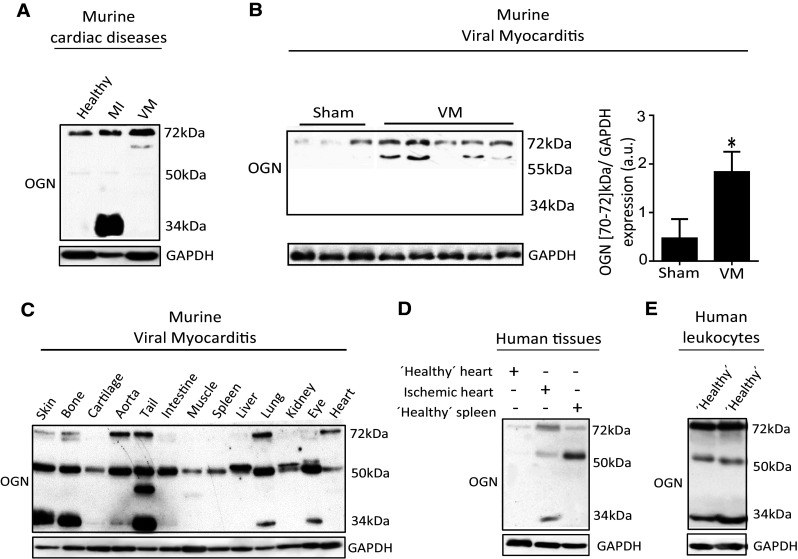
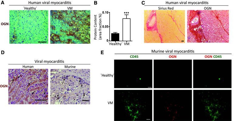
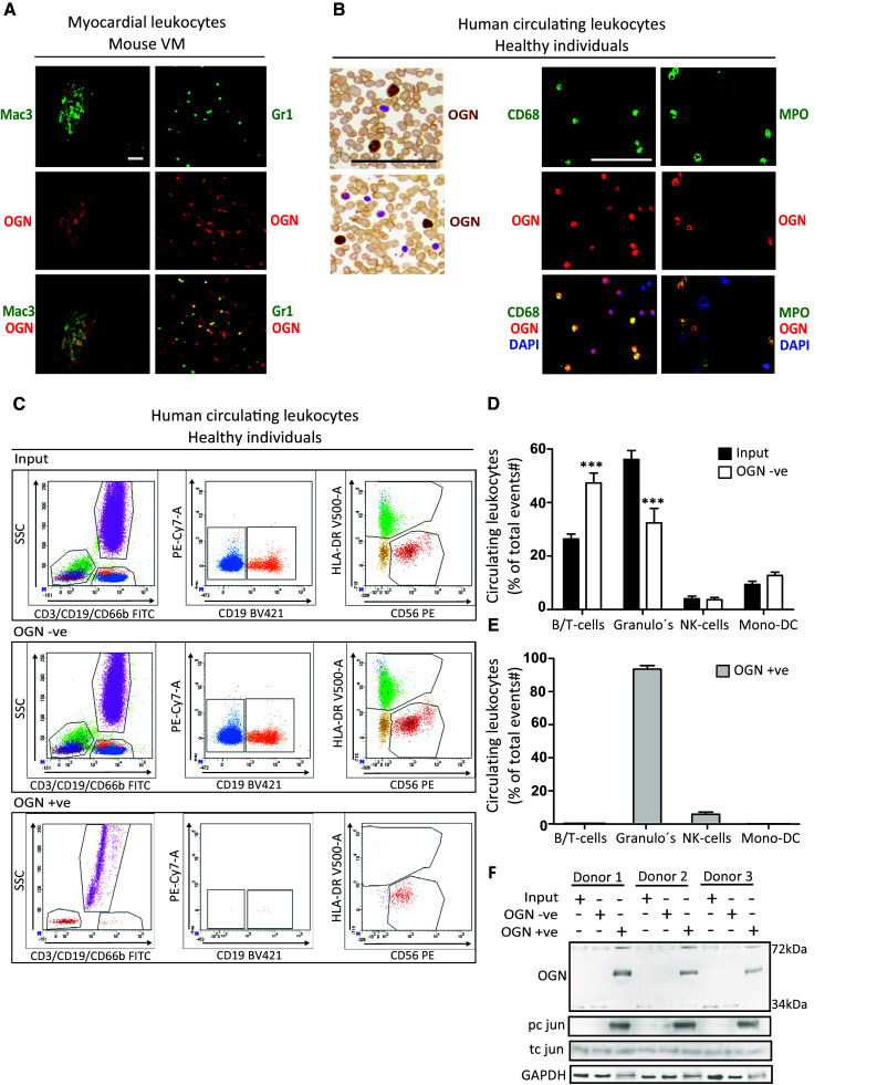
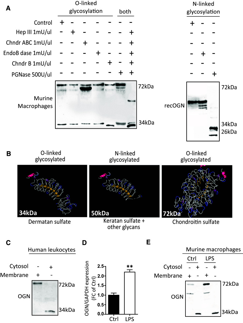
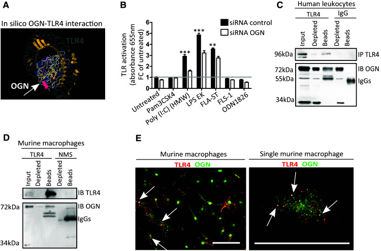
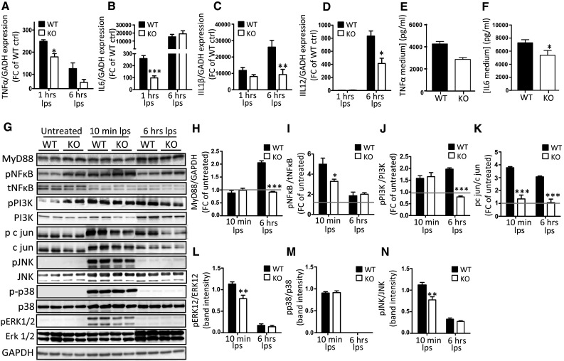
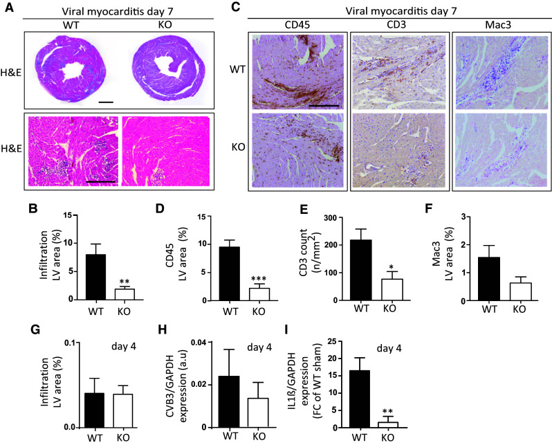
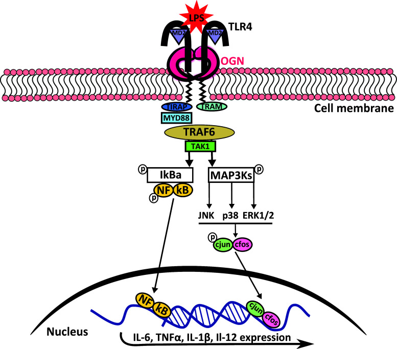
Methods and results: This study uncovered a previously unidentified 72-kDa variant of OGN that is predominant in cardiac human and mouse samples of viral myocarditis. Its absence in mice significantly decreased cardiac inflammation and injury in Coxsackievirus-B3-induced myocarditis. It also delayed mortality in lipopolysaccharide-induced endotoxemia going along with a reduced systemic production of pro-inflammatory cytokines. This 72-kDa OGN is expressed in the cell membrane of circulating and resident cardiac macrophages and neutrophils. Co-immunoprecipitation and OGN siRNA experiments revealed that this 72-kDa variant activates the toll-like receptor-4 (TLR4) with a concomitant increase in IL-6, TNF-α, IL-1β, and IL-12 expression. This immune cell activation by OGN occurred via MyD88 and increased phosphorylation of c-jun. Finally, the 72-kDa chondroitin sulfate is the result of O-linked glycosylation of the 32-kDa protein core of OGN. In contrast, the 34-kDa dermatan sulfate-OGN, involved in collagen cross linking, was also the result of O-linked glycosylation.

Conclusion: The current study discovered a novel 72-kDa chondroitin sulfate-OGN that is specific for innate immune cells. This variant is able to bind and activate TLR4. The absence of OGN decreases cytokine production by both circulating and cardiac leukocytes upon (systemic) LPS exposure, and reduces cardiac inflammation and injury in viral myocarditis.

Keywords: Glycosylation; Inflammation; Osteoglycin; TLR4; Viral myocarditis.


Conflict of interest statement

The authors have no conflicts of interest to disclose.

Figures

Fig. 1
Fig. 1
Murine and human tissues express different OGN variants in distinct cardiac diseases. a Western blot analysis for OGN revealed a small variant (34 kDa) in murine myocardial infarction, whereas in murine viral myocarditis, a large variant (72 kDa) was present (n ≥ 4). b Western blot analysis of cardiac tissues of mice 7 days after viral exposure was compared with healthy controls and quantified. Viral infection led to increased expression of a 70–72 kDa OGN variant compared with healthy controls (sham n = 3, VM n = 5, *p < 0.05). c Differential expression of the main OGN variants in various healthy murine tissues. d Western blot analysis for OGN in human ischemic myocardial tissue showing increased expression of the large (72 kDa) and the small (34 kDa) OGN variants relative to those in the control cardiac tissue. e Human buffy coat revealed a similar presence of the OGN variants with high abundance of the 72-kDa variant, which was barely detectable in human splenic tissues
Fig. 2
Fig. 2
Different OGN variants are correlated with fibrosis and inflammation in murine and human cardiac diseases. a Increased immunohistochemical OGN staining was found in patients diagnosed with myocarditis. b Quantification of OGN staining in patients diagnosed with myocarditis (n ≥ 14). c OGN expression in human myocarditis biopsies coincided with fibrosis as shown by Sirius red staining. d OGN staining was also found on leukocytes in human myocarditis biopsies as well as in the hearts of mice subjected to CVB3-induced myocarditis. e Immunofluorescence demonstrated the co-localization of OGN with CD45+ leukocytes in the hearts of mice subjected to virus myocarditis compared with healthy controls (scale bar healthy controls: 20 µm). All experiments were repeated at least twice. Scale bar 50 µm. VM viral myocarditis, MI myocardial infarction
Fig. 3
Fig. 3
72-kDa iOGN was found on cardiac and circulating innate immune cells. a Murine cardiac monocytes/macrophages (Mac-3) and neutrophils (Gr1) expressed OGN during CVB3-induced myocarditis. b Circulating human monocytes and neutrophils also expressed OGN. c Representative FACS plots of total human circulating leukocytes, OGN-negative (OGN −ve), and -positive (OGN +ve) circulating leukocytes. d, e FACS analysis revealed that the OGN-positive circulating leukocytes consisted mostly of neutrophils. f OGN-positive circulating immune cells had increased phosphorylation of c jun. n = 4, *p < 0.001; all experiments were repeated at least twice. Scale bar 50 µm
Fig. 4
Fig. 4
Glycosylation of the 32-kDa OGN core protein results in the production of different protein variants. a Enzyme treatment of macrophage protein lysates revealed the differential presence of glycans and glycosaminoglycans; treatment with chondroitinase ABC reduced the size of the 72-kDa protein variant, indicating that chondroitin and dermatan sulfate is attached, whereas treatment with chondroitinase B only slightly reduced the size of the 34-kDa variant, indicating that it only has dermatan sulfate attached. The simultaneous addition of all of the enzymes reduced the protein glycosylation of both the 34-kDa and 72-kDa variants entirely. Treatment of the 50-kDa OGN variant with PGNase reduced the size detected on western blot significantly, indicating that it is N-linked glycosylated. b Predicted structures of the OGN variants and their respective glycosylations. c Cell fractionation of fresh human buffy coat lysates revealed the presence of the 72-kDa OGN variant in the cell membrane, whereas the 34-kDa variant was found in the cytosol. d, e In mouse bone marrow-derived macrophages, LPS stimulation increased the expression of OGN in the cell membrane. All experiments were repeated at least twice
Fig. 5
Fig. 5
Membrane bound iOGN interacts with TLR4 on leukocytes. a Docking predictions of TLR4 and OGN reveal a potential interaction between these proteins via their leucine-rich repeats. b HEK-Blue™-mTLR cells containing an inducible SEAP reporter gene were stimulated with TLR-specific ligands (Pam3CSK4 for TLR1/2, Poly(I:C) (HMW) for TLR3, LPS-EK for TLR4, FLA-ST for TLR5, FLS-1 for TLR6/2, and ODN1826 for TLR9) with and without OGN knockdown. OGN knockdown reduced the activation of TLR3, -4 and -5. c, d OGN co-immunoprecipitated with TLR4 in human peripheral leukocytes and murine bone marrow-derived macrophages. e Confocal immunofluorescence revealing the co-localization of OGN with TLR4 in primary murine macrophages. n ≥ 4, *p < 0.001. All experiments were repeated at least twice. Scale bar 50 µm
Fig. 6
Fig. 6
OGN promotes TLR4 activation by enhancing MAPK-induced cytokine production. Bone marrow-derived macrophages (BMDMs) from WT and KO mice were isolated and stimulated with LPS (10 ng/ml). ad LPS stimulation resulted in the blunted induction of pro-inflammatory cytokines TNFα and IL-6 expression (1 h) as well as IL-1β and IL-12 at a later stage (6 h) (c, d). This difference in TLR4 activation was reflected in the lower TNFα and IL-6 expression levels in the medium of OGN-KO BMDMs (e, f). This increased cytokine production was the result of significant induction of MyD88 that was blunted in the OGN-KO BMDMs (g, h). g Western blot analysis and further quantification revealed blunted c-jun, ERK1/2 and JNK phosphorylation in the KO BMDMs, and blunted pNFkB induction (il). All experiments were repeated at least twice. n ≥ 3; *p < 0.05; **p < 0.01; ***p < 0.001
Fig. 7
Fig. 7
OGN increases cardiac inflammation in CVB3-induced murine myocarditis. Analysis of H&E staining in OGN-KO mice revealed significantly reduced cellular infiltration relative to WT mice in human CVB3-induced viral myocarditis (a, scale: 1 mm top and 50 µm bottom) and b quantification (n ≥ 11, **p < 0.01). c Decreased immune infiltration in OGN-KO mice was confirmed by the reduced presence of CD45 positive leukocytes (d), CD3 positive lymphocytes (e), and Mac3 positive macrophages (f). g Four days after viral inoculation, the amount of immune cell infiltration was still low and comparable in both genotypes (n ≥ 4). h Viral levels analyzed by RT-PCR were also comparable at this time. i Pro-inflammatory cytokine IL-1β RNA expression, on the other hand, was already significantly increased in WT compared with KO mice 4 days after viral exposure. Scale 50 µm
Fig. 8
Fig. 8
Schematic overview of the proposed iOGN:TLR4 signaling pathway. TLR4 stimulation with LPS results in receptor dimerization and subsequent high-affinity binding of the bridging adaptor molecules TRAM and TIRAP. The bound and activated MyD88 then activates IRAK4, TRAF6, TAK1, and IKK complexes, and TRIF signals through RIP1 to TRAF6/TAK1 and IKK. Both of these pathways result in increased pro-inflammatory cytokine expression. The phosphorylation of MAPKs by TAK1 is enhanced by OGN, resulting in increased c-jun phosphorylation with subsequent nuclear translocation of AP1 (the phosphorylated c-jun/c fos complex), which again resulted in increased pro-inflammatory cytokine expression, reinforcing the inflammatory response [47]

References

    1. Van Aelst LN, et al. Osteoglycin prevents cardiac dilatation and dysfunction after myocardial infarction through infarct collagen strengthening. Circ Res. 2015;116(3):425–436. doi: 10.1161/CIRCRESAHA.116.304599. - DOI - PubMed
    1. Rienks M, et al. Myocardial extracellular matrix: an ever-changing and diverse entity. Circ Res. 2014;114(5):872–888. doi: 10.1161/CIRCRESAHA.114.302533. - DOI - PubMed
    1. Funderburgh JL, et al. Mimecan, the 25-kDa corneal keratan sulfate proteoglycan, is a product of the gene producing osteoglycin. J Biol Chem. 1997;272(44):28089–28095. doi: 10.1074/jbc.272.44.28089. - DOI - PubMed
    1. Dunlevy JR, et al. Expression of the keratan sulfate proteoglycans lumican, keratocan and osteoglycin/mimecan during chick corneal development. Exp Eye Res. 2000;70(3):349–362. doi: 10.1006/exer.1999.0789. - DOI - PubMed
    1. Yajima T, Knowlton KU. Viral myocarditis: from the perspective of the virus. Circulation. 2009;119(19):2615–2624. doi: 10.1161/CIRCULATIONAHA.108.766022. - DOI - PubMed

Publication types

MeSH terms