

Aqueous immune mediators in malignant uveal melanomas in comparison to benign pigmented intraocular tumors
- PMID: 27878431
- PMCID: PMC5285432
- DOI: 10.1007/s00417-016-3541-5
Aqueous immune mediators in malignant uveal melanomas in comparison to benign pigmented intraocular tumors
Abstract
Background: To examine the usefulness of measuring immune mediators in aqueous humor samples for differentiating malignant uveal melanoma from benign pigmented intraocular tumors.
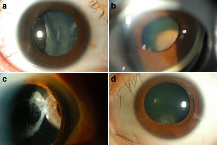
Methods: Thirteen eyes of 13 patients with uveal melanoma were studied, and 13 eyes of 13 patients with benign pigmented intraocular tumors served as controls. Undiluted samples of aqueous humor were collected, and a cytometric bead array was used to determine the aqueous humor concentrations of 35 immune mediators comprising 14 interleukins (IL), interferon-γ, interferon-γ-inducible protein-10, monocyte chemoattractant protein (MCP)-1, macrophage inflammatory protein (MIP)-1α, MIP-1β, regulated on activation normal T cell expressed and secreted, monokine induced by interferon-γ, basic fibroblast growth factor, Fas ligand, granzyme A, granzyme B, eotaxin, interferon-inducible T-cell alpha chemoattractant, fractalkine, granulocyte macrophage colony-stimulating factor, granulocyte colony-stimulating factor, vascular endothelial growth factor, angiogenin, tumor necrosis factor-α, lymphotoxin-α, and CD40L.
Results: Aqueous humor levels of angiogenin, IL-8, and MCP-1 were significantly higher in eyes with malignant melanoma than in those with benign tumors (p < 0.05).
Conclusions: Angiogenin, IL-8, and MCP-1 levels in aqueous humor may be potential markers for distinguishing malignant uveal melanoma from benign pigmented intraocular tumors, and may be a useful adjunct to histomorphology, diagnostic imaging, and other biomarkers for the diagnosis and appropriate clinical management of malignant uveal melanoma.
Keywords: Benign pigmented intraocular tumor; Immune mediator; Uveal melanoma.
Conflict of interest statement
All authors certify that they have no affiliations with or involvement in any organization or entity with any financial interest (such as honoraria; educational grants; participation in speakers‘ bureaus; membership, employment, consultancies, stock ownership, or other equity interest; and expert testimony or patent-licensing arrangements) or non-financial interest (such as personal or professional relationships, affiliations, knowledge or beliefs) in the subject matter or materials discussed in this manuscript. Funding No funding was received for this research. Ethical approval All procedures performed in studies involving human participants were in accordance with the ethical standards of the institutional and/or national research committee and with the 1964 Declaration of Helsinki and its later amendments or comparable ethical standards. Informed consent Informed consent was obtained from all individual participants included in the study.
Figures
References
Publication types
MeSH terms
Substances
LinkOut - more resources
Full Text Sources
Other Literature Sources
Medical
Molecular Biology Databases
Research Materials
Miscellaneous
