. 2017 May;21(5):955-967.
doi: 10.1111/jcmm.13035. Epub 2016 Nov 23.

Long non-coding RNA linc00261 suppresses gastric cancer progression via promoting Slug degradation

Affiliations

Long non-coding RNA linc00261 suppresses gastric cancer progression via promoting Slug degradation

Yingcong Yu et al. J Cell Mol Med. 2017 May.

Abstract

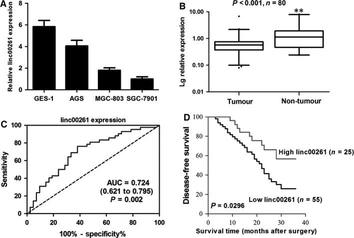
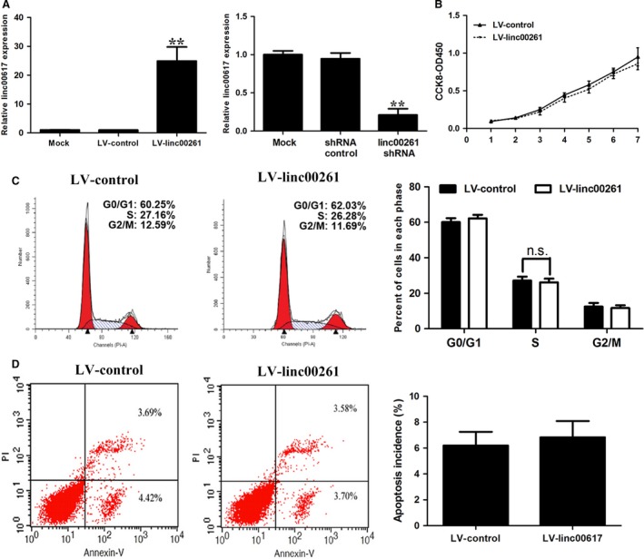
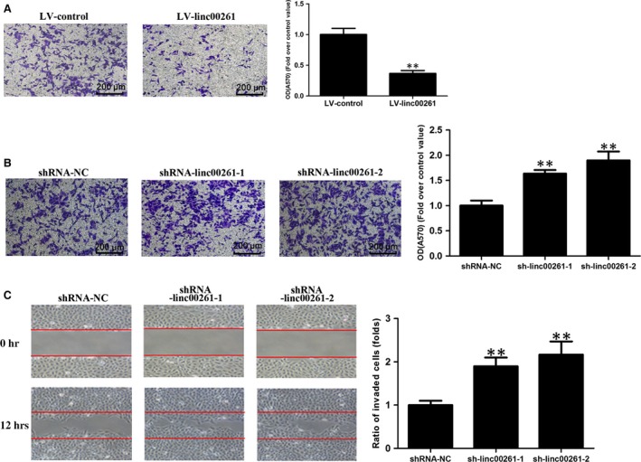
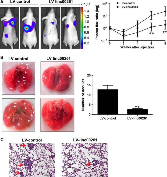
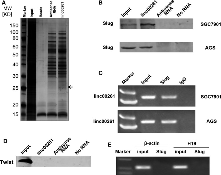
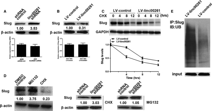
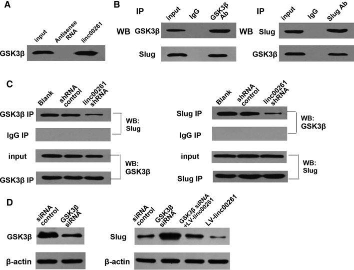
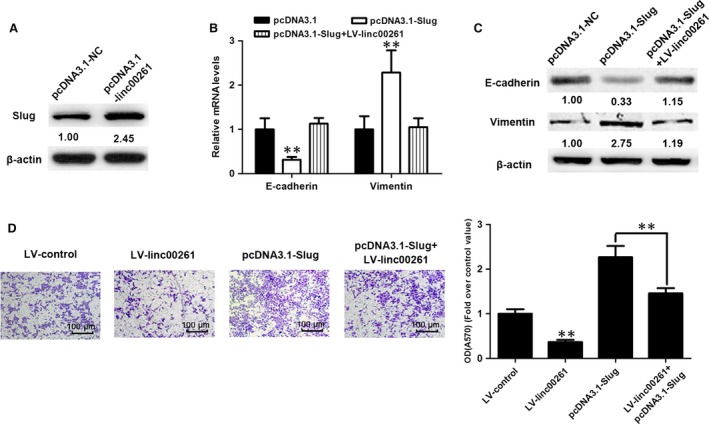
Gastric cancer (GC) remains a threat to public health with high incidence and mortality worldwide. Increasing evidence demonstrates that long non-coding RNAs (lncRNAs) play critical regulatory roles in cancer biology, including GC. Previous profiling study showed that lncRNA linc00261 was aberrantly expressed in GC. However, the role of linc00261 in GC progression and the precise molecular mechanism remain unknown. In this study, we report that linc00261 was significantly down-regulated in GC tissues and the expression level of linc00261 negatively correlated with advanced tumour status and clinical stage as well as poor prognostic outcome. In vitro functional assays indicate that ectopic expression of linc00261 suppressed cell invasion by inhibiting the epithelial-mesenchymal transition (EMT). By RNA pull-down and mass spectrum experiments, we identified Slug as an RNA-binding protein that binds to linc00261. We confirmed that linc00261 down-regulated Slug by decreasing the stability of Slug proteins and that the tumour-suppressive function of linc00261 can be neutralized by Slug. linc00261 may promote the degradation of Slug via enhancing the interaction between GSK3β and Slug. Moreover, linc00216 overexpression repressed lung metastasis in vivo. Together, our findings suggest that linc00261 acts a tumour suppressor in GC by decreasing the stability of Slug proteins and suppressing EMT. By clarifying the mechanisms underlying GC progression, these findings may facilitate the development of novel therapeutic strategies for GC.

Keywords: Slug; epithelial-mesenchymal transition; gastric cancer; invasion; long non-coding RNA.


Figures

Figure 1
Figure 1
linc00261 expression in GC cell lines, cancer tissues and its clinical significance. (A) qRTPCR analysis of linc00261 expression levels in gastric cancer cell lines (BGC‐823, MGC‐803 and SGC‐7901) compared with the normal gastric epithelium cell line (GES‐1). Data represent the mean ± S.D. from three independent experiments. (B) linc00261 expression was analysed by qRTPCR in GC samples and adjacent non‐tumour gastric tissues (n = 80). linc00261 expression level was normalized to that of GAPDH. Horizontal lines in the box plots represent the medians, the boxes represent the interquartile range, and the whiskers represent the 2.5th and 97.5th percentiles. The significant differences between samples were analysed using the Wilcoxon signed‐rank test. **P < 0.01. (C) ROC curve for prediction of gastric cancer using RTqPCR‐based lncRNALEGBC expression level. The AUC was 0.724, with 95% CI and P value indicated. (D) Kaplan–Meier analysis of disease‐free survival based on linc00261 expression in all the 80 patients.
Figure 2
Figure 2
Effect of linc00261 overexpression on gastric cancer cell proliferation, cell cycle and apoptosis. (A) The expression of linc00261 (mean ± S.D.) in stable SGC7901 cell clones infected with lentiviruses encoding linc00261. linc00261 expression (mean ± S.D.) in AGS cells that were stably transfected with shRNA against linc00261. (B) CCK8 assays were performed to determine the proliferation of SGC7901 cells. (C) Cell cycle analysis determined the relative cell numbers in each cell cycle phase after propidium iodide staining of linc00261‐up‐regulated SGC7901 cells. Numbers inside bars represent percentages of cells in each phase. (D) Annexin V/PI staining and flow cytometry analysis assessing apoptosis in SGC7901 cells after LV‐linc00261 transfection. Data represent the mean ± S.D. from three independent experiments. N.S., not significant. *P < 0.05; **P < 0.01.
Figure 3
Figure 3
Effect of linc00261 on the invasion of GC cells. (A) SGC7901 cells were transfected with LV‐control or LV‐linc00261. Matrigel invasion assay was performed to investigate the invasive ability of SGC7901 cells. Histological analysis of OD (570 nm) absorbance of crystal violet‐stained cells in Matrigel invasion assay. (B) AGS cells were transfected with shRNA control or shRNA linc00261. Matrigel invasion assay was performed to investigate the invasive ability of AGS cells. Histological analysis of OD (570 nm) absorbance of crystal violet‐stained cells in Matrigel invasion assay. Data represent the mean ± S.D. from three independent experiments. (C) Representative images and quantification of cell motility changes by linc00261 knockdown.*P < 0.05; **P < 0.01.
Figure 4
Figure 4
linc00261 suppresses EMT in GC cells. (A) Immunofluorescence images for EMT markers of linc00261‐overexpressing or control SGC7901 cells. Scale bar, 20 μm. Western blot analysis of phenotypic markers after linc00261 overexpression (SGC7901). (B) Immunofluorescence images for EMT markers of linc00261‐knockdown or control AGS cells. Scale bar, 20 μm. Western blot analysis of phenotypic markers after linc00261 silencing (AGS). Relative protein expression was identified (n = 3) and normalized to β‐actin. Data represent the mean ± S.D.
Figure 5
Figure 5
linc00261 suppresses lung metastasis in vivo. (A) Representative BLI plots of lung metastases in mice after injecting SGC7901 cells stably expressing linc00261 or a control vector; four mice were used each group. (B) Lungs were extracted at 8 weeks after tail vein injection, and the number of nodules was counted. Arrow indicated tumours on the surface of the lungs. (C) Haematoxylin and eosin‐stained images of lung tissue isolated from nude mice that received intravenous tail injections of SGC7901 cells stably expressing linc00261 or a control vector. Arrows indicate the metastasis nodules (original magnification ×100). Scale bars, 50 μM. Data represent the mean ± S.D. from three independent experiments. *P < 0.05; **P < 0.01. Arrows indicate the metastasis nodules.
Figure 6
Figure 6
linc00261 binds to Slug protein. (A) Representative SDSPAGE gel after Coomassie blue staining shows a prominent band at about 25 kD, representing mainly Slug proteins (see also supplemental Table 4). (B) RNA pull‐down assay performed in SGC7901 and AGS cells. Biotinylated linc00261 or antisense RNA was incubated with cell extracts, targeted with streptavidin beads and washed, and the associated proteins were resolved on a gel. Western blot analysis detected the specific association of Slug and linc00261 (n = 3). (C) RIP experiments were performed using the Slug antibody for immunoprecipitation (IP) and a primer to detect linc00261. RIP enrichment was determined relative to the input controls. (D) No interaction between linc00261 and Twist could be detected. RNA pull‐down assay performed in SGC7901 cells. Biotinylated linc00261 or antisense RNA was incubated with cell extracts, targeted with streptavidin beads and washed, and the associated proteins were resolved on a gel. Western blot analysis detected the specific association of Twist and linc00261. (E) RIP experiments were performed using Slug to immunoprecipitate RNA and a primer to detect H19 and β‐actin.
Figure 7
Figure 7
linc00261 decreases the protein level of Slug by inhibiting its stability. (A) The protein levels of Slug were detected in linc00261‐knockdown AGS cells by Western blot analysis. The mRNA levels of Slug were detected in linc00261‐knockdown AGS cells by qRTPCR (n = 3). (B) The protein levels of Slug were detected in linc00261‐up‐regulated SGC7901 cells. The mRNA levels of Slug were detected in linc00261‐overexpressing SGC‐7901 cells by qRTPCR (n = 3). (C) linc00261 overexpression enhanced Slug protein degradation. SGC‐7901 cells were transfected with Luc or linc00261. After treating the cells with cycloheximide (CHX, 0.5 μg/μl) for an indicated time, the expression of endogenous Slug protein was analysed by WB. The band intensity of Slug for each time‐point was quantified by ImageJ and plotted. Experiments were repeated for three times, and a representative experiment is presented. Error bars represent S.D. Every experimental group was compared with the control Lucsi group. (D) The comparison and quantification of Slug proteins in AGS cells with and without the protein synthesis inhibitor cycloheximide (CHX, 0.5 μg/μl) or the proteasome inhibitor MG‐132 (5 μM) for 24 hrs. linc00261 stable knockdown AGS cells and control cells were incubated with MG132 (5 μM) or CHX (0.5 μg/μl) for 24 hrs. The levels of Slug proteins were detected by Western blot analysis. (E) Stable linc00261 overexpressing SGC7901 cells and control cells were treated with MG132 (10 mM) for 6 hrs. Cell lysates were immunoprecipitated with a Slug‐specific antibody, followed by Western blotting with an antibody to ubiquitin. The bottom panel depicts the input of the cell lysates.
Figure 8
Figure 8
linc00261 promote the degradation of Slug via enhancing the interaction between GSK3β and Slug. (A) RNA pull‐down assay performed in AGS cells. Biotinylated linc00261 or antisense RNA was incubated with cell extracts, targeted with streptavidin beads and washed, and the associated proteins were resolved on a gel. Western blot analysis detected the specific association of GSK3β and linc00261 (n = 3). (B) Co‐immunoprecipitation detected the interaction of GSK3β and Slug in AGS cells. The 20% of input (cell lysate) and GSK3β or Slug immunoprecipitates were separated by SDSPAGE. The specific immunoprecipitation of GSK3β and Slug was confirmed by Western blot analysis (n = 3). (C) Immunoprecipitation assay was performed to detect the interaction between GSK3β and Slug after transfection of linc00261 shRNA in AGS cells. (D) Western blot analysis confirmed that GSK3β‐specific siRNA down‐regulated the protein level of GSK3β. Knockdown of GSK3β abolished the down‐regulation of protein level of Slug induced by linc00261 overexpression.
Figure 9
Figure 9
Slug plays a critical role in linc00261‐mediated EMT. (A) Western blot analysis was performed to confirm the successful up‐regulation of Slug protein levels. qRTPCR analysis (B) and Western blots (C) of EMT‐specific marker E‐cadherin and Vimentin from SGC‐7901 cells receiving the indicated vectors (D) linc00261 overexpression suppressed the invasion of SGC7901 cells and Slug‐overexpression attenuated the linc00261 overexpression‐induced suppression of SGC‐7901 cell invasiveness. Histological analysis of OD (570 nm) absorbance of crystal violet‐stained cells in transwell assay. Data represent the mean ± S.D. from three independent experiments.*P < 0.05. **P < 0.01.

References

    1. Ferro A, Peleteiro B, Malvezzi M, et al Worldwide trends in gastric cancer mortality (1980–2011), with predictions to 2015, and incidence by subtype. Eur J Cancer. 2014; 50: 1330–44. - PubMed
    1. Karimi P, Islami F, Anandasabapathy S, et al Gastric cancer: descriptive epidemiology, risk factors, screening, and prevention. Cancer Epidemiol Biomarkers Prev. 2014; 23: 700–13. - PMC - PubMed
    1. Dassen AE, Dikken JL, van de Velde CJ, et al Changes in treatment patterns and their influence on long‐term survival in patients with stages I‐III gastric cancer in The Netherlands. Int J Cancer. 2013; 133: 1859–66. - PubMed
    1. Nagarajan N, Bertrand D, Hillmer AM, et al Whole‐genome reconstruction and mutational signatures in gastric cancer. Genome Biol. 2012; 13: R115. - PMC - PubMed
    1. Zang ZJ, Cutcutache I, Poon SL, et al Exome sequencing of gastric adenocarcinoma identifies recurrent somatic mutations in cell adhesion and chromatin remodeling genes. Nat Genet. 2012; 44: 570–4. - PubMed

Publication types

MeSH terms

Substances