Review
. 2017 Jun;65(6):851-863.
doi: 10.1002/glia.23102. Epub 2016 Nov 23.

Corneal epithelial cells function as surrogate Schwann cells for their sensory nerves

Affiliations
Review

Corneal epithelial cells function as surrogate Schwann cells for their sensory nerves

Mary Ann Stepp et al. Glia. 2017 Jun.

Abstract

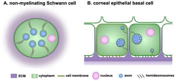
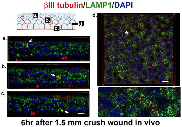
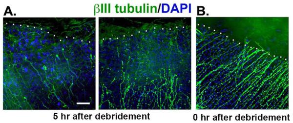
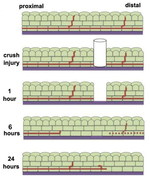
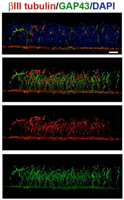
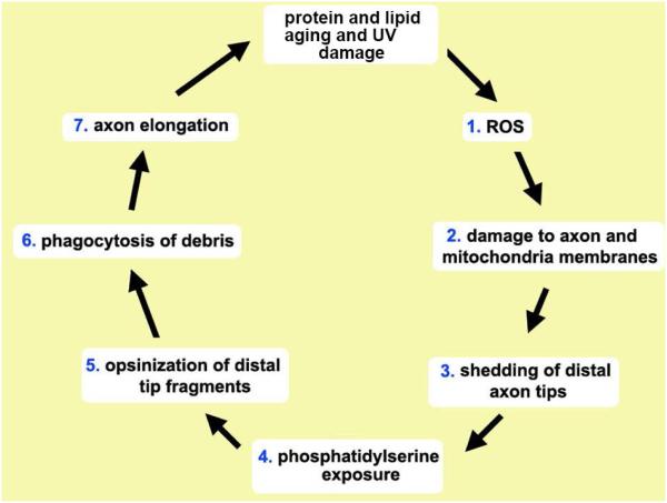
The eye is innervated by neurons derived from both the central nervous system and peripheral nervous system (PNS). While much is known about retinal neurobiology and phototransduction, less attention has been paid to the innervation of the eye by the PNS and the roles it plays in maintaining a functioning visual system. The ophthalmic branch of the trigeminal ganglion contains somas of neurons that innervate the cornea. These nerves provide sensory functions for the cornea and are referred to as intraepithelial corneal nerves (ICNs) consisting of subbasal nerves and their associated intraepithelial nerve terminals. ICNs project for several millimeters within the corneal epithelium without Schwann cell support. Here, we present evidence for the hypothesis that corneal epithelial cells function as glial cells to support the ICNs. Much of the data supporting this hypothesis is derived from studies of corneal development and the reinnervation of the ICNs in the rodent and rabbit cornea after superficial wounds. Corneal epithelial cells activate in response to injury via mechanisms similar to those induced in Schwann cells during Wallerian Degeneration. Corneal epithelial cells phagocytize distal axon fragments within hours of ICN crush wounds. During aging, the proteins, lipids, and mitochondria within the ICNs become damaged in a process exacerbated by UV light. We propose that ICNs shed their aged and damaged termini and continuously elongate to maintain their density. Available evidence points to new unexpected roles for corneal epithelial cells functioning as surrogate Schwann cells for the ICNs during homeostasis and in response to injury. GLIA 2017;65:851-863.

Keywords: Schwann cells; cornea; corneal nerves; epithelium; peripheral nervous system; wound response.


Figures

Figure 1
Figure 1. The corneal epithelial layer is densely innervated by subbasal nerves (SBNs)
A. This is a 21-panel projected and stitched spinning disk confocal image taken with a 25x objective showing the unwounded 8 week old Balb/c mouse flat mounted cornea stained to visualize the subbasal nerves using antibodies against βIII tubulin. The SBNs form a vortex at the apex of the cornea. The bar in A = 0.5 mm. B. Corneas from unwounded mice were stained to visualize the ICNs with βIII tubulin (red), β4 integrin (green), and nuclei with DAPI (blue) and imaged using a Zeiss 710 confocal microscope with a 60x oil objective. 3D confocal stacks were subjected to image processing using Volocity software and rotated to generate a cross section. The area identified by the asterisk was digitally enlarged and presented below. SBNs (red) localize adjacent to β4 integrin (green) at the basal and basolateral aspects of the corneal epithelial cells. β4 integrin expression is restricted primarily to the basal and basolateral membranes of the basal cells. Axons that project apically no longer interact with β4 integrin. Bars = 6 μm.
Figure 2
Figure 2. The SBNs contain variable numbers of individual axons bundled together
A. Transmission electron micrograph of two cross-sectioned single nerve fibers cut in a plane perpendicular to the orientation of SBNs shown in Figure 1A. These nerves are wrapped in the basal membranes of the basal cells. B. Higher magnification of a cross-sectioned SBN bundle with 8 individual axons. This bundle is wrapped within the basolateral membranes of adjacent basal cells. Note in both A and B numerous mitochondria in the axons as indicated by the M. In addition, note that SBNs do not interact directly with the epithelial basement membrane. The cell membranes of the corneal epithelial basal cells wrap around the nerves giving the appearance in cross section that the nerves are being endocytosed by the basal cells. Images shown are taken from a paper by Muller and colleagues (2003) with permission from the publisher. Bars = 1 μm.
Figure 3
Figure 3. Non-myelinating Schwann cells and corneal epithelial cells wrap their cell membranes around sensory axons
A. Schematic representation of a cross-section of a non-myelinating Schwann cell containing several axons. These cells are also referred to as Remak bundles or Remak Schwann cells. Axons are insulated from one another by the cytoplasm of the cell. Non-myelinating Schwann cells are embedded within the ECM under the basement membrane zone of the epidermis of the skin. B. Schematic representation of a cross section through two corneal epithelial basal cells whose cell membranes are wrapped around single axons or clusters containing several axons bundled together by ECM. Most axons are localized within the basolateral or apical membranes of the basal cells. Individual axons (blue) are the same diameter in both cell types. To make it easier to visualize the axons in the non-myelinating Schwann cell, the two schematic images are not shown at the same scale.
Figure 4
Figure 4. Intraepithelial corneal nerve density is greatest at the basolateral and apical aspects of the corneal basal cells
Using confocal stacks of images acquired for assessment of axon density, we determined in unwounded mouse corneas, axon density as a function of distance from the basement membrane zone (BMZ). We defined the BMZ as the site where β4 integrin, the integrin present within hemidesmosomes, had its maximum intensity. ~70% of all axon density is seen within 5 μm of the BMZ and includes the basal cells and the basal-most aspect of the suprabasal cells. Only 7-10% of all ICN density is found within the apical squames.
Figure 5
Figure 5. Corneal epithelial cells phagocytize axon fragments
3D confocal images using 100x oil immersion objectives were obtained to show fragments of ICNs (βIII-tubulin, red) accumulating within lysosomes (LAMP1, green) 6 hours after crush wounds to the cornea. When βIII-tubulin co-localizes with LAMP1+, structures are yellow. In the schematic, the letters a, b, and c correspond to the images presented below showing co-localization (arrows) of βIII-tubulin and LAMP1 in wing (a), suprabasal (b), and basal (c) corneal epithelial cells. These 3D images were generated using Volocity software. 3D images rotated and viewed en face in a section through the suprabasal cell layer in d. The area indicated by asterisk in d is magnified 3x and presented below. Nuclei are stained with DAPI (blue). Bars are 3 μm on the left and right, respectively.
Figure 6
Figure 6. Proximal stubs of the SBNs dieback towards the periphery within 5 hours after injury
Corneas wounded by 1.5 mm debridement injury and either sacrificed immediately or five hours after wounding. Tissues were stained to reveal the localization of the severed distal tips of the SBNs using an antibody against βIII tubulin (green) and the cell nuclei using DAPI (blue). A: Axons in corneas from mice sacrificed five hours after injury reveal significant retraction back from the leading edge. The white dotted line indicates the margin of the wound edge where epithelial cells are missing. The mean distance SBNs retracted was 39 μm but there is significant variation between corneas from different mice. SBNs, like myelinated PNS nerves, undergo dieback after wounding. B: Axons remain at the margin of the wound site in corneas from mice sacrificed within minutes of injury. Bar =10 μm.
Figure 7
Figure 7. Changes in subbasal nerves in the cornea after crush injury
At one hour after a crush wound, cells at the site of the injury die and the distal aspect of the axon is severed from the proximal aspect, which remains attached to the soma in the trigeminal ganglion. Between 5–18 hours after wounding, dieback of the proximal axon is observed and the distal axon loses it’s integrity. Axon fragments can be seen within lysosomes of the corneal epithelial cells. By 24 hours, axon regrowth can be observed.
Figure 8
Figure 8. SBNs express the regeneration-associated protein GAP43 during homeostasis
Unwounded corneas were stained to co-localize βIII tubulin (ρεδ) ανδ ΓΑΠ43(green); nuclei are indicated by staining with DAPI (blue) and used for whole mount imaging. Images were acquired with the 63x objective using confocal microscopy. Using Volocity, confocal image stacks are rotated to generate cross-sectional views. The field of view acquired in x and y is 135x135 μm; projection images show the full x or y field of view. The majority of the βIII tubulin+ SBNs in the unwounded cornea express GAP43 where it is localized within axons that extend apically. These data suggest that some axons are branching and growing more than others. Bar= 10 μm.
Figure 9
Figure 9. Proposed mechanism for light induced SBN distal axon tip shedding and phagocytosis by corneal epithelial cells
Protein and mitochondrial aging and light damage the activity of the enzymes responsible for maintaining (1) ROS balance inside axons and corneal epithelial cells. ROS leads to (2) DNA damage in SBN mitochondria and peroxidation of axonal proteins and lipids causing membrane fragmentation and leading to (3) the shedding of the SBN distal axon tips. Loss of membrane potential in SBN distal axon tips leads to (4) exposure of phosphatidylserine (PS) on the outer leaflet of their cell membranes. PS serves as a universal “eat me” signal and induces (5) opsinization of the axon fragments by galectin-3 and other glycoproteins present in the extracellular space between corneal epithelial basolateral membranes. Once opsinized, αvβ5 integrin mediated phagocytosis (6) takes place. To maintain subbasal nerve density, growth cones form on distal tips and SBNs elongate (7). In the retina, photoreceptor outer segments (POS) are shed and phagocytized by RPE cells via similar mechanism. Whether distal axon tip shedding and phagocytosis are subject to circadian regulation as seen in the shedding and phagocytosis of POS by RPE cells is not known.

References

    1. Acosta MC, Luna C, Quirce S, Gallar J. Corneal sensory nerve activity in an experimental model of UV keratitis. Invest Ophthalmol Vis Sci. 2014;55:3403–3412. C. - PubMed
    1. Afshari FT, Kwok JC, White L, Fawcett JW. Schwann cell migration is integrin-dependent and inhibited by astrocyte-produced aggrecan. GLIA. 2010;58:857–869. - PubMed
    1. Ahmad I, Fernando A, Gurgel R, Clark JJ, Xu L, Hansen MR. Merlin status regulates p75(NTR) expression and apoptotic signaling in Schwann cells following nerve injury. Neurobiol Dis. 2015;82:114–122. - PMC - PubMed
    1. Aota S-I, Nakajima N, Sakamoto R, Watanabe S, Ibaraki N, Okazaki K. Pax6 autoregulation mediated by direct interaction of Pax6 protein with the head surface ectoderm-specific enhancer of the mouse Pax6 gene. Dev Biol. 2003;257:1–13. - PubMed
    1. Ban D-X, Ning G-Z, Feng S-Q, Wang Y, Zhou X-H, Liu Y, Chen J-T. Combination of activated Schwann cells with bone mesenchymal stem cells: The best cell strategy for repair after spinal cord injury in rats. Regen Med. 2011;6:707–720. - PubMed

Publication types

MeSH terms

LinkOut - more resources