. 2017 Jan 1;312(1):E58-E71.
doi: 10.1152/ajpendo.00128.2016. Epub 2016 Nov 22.

High-fat diet-induced obesity regulates MMP3 to modulate depot- and sex-dependent adipose expansion in C57BL/6J mice

Affiliations

High-fat diet-induced obesity regulates MMP3 to modulate depot- and sex-dependent adipose expansion in C57BL/6J mice

Yuanyuan Wu et al. Am J Physiol Endocrinol Metab. .

Abstract

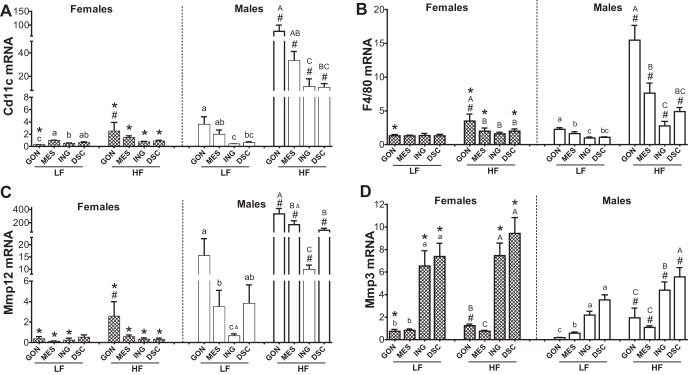
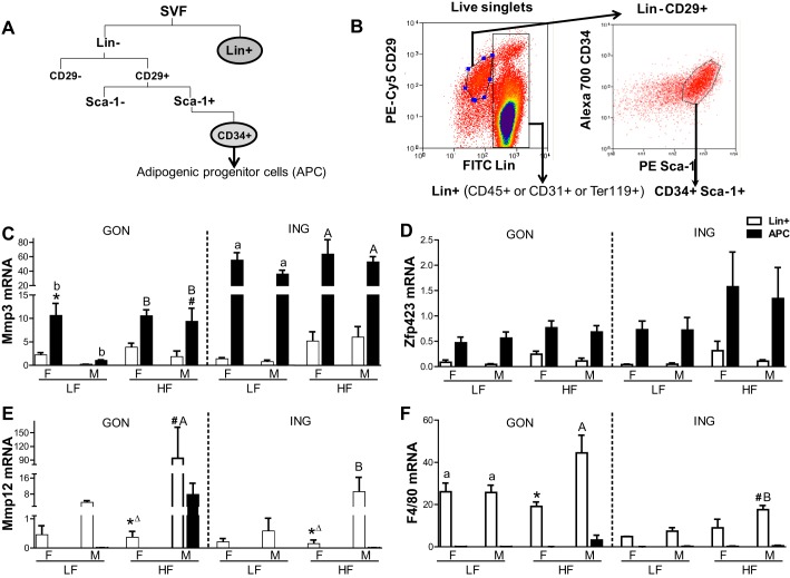
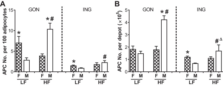
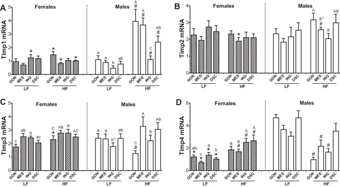
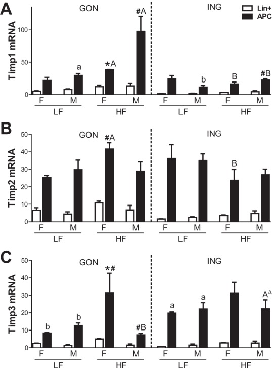
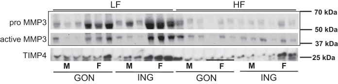
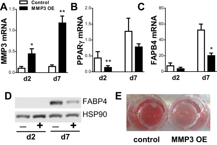
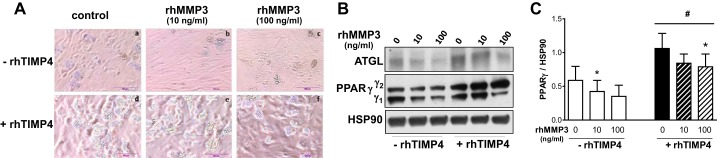
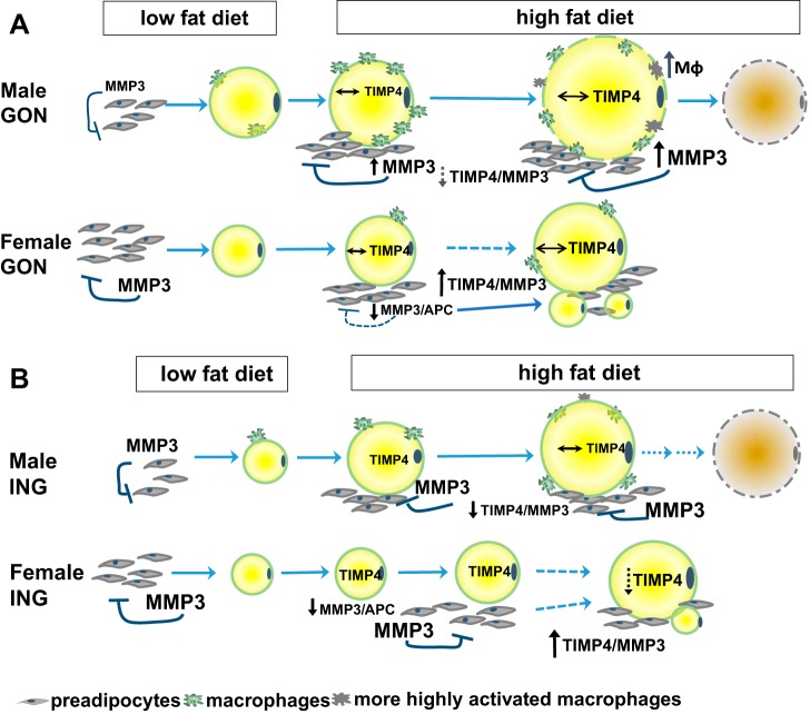
Increased adipocyte size is hypothesized to signal the recruitment of adipose progenitor cells (APCs) to expand tissue storage capacity. To investigate depot and sex differences in adipose growth, male and female C57BL/6J mice (10 wk-old) were challenged with high-fat (HF) or low-fat (LF) diets (D) for 14 wk. The HFD increased gonadal (GON) depot weight by adipocyte hypertrophy and hyperplasia in females but hypertrophy alone in males. In both sexes, inguinal (ING) adipocytes were smaller than GON, and depot expansion was due to hypertrophy. Matrix metalloproteinase 3 (Mmp3), an antiadipogenic factor, and its inhibitor Timps modulate the extracellular matrix remodeling needed for depot expansion. Mmp3 mRNA was depot different (ING > GON), higher in females than males and mainly expressed in APCs. In males, HFD-induced obesity increased tissue and APC Mmp3 mRNA levels and MMP3 protein and enzymatic activity. In females however, HFD significantly decreased MMP3 protein without affecting its mRNA levels. MMP3 activity also decreased (significant in ING). Timp4 mRNA was expressed mainly in adipocytes, and HFD-induced obesity tended to increase the ratio of TIMP4 to MMP3 protein in females, whereas it decreased it in males. Overexpression of Mmp3 in 3T3-L1 preadipocytes or rhMMP3 protein added to primary human preadipocytes inhibited differentiation, whereas rhTIMP4 improved adipogenesis and attenuated the inhibitory effect of rhMMP3. These data suggest that HFD-induced obesity downregulates APC MMP3 expression to trigger adipogenesis, and adipocyte TIMP4 may modulate this process to regulate hyperplastic vs. hypertrophic adipose tissue expansion, fat distribution, and metabolic health in a sex- and depot-dependent manner.

Keywords: adipocyte size; adipose progenitor; hyperplasia; matrix metalloproteinase 3; tissue inhibitor of matrix metalloproteinase 4.


Figures

Fig. 1.
Fig. 1.
Sex-, depot-, and diet-dependent effects on expression of Mmp12, Cd11c, F4/80, and matrix metalloproteinase 3 (Mmp3) in adipose tissue. Adipose tissues were obtained from male (open bars) and female (cross-hatched bars) mice after 14 wk of diet [n = 8–9 for all depots except dorsal (DSC; n = 5)] and expression levels of Cd11c (A), F4/80 (B), Mmp12 (C), and Mmp3 (D) were measured. Data for each depot were first analyzed by ANOVA for sex, diet, and their interaction, followed by post hoc t-tests if F-tests were significant. Sex effects for each gene (P < 0.01) were significant for all depots, except for F4/80 in inguinal (ING) for Mmp3 in mesenteric (MES). The diet effect was significant (P < 0.001) for all depots, with the exception of Mmp3 in MES (P = 0.06) and ING and DSC (P < 0.05). Furthermore, the sex × diet interaction was also significant for Cd11c, F4/80 (except for ING), and Mmp3 in gonadal (GON) and MES. *Significant sex effect by post hoc t-test; #significant diet effect by post hoc t-test (P < 0.05). Differences among depots within each sex and diet group were determined by 1-way ANOVA with repeated measures using paired values, followed by post hoc comparison of means by paired t-tests; F values were significant for all sex and diet groups. Bars with different letters show depot differences within diet groups [A,B,C,Dhigh fat (HF); a,b,c,dlow fat (LF; P < 0.05); BΔ, P = 0.06, vs. A (GON); cΔ, P = 0.06, compared with b (MES)].
Fig. 2.
Fig. 2.
Mmp3 is expressed in adipose progenitor cells (APCs); Mmp12 in Lin+ cells. A: scheme of the fluorescence-activated cell sorting (FACS) strategy. B: illustration of the gating strategy with representative example. Gene expression [quantitative PCR (qPCR)] measured in sorted cell populations [Mmp3 (C), Zfp423 (D), Mmp12 (E), and F4/80 (F); n = 3/group (2 chow fed and 1 LF fed in LF group]. *Sex effect (P < 0.05) by t-test; #diet effect by t-test. Depot differences within diet groups were indicated by different letters. ΔP < 0.06 by t-test.
Fig. 3.
Fig. 3.
APC numbers in GON and ING: depot, diet, and sex differences. Calculation of the size of APC pool expressed as APC number per 100 adipocytes (A) and per depot (B). When sex, diet, or sex × diet interactions were significant by MANOVA within depots, post hoc t-tests (P ≤ 0.05) were used to assess group differences: *Sex effects; #diet effects (P < 0.05); #ΔP = 0.06. The main effect of depot was also significant (not shown). Values are expressed as means ± SE; n = 3 for each group. F, females; M, males.
Fig. 4.
Fig. 4.
Sex-, depot-, diet-dependent effects on gene expression of tissue inhibitor of MMPs (Timps) in adipose tissue. qPCR [Timp1 (A), Timp2 (B), Timp3 (C), and Timp4 (D)] performed on the same samples and analyzed as described in Fig. 1. Bars with difference letters show depot differences within diet (P < 0.01). *Sex difference within diet, P < 0.05; #diet effect, P < 0.05 by post hoc t-test when main effects or interactions significant. BΔ, P = 0.07, compared with GON; bΔ, P = 0.06, compared with GON.
Fig. 5.
Fig. 5.
Expression of Timp 1, 2, and 3 in sorted cell populations. Timp mRNA levels were measured by qPCR in the cells described in Fig. 4. Symbols are as indicated in the legend to Fig. 4. Δ indicates P < 0.09 by T-test.
Fig. 6.
Fig. 6.
MMP3 and TIMP4 protein expression in gonadal and inguinal adipose tissues. Western blot data presented are based on loading same weight of adipose tissue/sample.
Fig. 7.
Fig. 7.
Overexpression of MMP3 in 3T3-L1 cells inhibits adipogenesis. Deoxycycline was removed at confluence, inducing overexpression of MMP3. AD: expression levels of MMP3 (A) and markers of differentiation peroxisome proliferator-activated receptor-γ (PPARγ; B), fatty acid-binding protein 4 (FABP4) mRNA (C), and FABP4 protein (D). E:Oil Red O staining of control and overexpressed adipocytes 7 days postdifferentiation. *P < 0.05 and **P < 0.01; n = 4–5. HSP90, heat shock protein 90.
Fig. 8.
Fig. 8.
Addition of TIMP4 blocks the antiadipogenic effects of MMP3 in human preadipocytes. Two-day postconfluent human preadipocytes were differentiated with recombinant human (rh)MMP3 (0, 10, and 100 ng/ml) in the absence or presence of rhTIMP4 (500 ng/ml) and maintained until day 12 of differentiation. A: representative images of 4 independent experiments. B and C: protein expression levels of adipose triglyceride lipase (ATGL) and PPARγ were measured on day 12 of differentiation (B), and PPARγ protein levels relative to HSP90 were quantified (C); n = 4–5. *P < 0.05, effects of rhMMP3; #P < 0.05, effects of rhTIMP4.
Fig. 9.
Fig. 9.
Model for sex differences in adipose tissue growth in response to HF feeding. A: in female GON LF-fed mice, there is a larger pool of preadipocytes than in males. In male GON, the high-fat diet (HFD) increases number of APCs, and their progression toward differentiation is restrained by high MMP3 expression, creating an autocrine loop that restrains adipogenesis. As activated macrophages accumulate in the tissue, inflammatory cytokines from activated macrophages also may contribute to the inhibition of adipogenesis in a paracrine loop, and growth proceeds by hypertrophy of existing adipocytes, which become dysfunctional and die. In female GON, the HFD causes a decrease in MMP3 expression that triggers an increase in adipocyte number by APC differentiation, but the number of APCs remains constant, presumably as they also proliferate. Although the HFD did not significantly affect TIMP4 protein at the time point studied, the ratio of TIMP4 to MMP3 protein tended to increase in females but decreased in males (as also observed in ING; B). We postulate that TIMP4 may serve as a signal of adipocyte enlargement that further inhibits MMP3 secreted by preadipocytes, permitting the differentiation of preadipocytes and the appearance of new, smaller adipocytes (hyperplasia) in females that can fill over time, resulting in healthy tissue expansion. B: In LF-fed mice, ING adipocytes are smaller than GON in both sexes. With HF feeding in males, tissue growth occurs by hypertrophy, and high MMP3 in preadipocytes restrains their recruitment. In females, HFD significantly decreased MMP3 protein expression, and TIMP4 did not change; the resulting higher TIMP4/MMP3 may contribute to the effect of the HFD to lower MMP3 activity (per mg tissue), which would facilitate their maturation and lipid filling. The resulting hyperplastic expansion is evidenced by an initial accumulation of new, smaller adipocytes (illustrated) that eventually fill with lipid. The timing of these changes likely varies among animals that have different-sized adipocytes at baseline and, therefore, depend on the time of overfeeding or a magnitude of the HFD challenge.

References

    1. Alexander CM, Selvarajan S, Mudgett J, Werb Z. Stromelysin-1 regulates adipogenesis during mammary gland involution. J Cell Biol 152: 693–703, 2001. doi:10.1083/jcb.152.4.693. - DOI - PMC - PubMed
    1. Cacicedo JM, Benjachareonwong S, Chou E, Yagihashi N, Ruderman NB, Ido Y. Activation of AMP-activated protein kinase prevents lipotoxicity in retinal pericytes. Invest Ophthalmol Vis Sci 52: 3630–3639, 2011. doi:10.1167/iovs.10-5784. - DOI - PMC - PubMed
    1. Chavey C, Mari B, Monthouel MN, Bonnafous S, Anglard P, Van Obberghen E, Tartare-Deckert S. Matrix metalloproteinases are differentially expressed in adipose tissue during obesity and modulate adipocyte differentiation. J Biol Chem 278: 11888–11896, 2003. doi:10.1074/jbc.M209196200. - DOI - PubMed
    1. Chun TH, Hotary KB, Sabeh F, Saltiel AR, Allen ED, Weiss SJ. A pericellular collagenase directs the 3-dimensional development of white adipose tissue. Cell 125: 577–591, 2006. doi:10.1016/j.cell.2006.02.050. - DOI - PubMed
    1. Faust IM, Johnson PR, Stern JS, Hirsch J. Diet-induced adipocyte number increase in adult rats: a new model of obesity. Am J Physiol Endocrinol Metab 235: E279–E286, 1978. - PubMed

Publication types

MeSH terms